Devices for covering ultrasound probes of ultrasound machines

a technology of ultrasound machine and probe, applied in the field of ultrasound imaging, can solve the problems of patient discomfort, adversely affecting the workflow efficiency of the technician performing the procedure, and the spread of ultrasonic gel into unwanted areas once applied

Inactive Publication Date: 2008-06-12
SIEMENS MEDICAL SOLUTIONS USA INC
View PDF20 Cites 111 Cited by
  • Summary
  • Abstract
  • Description
  • Claims
  • Application Information

AI Technical Summary

Problems solved by technology

The expenditures of time and effort associated with applying and removing the ultrasonic gel before and after the ultrasound procedure can adversely impact the workflow efficiency of the technician performing the procedure.
Applying and removing the ultrasonic gel can cause patient discomfort when the body surface is sensitive from an injury, a surgical procedure, etc.
Moreover, ultrasonic gel can spread into unwanted areas once applied.
Also, the quality of the ultrasound image can be adversely affected when an excessive amount of ultrasonic gel is applied to the interior of the sheath or the body surface of the patient.

Method used


Image

  • Devices for covering ultrasound probes of ultrasound machines
  • Devices for covering ultrasound probes of ultrasound machines
  • Devices for covering ultrasound probes of ultrasound machines

Examples


Embodiment Construction

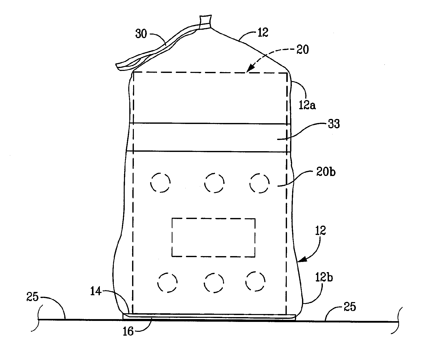
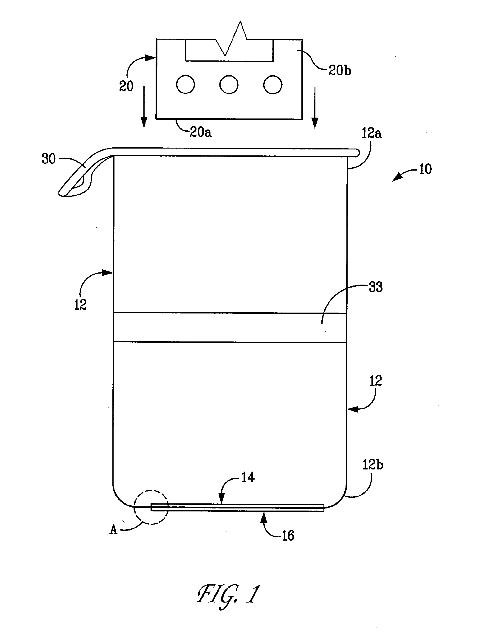
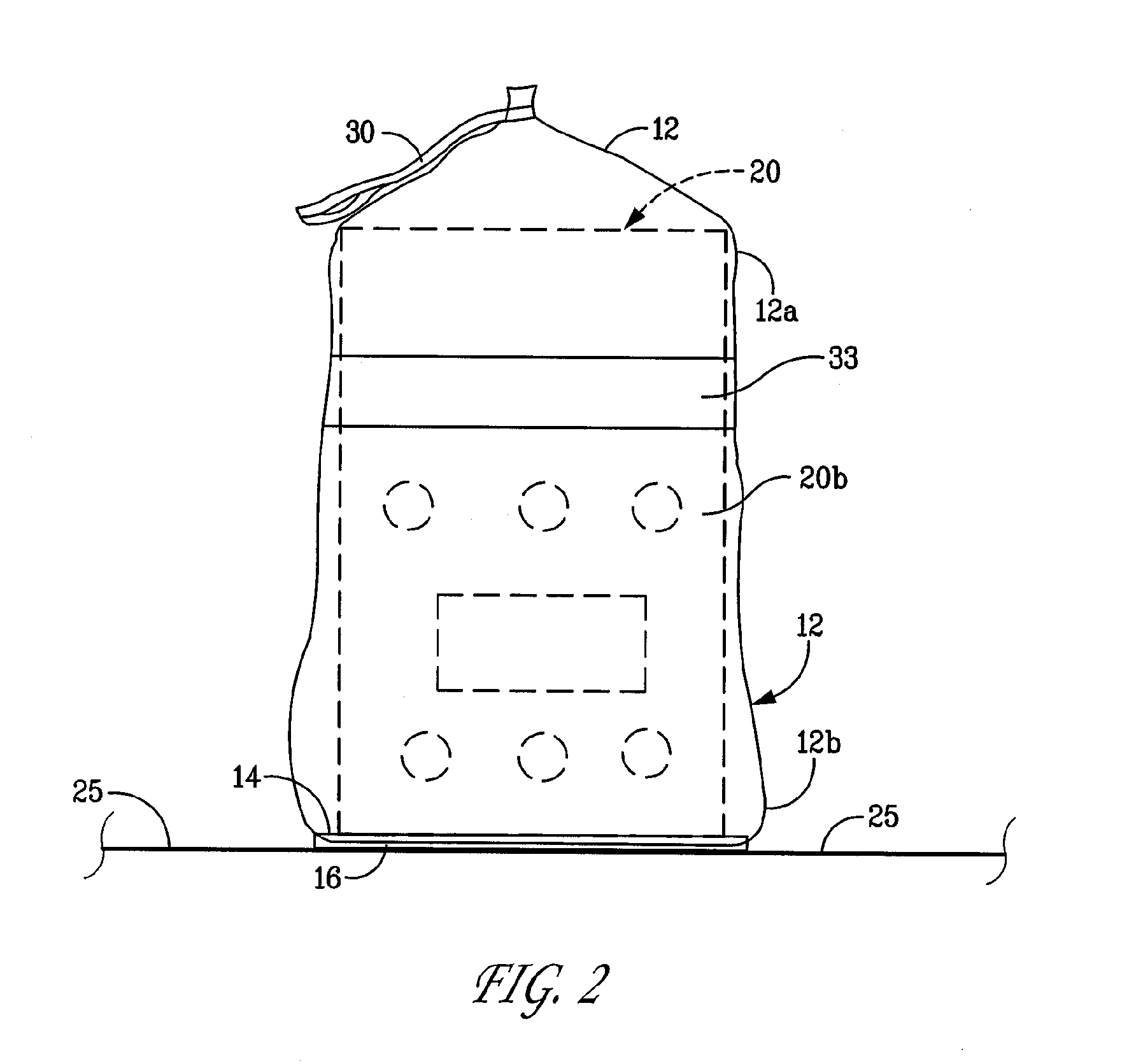
[0026]FIGS. 1-3 depict a preferred embodiment of a device 10 for covering an ultrasound probe 20 of an ultrasound machine. The device comprises a sheath 12, a first ultrasonic coupler 14, and a second ultrasonic coupler 16. The term “ultrasonic coupler,” as used throughout the specification and claims, refers to a device that transmits or transfers acoustical energy at ultrasonic frequencies, e.g., approximately 20 kHz to approximately 20 MHz or higher, with minimal attenuation. Typical imaging frequencies are in the range of approximately 1 MHz to approximately 20 MHz. An ultrasonic coupler can be formed from, for example, a liquid, a gel, or another substantially soft material, such as a pliable polymer matrix. Alternatively, an ultrasonic coupler can be formed from a water-containing polymer that is solidified by various methods.

[0027]The sheath 12 receives the ultrasound probe 20, as shown in FIG. 2. The ultrasound probe 20 is a wireless ultrasound probe that communicates over a...


PUM


Abstract

Preferred embodiments of devices for covering an ultrasound probe can include a membrane, such as a sheath, having one or more ultrasonic couplers attached thereto for transferring ultrasonic energy between the ultrasound probe and a body surface of a patient.

Description

FIELD OF THE INVENTION[0001]The present invention relates generally to ultrasound imaging. More particularly, the invention relates to devices and methods for covering ultrasound probes of ultrasound machines, and acoustically coupling the ultrasound probes to body surfaces of a patient.BACKGROUND OF THE INVENTION[0002]Ultrasound images are typically generated by placing an ultrasound probe of an ultrasound machine in contact with a body surface of a patient, and directing high-frequency acoustical energy at the body surface. The ultrasound probe senses the acoustical energy reflected from the body surface and the underlying portion of the patient's body. The reflected energy is converted to an electrical signal by the ultrasound probe. The signal is relayed to a processing and display unit of the ultrasound machine, where the signal is transformed into an image. The signal can be relayed by a cable connected to the ultrasound probe and the ultrasound machine, or by wireless means s...

Claims


Application Information

Patent Timeline
Patent Type & Authority Applications(United States)
IPC IPC(8): A61B8/00
CPCA61B8/4281A61B8/4472A61B8/4422
Inventor WEYMER, RAYMOND F.CANNON, MICHAEL G.ENGLE, LAWRENCE A.KLESSEL, JODI SCHWARTZLANNUTTI, ANTHONY P.RANDALL, KEVIN S.URBANO, JOSEPH A.
Owner SIEMENS MEDICAL SOLUTIONS USA INC
Why Eureka
Try Eureka