Arrangement and imaging of biological samples

Inactive Publication Date: 2008-06-26
THE BOARD OF TRUSTEES OF THE LELAND STANFORD JUNIOR UNIV
View PDF25 Cites 27 Cited by
  • Summary
  • Abstract
  • Description
  • Claims
  • Application Information

AI Technical Summary

Benefits of technology

[0010]According to other example embodiments, the present invention is directed to a system for imaging a biological sample. In one such embodiment, a system includes a fluorescence microscope arrangement to image a tissue specimen that is sliced and arranged in an array (e.g., on a glass microscope slide, a glass coverslip or other solid substrate). Data corresponding to the ordering of the tissue slices is stored in a three-dimensional database

Problems solved by technology

IFM, IEM and XFP imaging methods present challenges, however, and many important aspects of cell and tissue molecular architecture are difficult to explore using previous approaches.
For instance, while XFPs have provided unique opportunities for imaging live cell dynamics, they require the expression of transgenes and are thus impractical for the study of human clinical specimens.
XFPs also entail substantial probability of perturbing the molecular architectures of inte

Method used


Image

  • Arrangement and imaging of biological samples
  • Arrangement and imaging of biological samples
  • Arrangement and imaging of biological samples

Examples


Embodiment Construction

[0022]The present invention is believed to be applicable to a variety of different types of processes, devices and arrangements for imaging, and in particular, to approaches to serially imaging slices of a biological specimen for three-dimensional views of the specimen. While the present invention is not necessarily so limited, various aspects of the invention may be appreciated through a discussion of examples using this context.

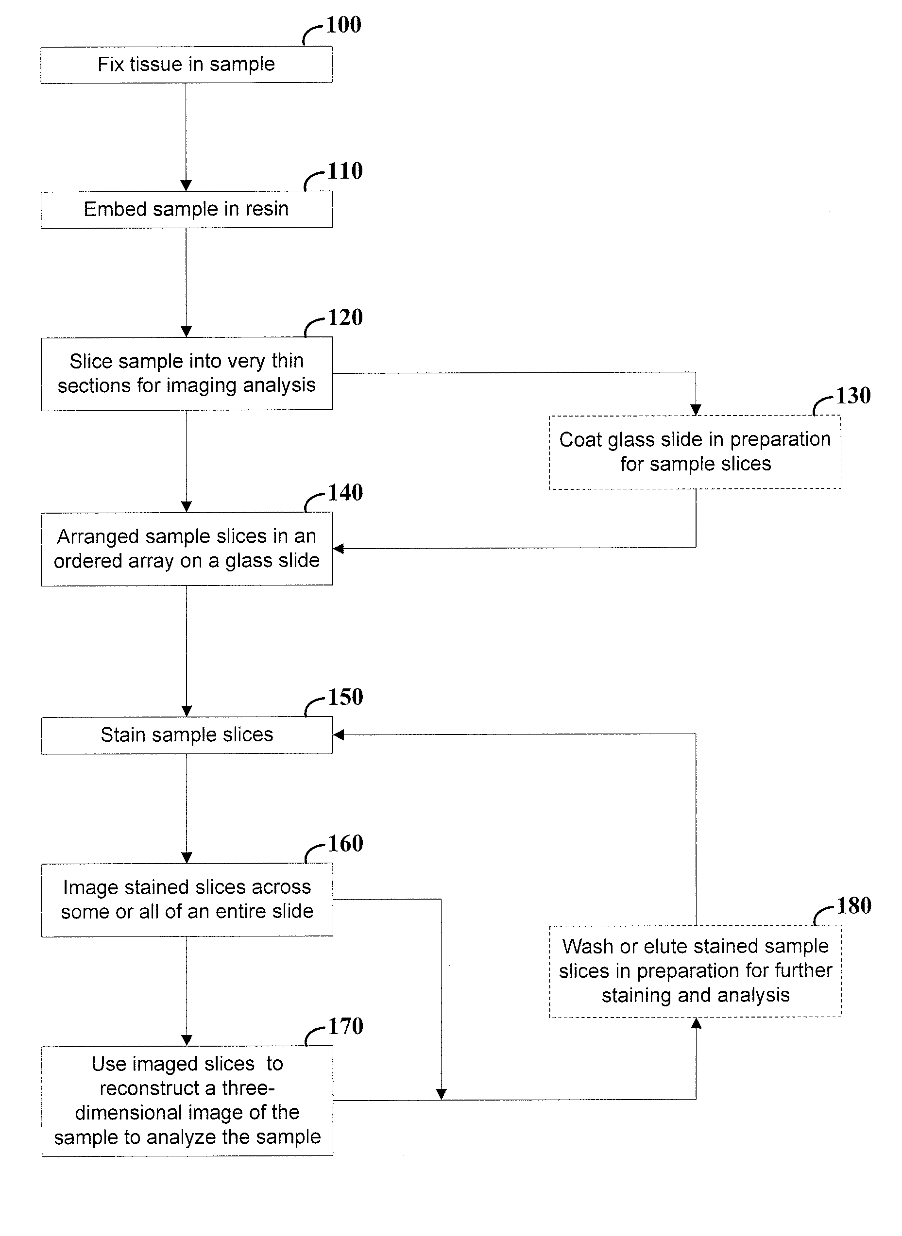
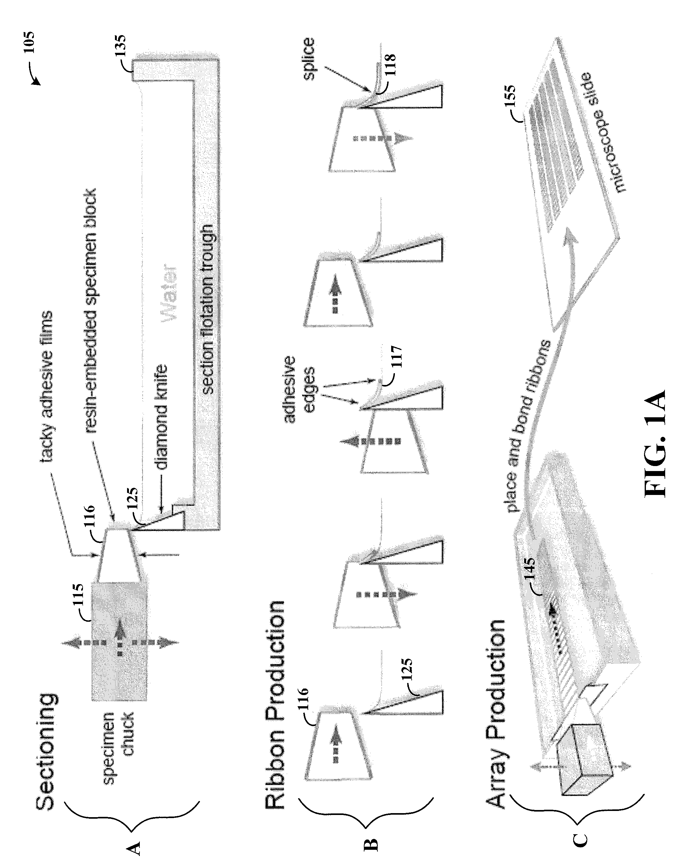
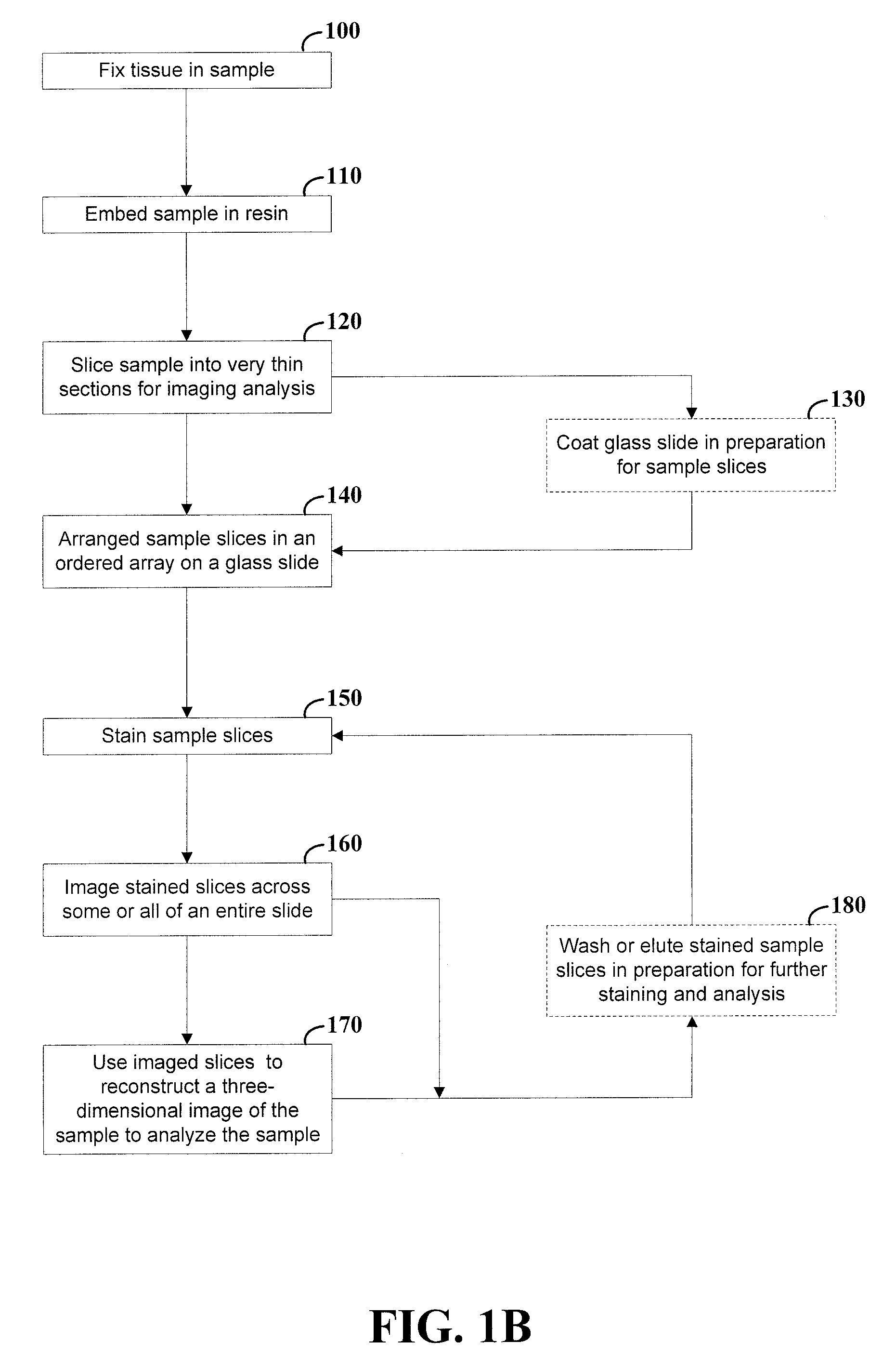
[0023]According to an example embodiment of the present invention, the molecular architecture of cells and tissues are analyzed by physically sectioning a sample (e.g., a specimen) to make very thin sections of the sample. The thickness of the sections varies with different embodiments; in some applications, the section thickness is in a range of tens to hundreds of microns, and in other applications, the section thickness is similar to the thickness of sections used for electron microscopy (e.g., in a range of about 50-70 nm). A large number of the thin se...


PUM


Abstract

High-resolution three-dimensional imaging of a specimen is facilitated. According to an example embodiment of the present invention, a series of very thin slices from a specimen are serially and robustly arranged on an imaging device such as a microscope slide. The slices are imaged and the images are used to reconstruct a three-dimensional image having high resolution at depths into the specimen. The serial arrangement of the slices facilitates the proper ordering of images for reconstruction. Further, the robust nature of the slice arrangement facilitates treatment of the slices and, in some applications, multiple treatments with corresponding imaging sequences for each treatment. Various embodiments are directed to methods and arrangements for three-dimensional characterization of biological specimen and to data that is accessible and/or executable by a computer for linking different images together in order to characterize such biological specimen in three dimensions.

Description

RELATED PATENT DOCUMENTS[0001]This patent document claims the benefit, under 35 U.S.C. § 119(e), of U.S. Provisional Patent Application No. 60 / 876,046 filed on Dec. 20, 2006 and entitled: “Arrangement and Imaging of Biological Samples,” which is fully incorporated herein by reference.FIELD OF THE INVENTION[0002]The present invention relates generally to the analysis of samples and, more particularly, to the analysis of arranged slices of biological samples.BACKGROUND OF THE INVENTION[0003]Imaging has played an important part in the analysis of specimen, and in particular, in the analysis of biological specimen. The tools most commonly used for imaging biological molecular architectures are immunofluorescence microscopy (IFM), immunoelectron microscopy (IEM) and genetically encoded fluorescent protein markers and reporters (XFPs). These powerful and widely used tools have led to discoveries that point to the dependence of cell and tissue function upon the very finest details of molec...

Claims


Application Information

Patent Timeline
IPC IPC(8): G06K9/00
CPCG06K9/00134G02B21/367G06T7/50H04N13/204G06V20/693G01N1/30G02B21/365G06T7/0012
Inventor MICHEVA, KRISTINA D.SMITH, STEPHEN J.
Owner THE BOARD OF TRUSTEES OF THE LELAND STANFORD JUNIOR UNIV
Why Eureka
Try Eureka