
Method for Adaptive Radiation Treatment of Breast Tissue Surrounding a Cancer Resection Cavity of Arbitrary Shape

a radiation treatment and breast tissue technology, applied in radiation therapy, radiation therapy, x-ray/gamma-ray/particle irradiation therapy, etc., can solve the problem that isotopes cannot be controlled in any practical sense, and achieve the effect of improving radiation treatment accuracy, less discomfort and trauma, and increasing cable length

Active Publication Date: 2008-06-26
XOFT INC
View PDF10 Cites 13 Cited by
  • Summary
  • Abstract
  • Description
  • Claims
  • Application Information

AI Technical Summary

Benefits of technology

[0012]By the procedures of the present invention, the need for external imaging is eliminated and radiation treatment is adapted to target tissues surrounding arbitrarily shaped cavities with local dose control. Such a procedure may be applied intraoperatively, or may be applied post-operatively over a fractionated course of treatments.
[0014]A treatment plan is calculated starting from three dimensional applicator / cavity mapping data and the treatment plan is executed preferably using the same applicator, the same applicator position and the same ionizing radiation sources as are used in the mapping, controlled by a processor. In a sense, the mapping step of the inventive procedure comprises a low-dose “trial treatment”, providing accurate, useful data, with verification of the effects of each of the radiation sources and dwell positions in the applicator guides, including areas where radiation needs to be modulated or is not to be delivered at all, thus enabling optimized planning and enabling accurate execution of the treatment plan subsequently.
[0016]Rotating x-ray sources are well known, but generally the methods used to effect rotation are directed to large, external x-ray sources, not miniature sources. Miniature sources are usually connected to a high-voltage electrical and / or optic cable, more or less in the manner of a source on the end of a catheter. The problems which would be encountered in making rotating high-voltage or optic connections are substantial. It is far easier to use solid cable connections and twist the cable rotationally, perhaps increasing cable length somewhat to accommodate sufficient accumulated rotation, or to resort to oscillatory rotation such that the cable is first wound and then unwound within a more limited range, but still sufficient to accommodate the rotational requirements of the treatment plan.
[0017]In order to use a directional source effectively in an arbitrary cavity, the cavity must be established in a configuration that can be maintained for the duration of the radiotherapy prescription, and then mapped such that optimized dose planning can proceed (or re-mapping can take place, accompanied by recalculating the delivery procedure or by real-time monitoring of dose received and real-time adjustment accordingly). If the radiotherapy prescription is carried out intraoperatively, the apparatus and procedure of the invention not only greatly improve accuracy in radiation treatment, but also subjects the patient to far less discomfort and trauma as compared to typical procedures. By the disclosed procedure the patient, while anesthetized in the operating room, is operated on to remove the tumor, the tumor is investigated as to pathology, by a very rapid process, decision is made as to any need for further excision (which is done if needed), the physician prescribes radiation dosage for a volume of tissue surrounding the excision cavity, the shape of the cavity is mapped and recorded by internal measurements, calculations of radiation to be delivered at various sites in the cavity are made by use of the mapping data to thus prepare a treatment plan, and the treatment plan is carried out, all without waking or moving the patient, and all within a reasonable time. In the fractionated procedure, on the other hand, the patient is sent home between the various steps and fractions as is either convenient or efficacious for the duration of the radiotherapy.

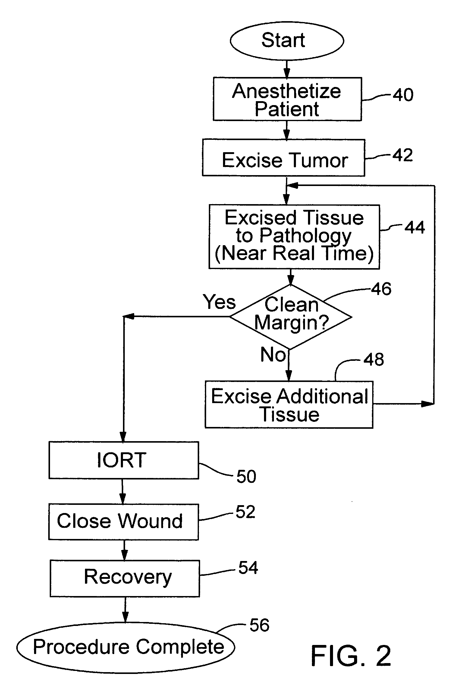
Problems solved by technology

In contrast, isotopes cannot be controlled in any practical sense.

Method used


Image

  • Method for Adaptive Radiation Treatment of Breast Tissue Surrounding a Cancer Resection Cavity of Arbitrary Shape
  • Method for Adaptive Radiation Treatment of Breast Tissue Surrounding a Cancer Resection Cavity of Arbitrary Shape
  • Method for Adaptive Radiation Treatment of Breast Tissue Surrounding a Cancer Resection Cavity of Arbitrary Shape

Examples


Embodiment Construction

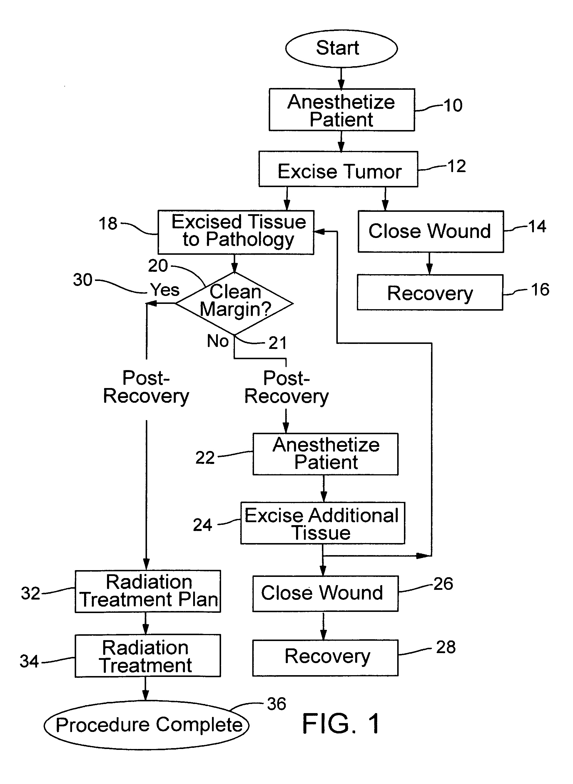
[0039]FIG. 1 shows typical brachytherapy practice where pathology of the excised tissue is performed post-operatively, particularly after a tumor has been excised from the breast of a patient; FIG. 1 represents prior art as well as the invention, with further details set out below. The patient is anesthetized as indicated at 10, and the tumor is excised as indicated at 12 in the drawing. Following excision, the surgical wound is closed as noted at 14, and the patient recovers as shown in the block 16 and is sent home. Optionally, a place holding balloon in accordance with the teaching of co-pending provisional patent application No. 60 / 851,687 may be implanted in the resection cavity before closing, in anticipation of subsequent radiotherapy following satisfactory pathology. The specification of application No. 60 / 851,687 is included herein by reference in its entirety.

[0040]Meanwhile, the excised tissue is sent to pathology as shown at 18, and the pathology of the tissue is determi...


PUM


Abstract

Radiation treatment methods for breast cancer following resection, both intraoperative and post operative, involve mapping preferably using an electronic x-ray source, and can include rotation of a direction source. By using a very rapid, near-instantaneous method of pathology of tissue margins following resection, the invention carries out intraoperative radiation treatment, preferably with the patient remaining anesthetized. Whether or not radiation is intraoperative, the invention preferably includes a mapping procedure using an electronic source that is progressed through the resection cavity while radiation dose is sensed at one or more points, to determine the shape of the resection cavity.

Description

[0001]This application is a continuation-in-part of application Ser. No. 10 / 464,160, filed Jun. 18, 2003, which is incorporated herein by reference.BACKGROUND OF THE INVENTION[0002]This invention concerns the treatment of breast cancer or otherwise-sited cancer, and especially an efficient brachytherapy procedure for radiation treatment of tissue surrounding an arbitrarily shaped cavity resulting from following surgical tumor excision. Current brachytherapy methods of treating cancer of the breast, as well as cancer found in other areas of the human body with the patient under anesthesia, the tumor is include surgical excision of the tumor (with some surrounding tissue) and then typically, after the surgical wound is closed, the patient is sent home pending determination of pathology of the excised tumor margin. Once clean margins are established, a radiation treatment plan is developed and the patient, in a series of later visits, is subjected to radiation treatment in the volume o...

Claims


Application Information

Patent Timeline
IPC IPC(8): A61N5/10
CPCA61N5/1015A61N5/1048A61N5/103
Inventor JERVIS, JAMES E.LOVOI, PAUL A.
Owner XOFT INC
Learn More