Apparatus and method for delivering a biocompatible material to a surgical site

a biocompatible material and surgical site technology, applied in the field of minimally invasive apparatus and methods for delivering biocompatible materials to surgical sites, can solve the problems of loss of mechanical strength, limited healing of damaged cartilage, and increased risk of injury to cartilage, so as to reduce or prevent the leakage of biocompatible materials

Inactive Publication Date: 2008-06-26
ZIMMER ORTHOBIOLOGICS
View PDF45 Cites 1 Cited by
  • Summary
  • Abstract
  • Description
  • Claims
  • Application Information

AI Technical Summary

Benefits of technology

[0007]Apparatus and method of delivering a biocompatible material to a surgical site that confines the biocompatible material to a desired location at the surgical site. The apparatus and method may also reduce or prevent the leakage of the biocompatible material to the surrounding tissue.

Problems solved by technology

In addition, as a person ages, articular cartilage loses mechanical strength, rendering the cartilage even more susceptible to trauma.
Because articular cartilage tissue is aneural, i.e., having few or no nerves, and avascular, i.e., having few or no blood vessels, the healing of damaged cartilage is limited.
While the use of hydrogels has generally been successful to effect joint repair, their use does have some drawbacks.
One such drawback is that while the hydrogel is in a liquid form, such as when delivering the hydrogel to a surgical site through the minimally invasive device, it has a relatively low viscosity.
Consequently, the hydrogel flows easily and is therefore difficult to contain at the treatment site.
Moreover, leakage of the hydrogel into or onto the tissue surrounding the surgical site may not be desirable in some surgical procedures.
As a result, the use of hydrogels to effect joint repair has been heretofore limited.

Method used


Image

  • Apparatus and method for delivering a biocompatible material to a surgical site
  • Apparatus and method for delivering a biocompatible material to a surgical site
  • Apparatus and method for delivering a biocompatible material to a surgical site

Examples


Embodiment Construction

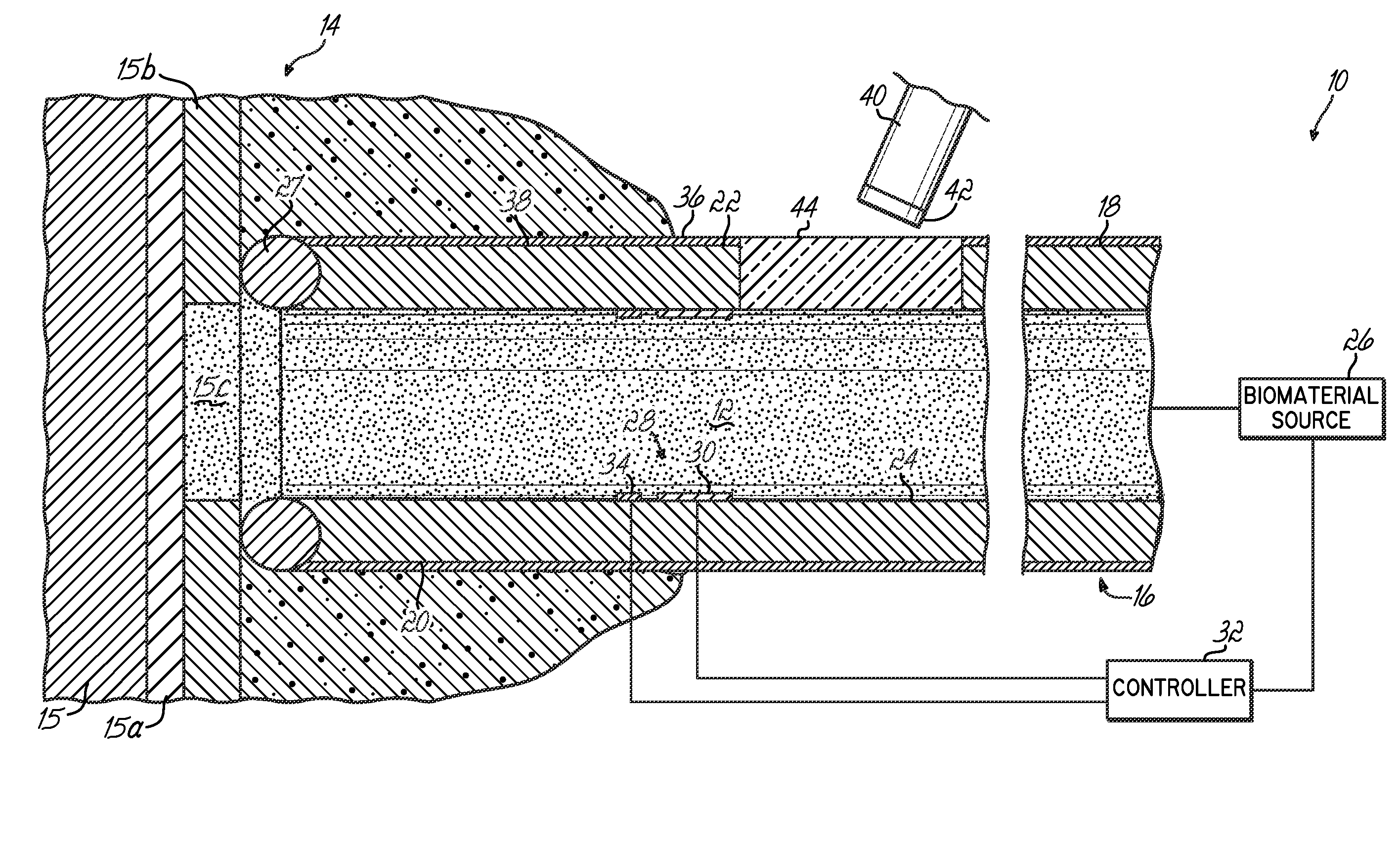
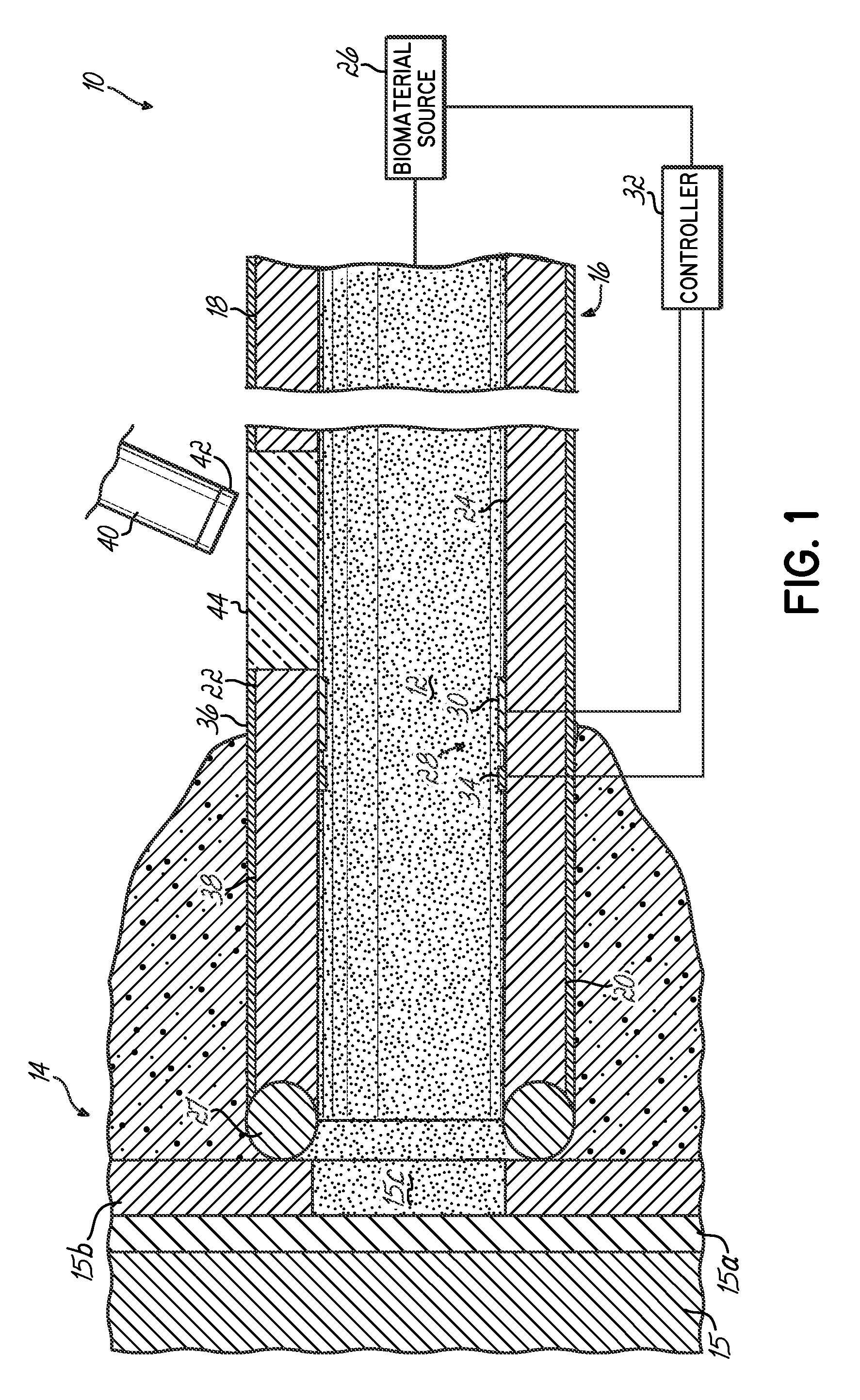
[0018]Referring to FIG. 1, one embodiment of a device 10 for delivering a curable biocompatible material 12 to a surgical site 14 is schematically illustrated. The device 10 may be used in various surgical procedures, including orthopedic surgical procedures to effect repair of the musculoskeletal system. In one embodiment, the device 10 may be used in minimally invasive procedures, such as arthroscopic and endoscopic procedures, to effect repair of a joint. In one embodiment, the device 10 may be used in orthopedic surgical procedures in general or to repair the cartilage within a diarthrodial joint, such as the knee. By way of example, as shown in FIG. 1, the surgical site 14 may include bone 15, subchondral bone 15a, and cartilage 15b wherein cartilage 15b includes a defect 15c that is to be repaired using embodiments of the invention. The invention, however, is not so limited, as those of ordinary skill in the art will recognize a wide range of surgical applications that may ben...


PUM


Abstract

A device for delivering a biocompatible material to a surgical site includes a cannula having proximal and distal portions and at least a first interior lumen disposed therebetween through which the biocompatible material is delivered. The device further includes an initiation member for initiating cross-linking of the biocompatible material while the biocompatible material is within the cannula. The cannula may include a heating element to thermally initiate cross-linking. Alternately, the cannula may include a second lumen for transmitting light from a light source. A movable blocking element controls the amount of light that passes into the first lumen. A method of delivering a curable biocompatible material to a surgical site includes positioning a distal portion of a cannula adjacent the surgical site and introducing the biocompatible material through a first lumen of the cannula. Cross-linking of the biocompatible material is then initiated while the biocompatible material is within the cannula.

Description

FIELD OF THE INVENTION[0001]An apparatus and method for surgical procedures, more particularly a minimally invasive apparatus and method for delivering a biocompatible material to a surgical site during various orthopedic procedures.BACKGROUND OF THE INVENTION[0002]The musculoskeletal system is subject to injury caused by traumatic events as well as by a number of diseases. Repair of connective tissue of the musculoskeletal system is commonly performed. By way of example, articular cartilage is a type of hyaline cartilage that lines the surfaces of the opposing bones in a diarthrodial joint (e.g., knee, hip, shoulder, etc.). Its primary function is to permit smooth, near frictionless movement during articulation between bones of the joint by providing a low-friction interface between the contacting cartilage surfaces of the joint. Articular cartilage is also load bearing, and serves to transmit and distribute compressive joint loads to the underlying subchondral bone.[0003]Articular...

Claims


Application Information

Patent Timeline
Patent Type & Authority Applications(United States)
IPC IPC(8): A61M5/00A61M31/00
CPCA61B17/00491A61B17/8836A61B17/3472
Inventor BEHRAVESH, ESSYYAO, JIAN Q.HUANG, XIAOZAPOROJAN, VICTORGAO, JIZONG
Owner ZIMMER ORTHOBIOLOGICS
Why Eureka
Try Eureka