
Osteochondral implant using a growth factor concentration gradient for repair of bone and cartilage tissue

a growth factor and concentration gradient technology, applied in the field of medical implants, can solve the problems of difficult treatment of damaged cartilage, difficult to remedy osteochondral defects, and difficult to repair, and achieve the effect of promoting the growth of both new bone and cartilage tissu

Inactive Publication Date: 2008-06-26
WARSAW ORTHOPEDIC INC
View PDF24 Cites 31 Cited by
  • Summary
  • Abstract
  • Description
  • Claims
  • Application Information

AI Technical Summary

Benefits of technology

[0022]The present invention overcomes the drawbacks of the prior art by providing a novel osteochondral implant that comprises a growth factor concentration gradient. The implant is inserted into a surgically prepared defect that extends from the surfaces of hyaline cartilage into sub-chondral bone tissue and promotes growth of both new bone and cartilage tissue.

Problems solved by technology

Trauma or frequent strain on articular joints can cause lesions to articular (hyaline) cartilage and fractures to the sub-chondral bone.
These lesions, often refereed to as osteochondral defects, are difficult to remedy.
Treatment of damaged cartilage is hindered by finding suitable implant materials and by hyaline cartilage's low reparative capabilities.
Hyaline cartilage defects heal in a manner where the repaired tissue lacks the structural and physical properties of healthy cartilage (fibrocartilage) and will degenerate over time.
This is due to the small population of type II collagen forming chondrocytes cells in hyaline cartilage and that hyaline cartilage is avascular, lacking nerves, blood vessels and lymphatic systems, which limits the healing and repair of cartilage defects.
This procedure can be compromised if the transplanted tissue is diseased, if there is damage to the collagen forming chondrocytes, or a wearing of the graft over time.
A drawback to this procedure is that there must be a fresh donor, the tissue must be stored at low temperatures and used within a month to ensure a greater than 50% cell viability.
However these techniques do little to promote the simultaneous growth of new articular cartilage and new bone tissue to repair osteochondral defects.
These methods provide temporary relief of pain but do little for the formation of new cartilage tissue.

Method used


Image

  • Osteochondral implant using a growth factor concentration gradient for repair of bone and cartilage tissue
  • Osteochondral implant using a growth factor concentration gradient for repair of bone and cartilage tissue
  • Osteochondral implant using a growth factor concentration gradient for repair of bone and cartilage tissue

Examples


embodiment

Implant—General Embodiment

[0045]FIG. 1

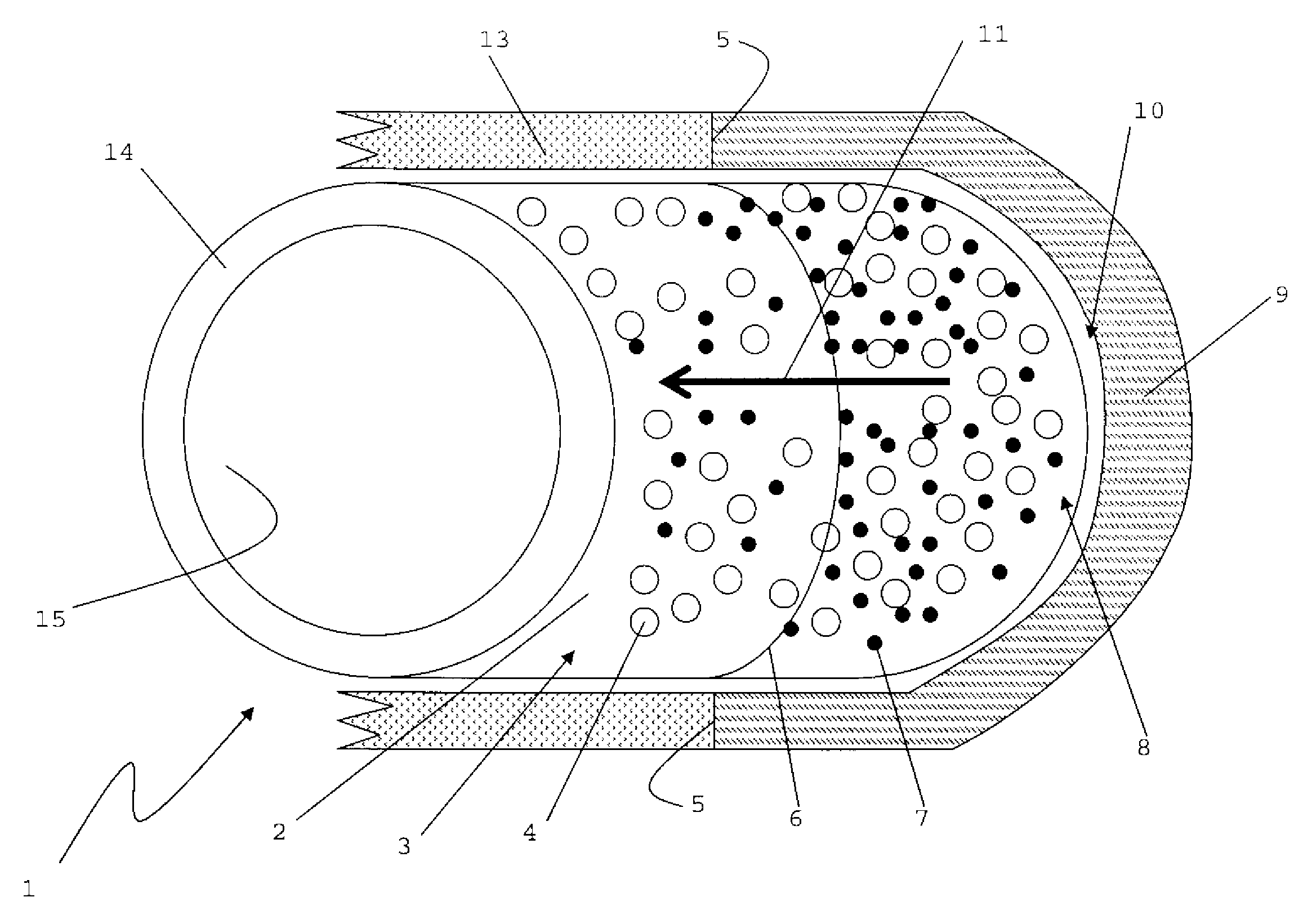
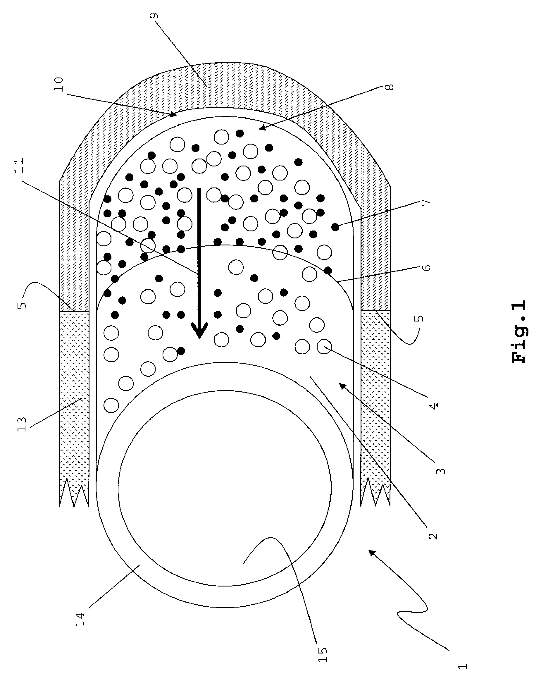
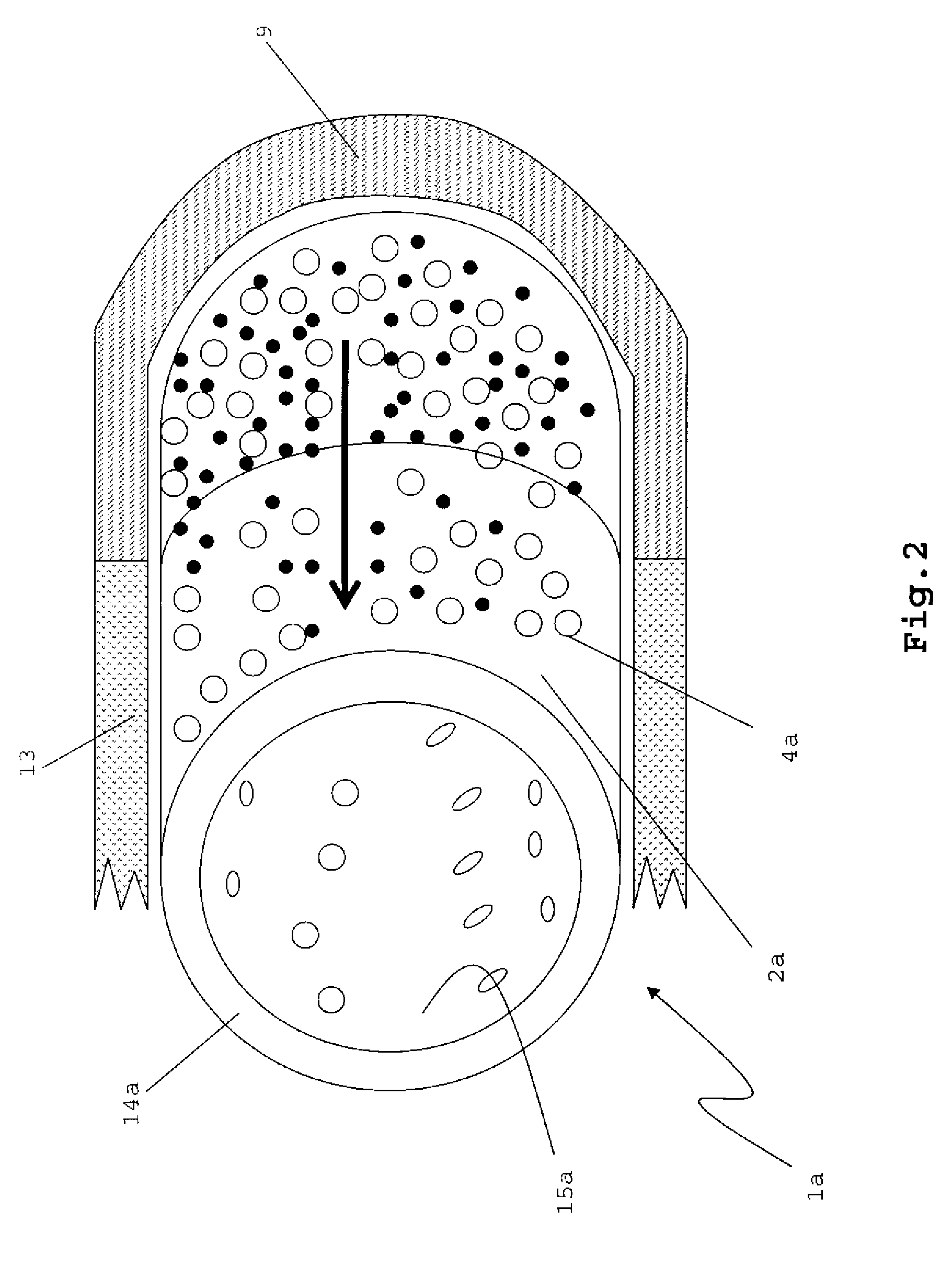
[0046]FIG. 1 shows a hollow cylindrical implant 1. The implant 1 may have a uniform size, with circular end surfaces that are connected by a single continuous cylindrical surface between the end surfaces. The cylindrical implant 1 may have a ring-like cross-section, with an external diameter from about 1 cm to about 1.5 cm, and an internal diameter that is from 40% to about 80% of the external diameter.

[0047]The implant 1 has an external surface 2 comprising a plurality of pores 4 and a therapeutically effective amount of a growth factor 7. An internal surface 15 of the implant 1 may be substantially non-porous, and may or may not have growth factors 7. The pores 4 may be, for example, from 50 microns to 1000 microns in size, and may or may not be substantially evenly disposed across the external surface 2.

[0048]One of ordinary skill in the art should appreciate that the implant 1 may be molded into various shapes and sizes, for example plug, to...


PUM


Abstract

An osteochondral device is provided comprising an implant having a growth factor concentration gradient. The implant can be porous with a higher concentration of growth factors dispersed in the portion of the implant where new bone tissue is needed and a lower concentration of growth factors dispersed in the portion of the implant where new cartilage tissue is needed.

Description

FIELD OF INVENTION[0001]The present invention generally relates to a medical implant and methods for promoting new bone and cartilage growth using a growth factor concentration gradient.BACKGROUND OF THE INVENTION[0002]Trauma or frequent strain on articular joints can cause lesions to articular (hyaline) cartilage and fractures to the sub-chondral bone. If the injury is not treated, it can progress into degenerative diseases, for example osteoarthritis, osteoporosis, Paget's disease, or osteohalisteresis. These lesions, often refereed to as osteochondral defects, are difficult to remedy. Treatment of damaged cartilage is hindered by finding suitable implant materials and by hyaline cartilage's low reparative capabilities.[0003]Whereas damaged sub-chondral bone is successfully healed by osteoclasts and osteblasts, cells that resorb and deposit bone minerals, hyaline cartilage lesions generally do not form new tissue. Hyaline cartilage defects heal in a manner where the repaired tissu...

Claims


Application Information

Patent Timeline
Patent Type & Authority Applications(United States)
IPC IPC(8): A61F2/28
CPCA61L27/54A61L2300/414A61L27/56
Inventor PECKHAM, STEVEN M.
Owner WARSAW ORTHOPEDIC INC
Learn More