Novel Anti-Plgf A Antibody

a technology of human anti-plgf and antibody, which is applied in the direction of peptides, drug compositions, and fused cells, can solve the problems of difficult to generate large amounts of human anti-human antibodies by conventional hybridoma technology, severely limit human therapy use, and large unknown molecular players, etc., to reduce tumor growth and tumor size, and inhibit p1gf

Inactive Publication Date: 2008-08-14
VLAAMS INTERUNIVERSITAIR INST VOOR BIOTECHNOLOGIE VZW +2
View PDF5 Cites 30 Cited by
  • Summary
  • Abstract
  • Description
  • Claims
  • Application Information

AI Technical Summary

Benefits of technology

[0017]Further uses of the antibodies and antigen-binding fragments of the present invention relate to the immunological detection of P1GF in human samples, as labeled targeting moieties in diagnostic methods and for the screening of compounds with an additive effect to P1GF inhibition in the treatment of cancer.
[0018]The present invention is based on th

Problems solved by technology

Evidence is emerging that distinct molecular mechanisms may mediate growth of blood vessels during pathological conditions, but the molecular players remain largely undetermined.
Antibodies generated in animals have characteristics which may severely limit their use in human therapy.
While the use of human monoclonal antibodies would address this limitation, it has proven difficult to

Method used


Image

  • Novel Anti-Plgf A Antibody
  • Novel Anti-Plgf A Antibody
  • Novel Anti-Plgf A Antibody

Examples


example 1

Production and Characterization of Murine Anti-Human P1GF Antibodies (16D3)

1. Immunization of Mice and Fusion

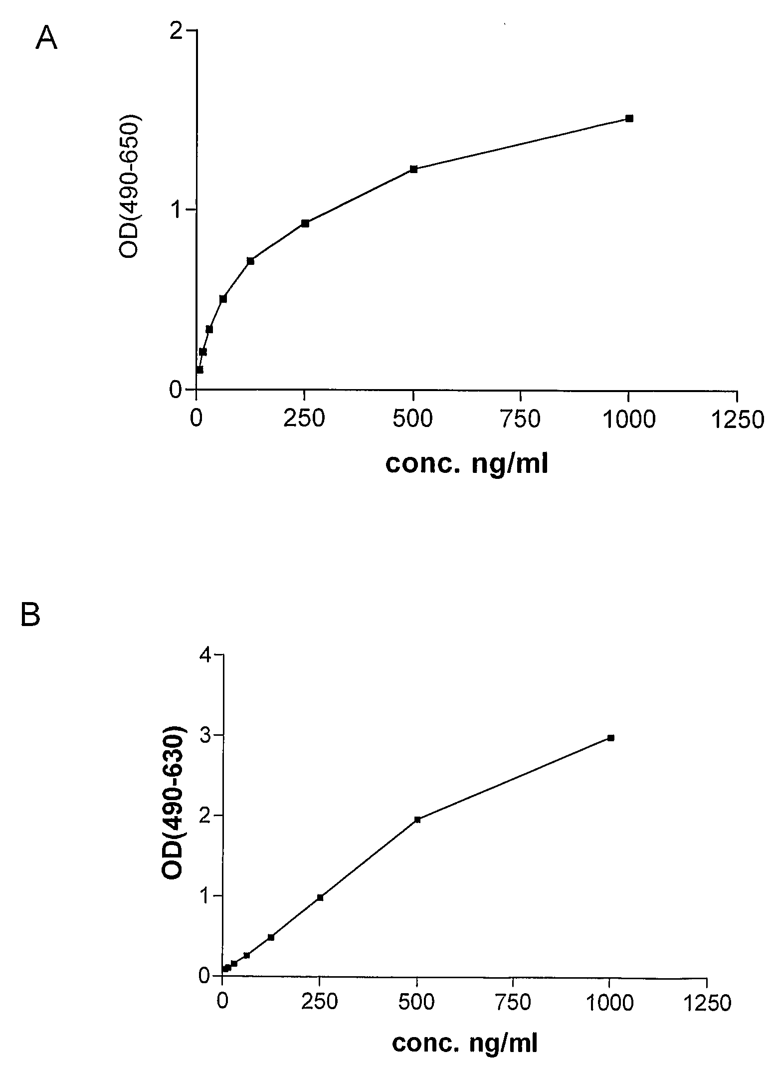
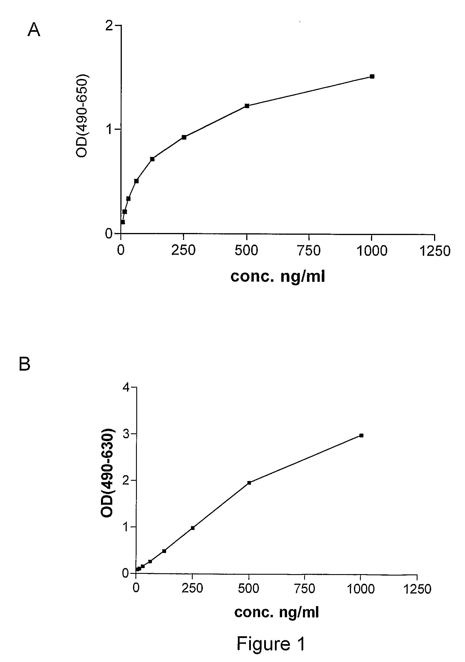
[0090]Monoclonal antibodies against human P1GF were produced essentially as described by Galfré and Milstein (Galfré F, Milstein C. Preparation of monoclonal antibodies: strategies and procedures. Method Enzymol 1981; 73: 3-46). Briefly, P1GF knock-out mice (Luttun et al., 2002, Biochem Biophys Res Comm 295(2):428-34, Carmeliet et al. (2001), Nat Med 7:575-593 or WO01 / 85796) were immunized by subcutaneous injection of 50 μg recombinant human P1GF-2 (produced in Pichia) in Complete Freund's adjuvant, followed two weeks later by subcutaneous injection with 50 μg recombinant human P1GF in incomplete Freund's adjuvant. Blood samples (about 100 μl) were collected from the tails of the mice after 10 days. The sera were tested for anti-huP1GF antibodies by ELISA using microtiter plates coated with human P1GF as capture, application of diluted sera (from 1 / 500 to 1 / 8000), and horsera...

example 2

Production and Characterization of Humanized Anti-Human P1GF Antibodies (16D3)

1. Construction of Humanized 16D3

Humanization of the Variable Regions

[0102]Mutations: The following mutations were made in order to humanize the variable parts of the mouse antibody 16D3:[0103]Heavy chain:[0104]I2V[0105]P9A[0106]K40A[0107]T111L[0108]Light chain:[0109]S5T[0110]S9D[0111]A15L[0112]K18R[0113]R22N[0114]L89V[0115]The mutations were made by using the QuickChange® Multi Site-Directed Mutagenesis Kit (Stratagene®). 1 to 5 primers containing the mutations could be used in 1 PCR. After transformation colonies were picked and the DNA sequenced to determine the clone with the most point mutations. This clone was taken to repeat this procedure till all the mutations were present in the sequence.

Linking the Variable Regions to a Human IgG Constant Region:

[0116]By performing a PCR the humanized variable parts of the heavy and light chain were obtained to which the appropriate restriction sites were added....

example 3

In Vivo Investigation of the Inhibition of Tumor Growth with Anti-PLGF Antibodies

1. General Description of Materials and Methods

Cell Culture

[0123]The murine pancreatic cell lines Panc02 and the human pancreatic cell line DanG are a kind gift of S. Rosewicz (Charité-Universitätsmedizin Berlin, Germany). The human breast MDA-MB, colon LOVO and melanoma Mel2a cell line were obtained from the American Type Culture Collection (ATCC; Manassas, Va., USA). All cells were cultured as recommended. Murine 16D3 anti-P1GF antibodies were used for all in vivo experiments, to avoid immune reaction to the antibody.

Animal Experiments

[0124]All animal procedures were fully approved by the institutional animal care and use committee. Female NMRI nu / nu mice were housed under semi-sterile conditions and were routinely used at 8-10 weeks of age (approximate weight, 25 g). For the preparation of tumor source, tumor cells were trypsinized to prepare single-cell suspension, washed with PBS. The centrifuged p...


PUM


Abstract

The present invention provides novel monoclonal antibodies directed to P1GF and fragments and derivatives thereof, more particularly to humanized antibodies and fragments thereof for use in the treatment and/or prevention of pathological angiogenesis.

Description

FIELD OF THE INVENTION[0001]The present invention relates to novel antibodies and fragments and derivatives thereof particularly suited for the inhibition of angiogenesis in pathological conditions. The invention also relates to cell lines producing the specific antibodies. The present invention provides pharmaceutical compositions comprising the antibodies, fragments and / or derivatives of the invention and provides methods of preventing and treating angiogenesis and / or vascular leakage where these are undesired such as in tumour formation, diseases of the eye, pulmonary hypertension and inflammatory disorders and methods of preventing and treating bone disorders such as osteoporosis.BACKGROUND OF THE INVENTION[0002]Abnormal blood vessel formation contributes to the pathogenesis of numerous diseases with high morbidity and mortality. Elucidation of the mechanisms underlying vascular growth might allow the development of therapeutic strategies to stimulate vascular growth in ischemic...

Claims


Application Information

Patent Timeline
IPC IPC(8): A61K39/395C07K16/18G01N33/567A61P43/00A01K67/027C12N5/06
CPCA61K2039/505C07K16/22A61K45/06A61K39/3955C07K2317/24A61P11/00A61P19/00A61P27/02A61P29/00A61P35/00A61P41/00A61P43/00A61P7/04A61P9/00A61P9/12A61P9/14C07K16/18C07K16/00
Inventor STASSEN, JEAN-MARIECARMELIET, PETERCOLLEN, DESIRE
Owner VLAAMS INTERUNIVERSITAIR INST VOOR BIOTECHNOLOGIE VZW
Why Eureka
Try Eureka