Cytotoxicity mediation of cells evidencing surface expression of CD44

a cytotoxicity and surface expression technology, applied in the field of cancer diagnosis and treatment, can solve the problems of insufficient evidence, inconsistent findings, and increased likelihood of tumor invasiveness and induction of angiogenesis through the ecm

Inactive Publication Date: 2007-03-13
F HOFFMANN LA ROCHE & CO AG
View PDF22 Cites 15 Cited by
  • Summary
  • Abstract
  • Description
  • Claims
  • Application Information

AI Technical Summary

Benefits of technology

[0042]In the preventative in vivo model of human breast cancer, H460-16-2 treatment was significantly (p<0.0001) more effective in suppressing tumor growth during the treatment period than an isotype control antibody, which was identical to H460-16-2 in structure and size but incapable of binding MB-231 cells. At the end of the treatment phase, mice given H460-16-2 had tumors that grew to only 1.3 percent of the control group. During the post treatment follow-up period, the treatment effects of H460-16-2 were sustained and the mean tumor volume in the treated groups continued to be significantly smaller than controls until the end of the measurement phase. Using survival as a measure of antibody efficacy, it was estimated that the risk of dying in the H460-16-2 treatment group was about 71 percent of the antibody buffer control group (p=0.028) at 70 days post-treatment. These data demonstrated that H40-16-2 treatment conferred a survival benefit compared to the control-treated groups. H460-16-2 treatment appeared safe, as it did not induce any signs of toxicity, including reduced body weight and clinical distress. Thus, H460-16-2 treatment was efficacious as it both delayed tumor growth and enhanced survival compared to the control-treated groups in a well-established model of human breast cancer.
[0044]In the post-treatment period, H460-16-2 showed a significant survival benefit (p<0.02) as the risk of dying in the H460-16-2 group was about half of that in the isotype control antibody group at >70 days after treatment. The observed survival benefit continued past 120 days post-treatment where 100 percent of the isotype control and cisplatin treated mice had died compared to 67 percent of the H460-16-2 treatment group. H460-16-2 maintained tumor suppression by delaying tumor growth by 26 percent compared to the isotype control antibody group. At 31 days post treatment, H460-16-2 limited tumor size by reducing tumor growth by 48 percent compared to the isotype control group, which is comparable to the 49 percent reduction observed at the end of the treatment. In the established tumor model of breast cancer, these results indicate the potential of H460-16-2 to maintain tumor suppression beyond the treatment phase and demonstrates the ability of the antibody to reduce the tumor burden and enhance survival in a mammal.
[0051]In all, this invention teaches the use of the H460-16-2 antigen as a target for a therapeutic agent, that when administered can reduce the tumor burden of a cancer expressing the antigen in a mammal, and can also lead to a prolonged survival of the treated mammal. This invention also teaches the use of a CDMAB (H460-16-2), and its derivatives, to target its antigen to reduce the tumor burden of a cancer expressing the antigen in a mammal, and to prolong the survival of a mammal bearing tumors that express this antigen. Furthermore, this invention also teaches the use of detecting the H460-16-2 antigen in cancerous cells that can be useful for the diagnosis, prediction of therapy, and prognosis of mammals bearing tumors that express this antigen.
[0052]If a patient is refractory to the initial course of therapy or metastases develop, the process of generating specific antibodies to the tumor can be repeated for re-treatment. Furthermore, the anti-cancer antibodies can be conjugated to red blood cells obtained from that patient and re-infused for treatment of metastases. There have been few effective treatments for metastatic cancer and metastases usually portend a poor outcome resulting in death. However, metastatic cancers are usually well vascularized and the delivery of anti-cancer antibodies by red blood cells can have the effect of concentrating the antibodies at the site of the tumor. Even prior to metastases, most cancer cells are dependent on the host's blood supply of their survival and anti-cancer antibody conjugated to red blood cells can be effective against in situ tumors as well. Alternatively, the antibodies may be conjugated to other hematogenous cells, e.g. lymphocytes, macrophages, monocytes, natural killer cells, etc.

Problems solved by technology

Also, the intracellular degradation of HA by lysosomal hylauronidases after internalization of the CD44-HA complex can potentially increase the likelihood of tumor invasiveness and induction of angiogenesis through the ECM.
However, inconsistent findings were observed in a majority of tumor types and this is probably due to a combination of reagents, technique, pathological scoring and cell type differences between researchers.
Taken together, there is no evidence that antibodies such as these are suitable for use as cancer therapeutics since they either are not directed against cancer (e.g. activate lymphocytes), induce cell proliferation, or when used with cytotoxic agents inhibited drug-induced death of cancer cells.
Survival of the treated animals was concomitantly increased.
The antibody was only effective if administered before lymph node colonization, and was postulated to interfere with cell proliferation in the lymph node.
None of these studies demonstrated human utility for this antibody.
Further limiting the application of BIWA 1 is the immunogenicity of the murine antibody (11 of 12 patients developed human anti-mouse antibodies (HAMA)), heterogenous accumulation throughout the tumor and formation of antibody-soluble CD44 complexes.
Although deemed to be acceptable in both safety and tolerablility for the efficacy achieved these studies have higher rates of adverse events compared to other non-radioisotope conjugated biological therapies in clinical studies.
Although there were some effects on tumor size these effects did not fulfill the criteria for objective responses to treatment.
Thus, it can be seen that although Mab U36 is a highly specific antibody the disadvantage of requiring a radioimmunoconjugate to achieve anti-cancer effects limits its usefulness because of the toxicity associated with the therapy in relation to the clinical effects achieved.
These results, though they are encouraging and support the development of anti-CD44 monoclonal antibodies as potential cancer therapeutics, demonstrate limited effectiveness, safety, or applicability to human cancers.
More specifically the inventors point out that this antibody does not recognize human cancers.

Method used


Image

  • Cytotoxicity mediation of cells evidencing surface expression of CD44
  • Cytotoxicity mediation of cells evidencing surface expression of CD44
  • Cytotoxicity mediation of cells evidencing surface expression of CD44

Examples


example 1

Identification of Binding Proteins by Western Blotting

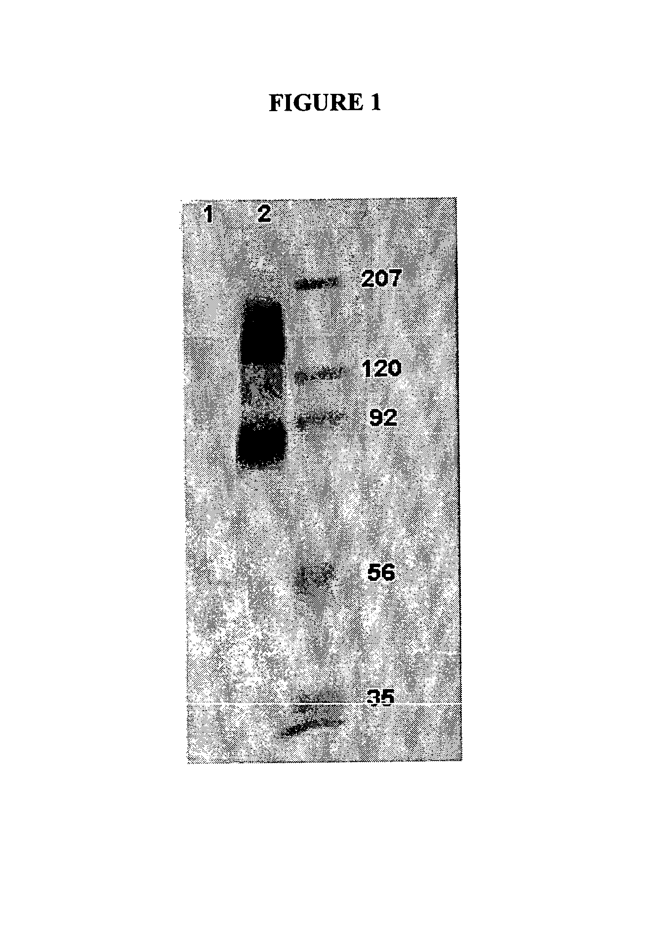
[0081]To identify the antigen(s) recognized by the antibody H460-16-2, cell membranes expressing this antigen were subjected to gel electrophoresis, and transferred to membranes. Western blotting was used to determine proteins detected by this antibody.

1. Membrane Preparation

[0082]Previous work demonstrated binding by FACS of H460-16-2 to the breast cancer cell lines MDA-MB-231 (MB-231) and MDA-MB-468 (MB-468). Accordingly, membrane preparations from these two cell lines were used for antigen identification. Total cell membranes were prepared from confluent cultures of MB-231 or MB-468 breast cancer cells. Media was removed from flasks, and the cells were washed 3 times with PBS. After the final wash, cells were dissociated with Dissociation Buffer (Gibco-BRL; Grand Island, N.Y.) for 5 min at 37° C. Cells were collected and centrifuged at 1200 rpm for 10 minutes at 4° C. After centrifugation, cell pellets were resuspended in 1 mL...

example 2

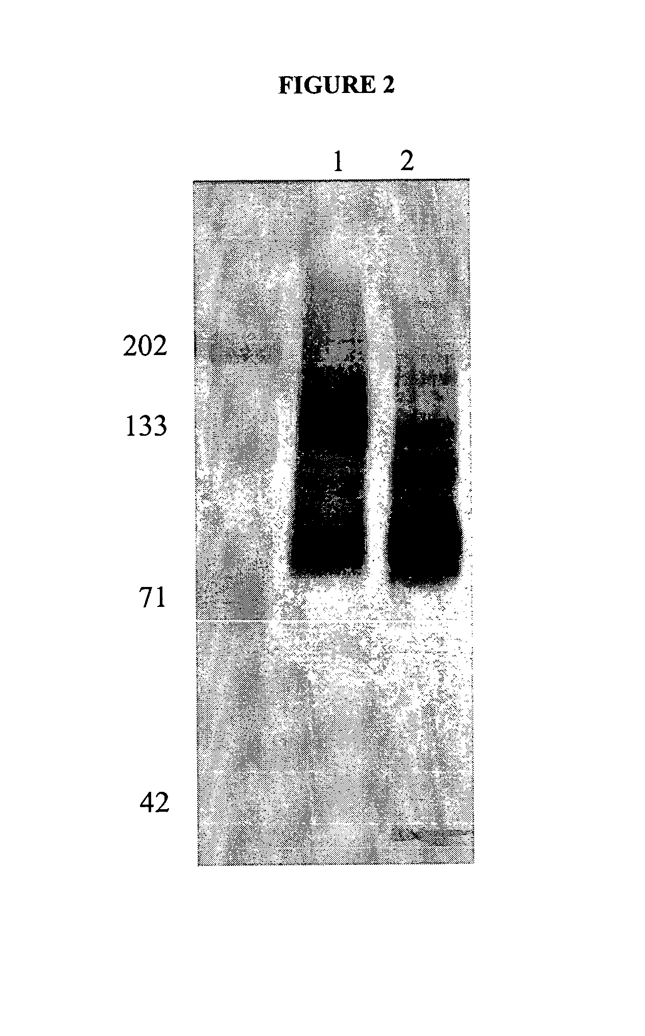
Determining Glycosylation of Antigens Bound by H460-16-2

[0089]In order to determine if the antigen(s) recognized by the antibody H460-16-2 were glycoproteins, MB-231 membranes were incubated with PNGase F, Endo-o-glycosidase, and sialidase A according to manufacturer's protocol (DeglycoPro deglycosylation kit; Prozyme, San Leandro, Calif.) for 24 hr at room temperature or at 37° C. Membranes were separated by 1-D polyacrylamide gel electrophoresis as described, and then Western blotting was carried out as described with H460-16-2. The results of the Western blot are shown in FIG. 5. In MB-468 membranes that were not treated with glycosidases, H460-16-2 recognized the expected 85-95 kD band (FIG. 5, Panel A, Lane 2). In membranes treated with glycosidases at 25° C., there is a distinct shift of this band to a lower molecular weight (Lane 4). In membranes treated with glycosidases at 37° C., the binding of 14460-16-2 is eliminated (Lane 3). In order to determine the completeness of de...

example 3

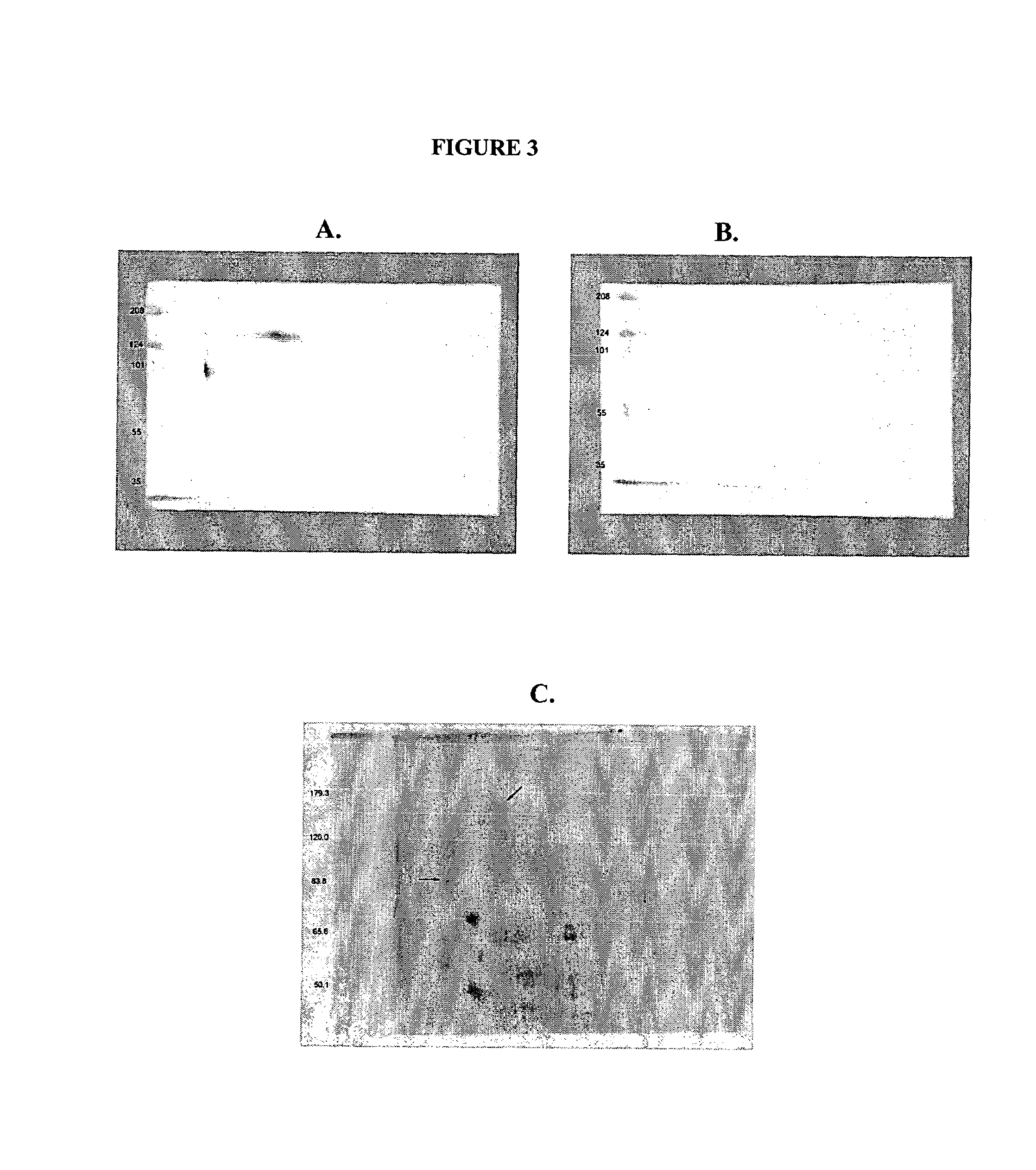
Identification of Antigens Bound by H460-16-2

1. Immunoprecipitation

[0091]1 mL of Protein G Dynabeads (DYNAL) was washed 3 times with 0.1 M sodium phosphate buffer, pH 6.0. 2500 μg of H460-16-2 was added to the washed beads in a total volume of 500 μl. The mixture was incubated with gentle mixing for 1 hr. Unbound antibody was removed and the H460-16-2-coated beads were washed 3 times in 2.5 mL volumes of 0.1 M sodium phosphate buffer, pH 7.4 containing 0.1 percent Tween-20. The H460-16-2-coated beads were washed 2 times in 5 mL of 0.2 M triethanolamine, pH 8.2 followed by the addition of 5 mL. H460-16-2 was chemically crosslinked to the beads by gentle mixing in the presence of 5 mL of 0.2 M triethanolamine, pH 8.2 containing 20 mM dimethyl pimelimidate for 30 min. The reaction was stopped by the addition of 5 mL of 50 mM Tris, pH 7.5. After 15 min incubation, the H460-16-2 crosslinked beads were washed 3 times in PBS containing 0.1 percent Tween-20. The H460-16-2-crosslinked beads ...


PUM

PropertyMeasurementUnit
temperatureaaaaaaaaaa
total volumeaaaaaaaaaa
pHaaaaaaaaaa

Abstract

This invention relates to the diagnosis and treatment of cancerous diseases, particularly to the mediation of cytotoxicity of tumor cells; and most particularly to the use of cancerous disease modifying antibodies (CDMAB), optionally in combination with one or more chemotherapeutic agents, as a means for initiating the cytotoxic response. The invention further relates to binding assays which utilize the CDMABs of the instant invention.

Description

REFERENCE TO RELATED APPLICATIONS[0001]This application is a continuation-in-part of application Ser. No. 10 / 603,000, filed June 23, 2003, which is a continuation-in-part of application Ser. No. 09 / 727,361, filed Nov. 29, 2000, now U.S. Pat. No. 6,657,048, issued Dec. 2, 2003, which is a continuation-in-part of application Ser. No. 09 / 415,278, filed Oct. 8, 1999, now U.S. Pat. No. 6,180,357 B1, the contents of each of which are herein incorporated by reference.FIELD OF THE INVENTION[0002]This invention relates to the diagnosis and treatment of cancerous diseases, particularly to the mediation of cytotoxicity of tumor cells; and most particularly to the use of cancerous disease modifying antibodies (CDMAB), optionally in combination with one or more chemotherapeutic agents, as a means for initiating the cytotoxic response. The invention further relates to binding assays which utilize the CDMAB of the instant invention.BACKGROUND OF THE INVENTION[0003]Raising monoclonal antibodies aga...

Claims


Application Information

Patent Timeline
Patent Type & Authority Patents(United States)
IPC IPC(8): A61K39/395G01N33/574C07K16/00C12P21/04C12P21/08A61K49/00C07K16/28C07K16/30G01N33/569
CPCA61K49/0041A61K49/0058C07K16/00C07K16/2884C07K16/30G01N33/57484G01N33/56972G01N33/57415G01N33/57419G01N33/57423G01N33/57449C07K16/3015A61K2039/505A61P35/00
Inventor YOUNG, DAVID S. F.HAHN, SUSAN E.FINDLAY, HELEN P.
Owner F HOFFMANN LA ROCHE & CO AG
Why Eureka
Try Eureka