
Compositions and methods for monosynaptic transport

a monosynaptic and transport technology, applied in the field of monosynaptic transport, can solve the problems of not being able to identify neurons in a wholesale fashion, unable to understand how neural circuits generate perception and behavior, and unable to achieve the effect of specific labelling and/or manipulation

Active Publication Date: 2008-08-14
SALK INST FOR BIOLOGICAL STUDIES
View PDF3 Cites 3 Cited by
  • Summary
  • Abstract
  • Description
  • Claims
  • Application Information

AI Technical Summary

Benefits of technology

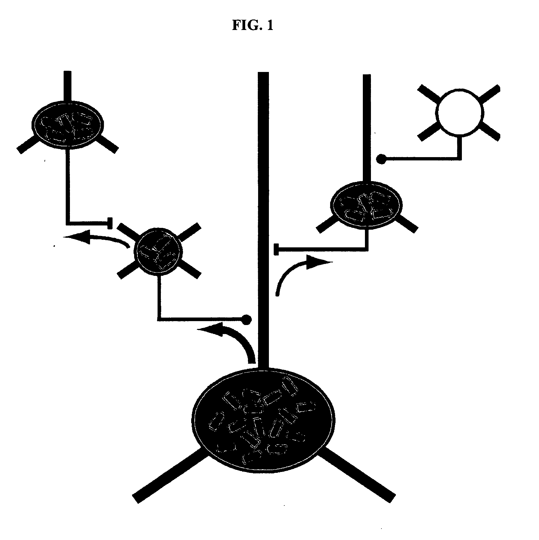
[0009]Disclosed herein are methods of, and compositions (including kits) for, expressing a heterologous nucleic acid sequence, such as a sequence encoding a detectable protein, in a primary neuron (or plurality of primary neurons) and other neurons that are monosynaptically connected to the primary neuron (or plurality of primary neurons). In particular examples such methods involve viruses (such as rabies viruses) defective for transsynaptic transport (TST-defective virus) and in situ complementation of the defect in a manner that permits only monosynaptic transport of the TST-defective virus. The TST-defective virus and, therefore, any heterologous nucleic acid sequence it carries in its genome, are not transmitted to neurons that are not monosynaptically connected to the primary neuron (or plurality of primary neurons). Also disclosed are methods of targeting a TST-defective virus to a genetically defined primary neuron (or plurality of primary neurons). The disclosed technology enables far more specific labelling and / or manipulation of neural networks than has previously been possible.
[0010]The foregoing and other features will become more apparent from the following detailed description of several embodiments, which proceeds with reference to the accompanying figures.

Problems solved by technology

Recent advances in the knowledge of the complexity and specificity of neural circuits suggest that understanding how neural circuits generate perception and behavior will be nearly impossible with presently available techniques.
Historically, the identification of neural networks has involved labor-intensive and technologically challenging methods, such as examining sections of neural tissue with light or electron microscopy (Gilbert, Ann. Rev. Neurosci., 6:217-47, 1983; Douglas and Martin, Ann. Rev. Neurosci., 27:419-51, 2004; Gray, Nature, 183(4675):1592-3, 1959; Timofeeva et al., J. Neurosci., 25(40):9135-43, 2005), simultaneous patch recording from pairs of neural cells (Mercer, et al., Cereb.
None of these methods permit a wholesale way of identifying neurons that are connected either to some other cell group or, especially, to a single cell.
However, no such method has been sensitive enough to label cells synaptically connected to a single cell of origin.
No technique to date has been capable of identifying en masse neurons that are connected directly to a primary neuron (or population of primary neurons) of interest.
Furthermore, no tracer has proven potent enough to label any connected neurons whatsoever when starting from a single cell.

Method used


Image

  • Compositions and methods for monosynaptic transport
  • Compositions and methods for monosynaptic transport
  • Compositions and methods for monosynaptic transport

Examples


example 1

Construction and Function of a Rabies Virus Glycoprotein Deletion Mutant that Encodes a Marker Gene

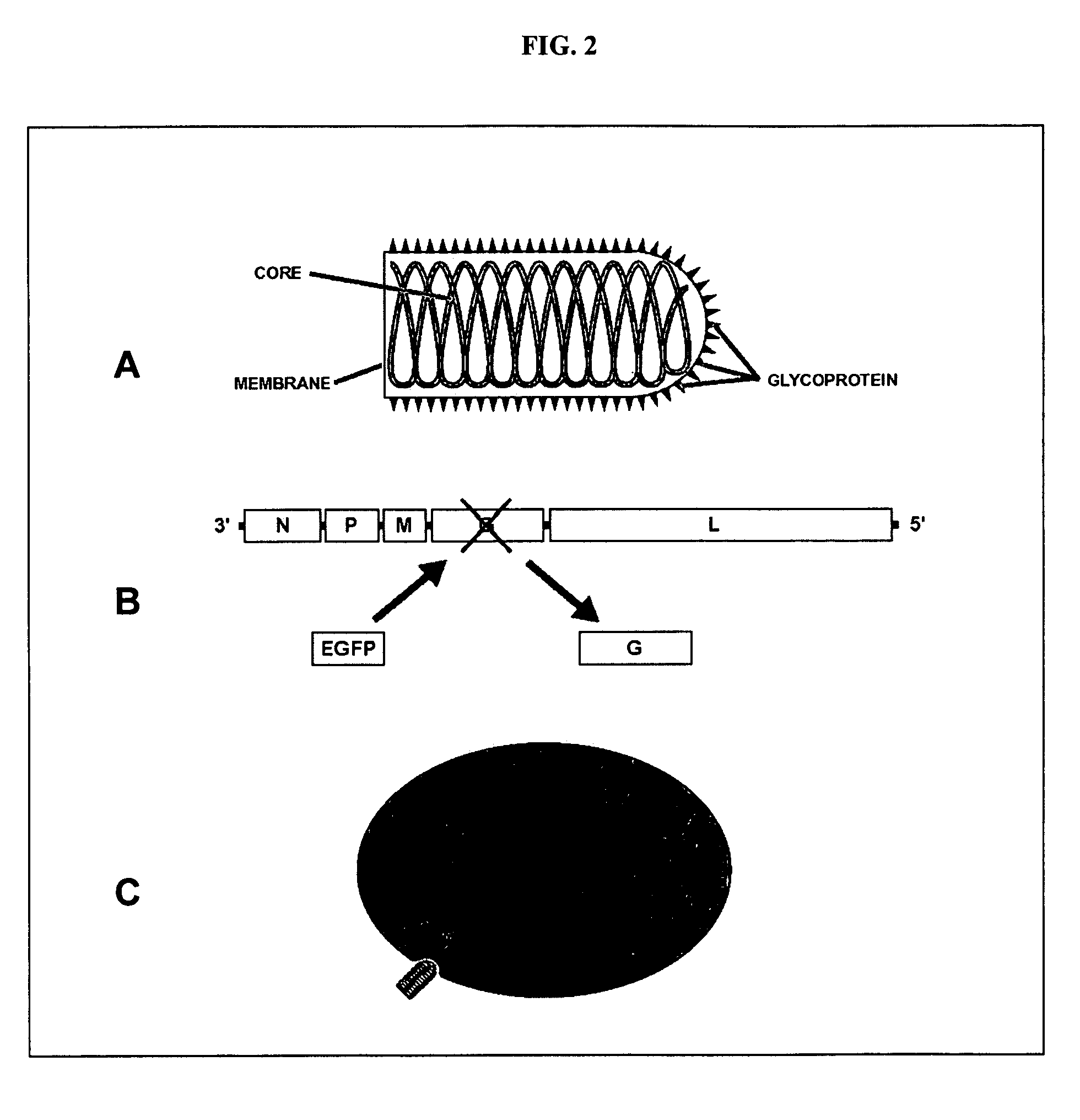
[0127]Rabies virus is a neurotropic virus that infects neurons via axon terminals, replicates in the cytoplasm, and spreads between synaptically coupled neurons in an exclusively retrograde direction. A single infectious unit of rabies virus introduced into a brain is sufficient to cause a full-scale case of rabies (Lafay et al., Virol., 183(1):320-30, 1991, Ito et al., J. Virol., 75(19):9121-9128, 2001).

[0128]Mebatsion et al. (Cell, 84(6)941-51, 1996) previously described a rabies virus with the envelope glycoprotein gene deleted from its genome (G− rabies virus). The G− rabies virus was grown in cells where the glycoprotein was provided in trans for incorporation into the viral particles' membranes. The G− rabies virus normally infected neurons with which it came in contact. Because the glycoprotein plays no role in transcription and replication, the G− rabies virus replicated its vi...

example 2

Specific Targeting of and Monosynaptic Labeling by a Rabies Virus Glycoprotein Deletion Mutant

[0145]This Example provides a representative method involving a rabies virus monosynaptic tracer, which tracer can be specifically targeted to a particular population of originating neurons and is transmitted substantially only to neurons monosynaptically connected to the originating neurons. Although this example describes a tracer that includes the envelope protein of the ASLV-A virus (EnvA), one skilled in the art will appreciate that other envelope proteins can be used (such as EnvB), and that other viruses can be used in place of the rabies virus.

[0146]The interaction between subgroup A avian sarcoma and leukosis virus (ASLV-A) and its receptor(s) is highly specific. The envelope protein of the ASLV-A virus (EnvA) can direct virus infection specifically into cells that express the cognate TVA viral receptor (Young et al., J. Virol., 67(4):1811-6, 1993; Bates et al., Cell, 74(6):1043-51...

example 3

Monosynaptically Labeled Cells are Functionally Connected

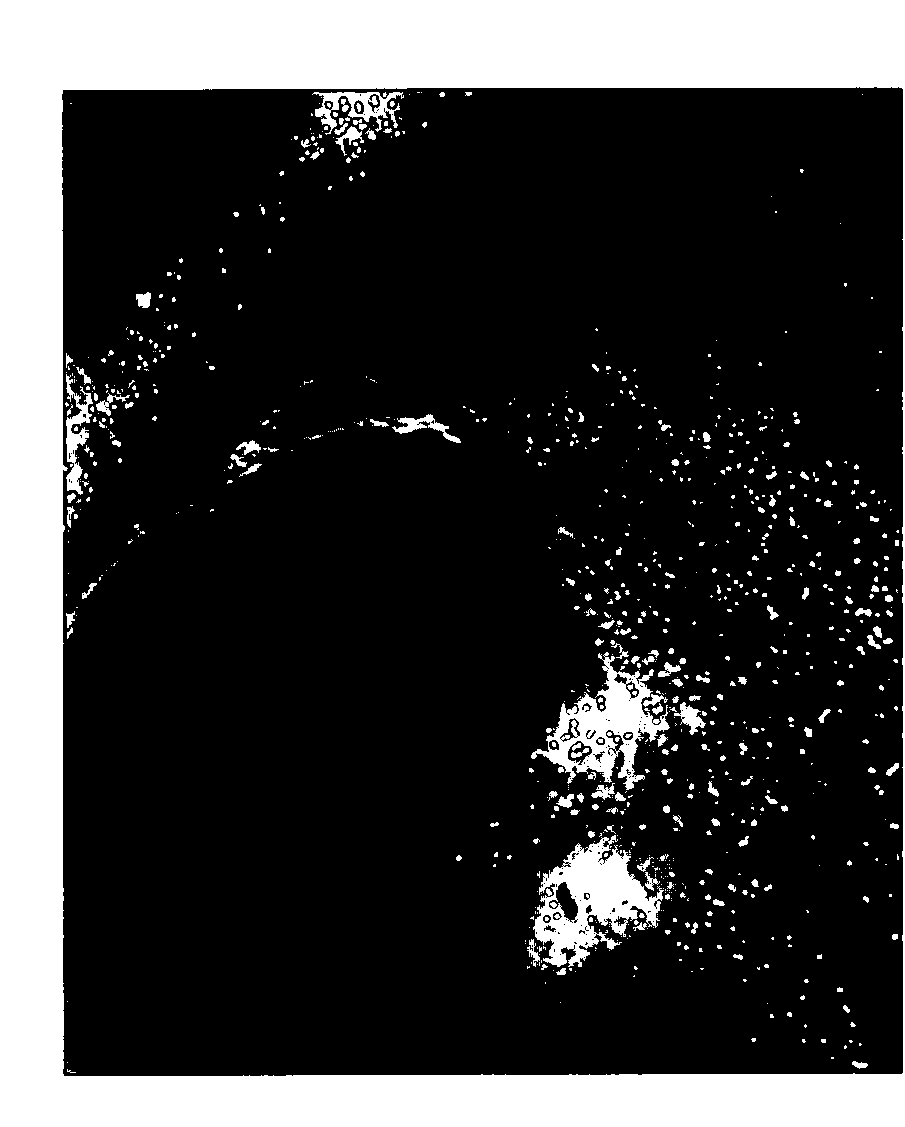
[0153]This Example demonstrates that neurons labeled as described in Example 3 are functionally connected. Thus, the observed labeling was substantially transsynaptic and not, for instance, nonspecific infection of neighboring cells.

[0154]Paired whole-cell recordings were made from putatively pre- and postsynaptic cells. Cells fluorescent in both red and green channels (red / green cells) were recorded under voltage clamp while nearby green-only cells, held in current clamp, were depolarized to fire action potentials (see, for instance, FIGS. 6A-D and F-I). Synaptic currents in the voltage-clamped red / green cell that were simultaneous with the green cell's action potentials indicated a monosynaptic connection as seen in the example traces in FIGS. 6E and 6J. Non-fluorescent cells at similar distances from the red / green cells were also stimulated as controls. Because the gene gun typically transfected many more than one neuron wi...


PUM

PropertyMeasurementUnit
volumesaaaaaaaaaa
volumesaaaaaaaaaa
pHaaaaaaaaaa

Abstract

Disclosed herein are methods of expressing a heterologous nucleic acid sequence, such as a sequence encoding a detectable protein, in a primary neuron (or plurality of primary neurons) and other neurons that are monosynaptically connected to the primary neuron (or plurality of primary neurons). Such methods involve viruses (such as, rabies viruses) defective for transsynaptic transport (TST-defective virus) and in situ complementation of the defect in a manner that permits only monosynaptic transport of the TST-defective virus. The TST-defective virus and, therefore, any heterologous nucleic acid sequence it carries in its genome, are not transmitted to neurons that are not monosynaptically connected to the primary neuron (or plurality of primary neurons). Also disclosed are methods of targeting a TST-defective virus to a genetically defined primary neuron (or plurality of primary neurons). The disclosed technology enables far more specific labelling and / or manipulation of neural networks than has previously been possible.

Description

CROSS REFERENCE TO RELATED APPLICATIONS[0001]This application claims the benefit of U.S. Provisional Application No. 60 / 884,462, filed Jan. 11, 2007, which application is incorporated herein in its entirety.STATEMENT OF GOVERNMENT SUPPORT[0002]This invention was made with United States government support pursuant to grant no. MH63912 from the National Institute of Mental Health (NIMH), grant no. EY10742 from the National Eye Institute (NEI), grant no. DA018828 from the National Institute on Drug Abuse (NIDA), and grant no. CA70810 from the National Cancer Institute (NCI); the United States government has certain rights in the invention.FIELD OF THE DISCLOSURE[0003]This disclosure concerns methods and compositions (such as kits and their components) useful for identifying, monitoring, or affecting the function of neurons monosynaptically connected to a single primary neuron or plurality of primary neurons of interest.BACKGROUND[0004]Recent advances in the knowledge of the complexity ...

Claims


Application Information

Patent Timeline
Patent Type & Authority Applications(United States)
IPC IPC(8): C12Q1/70C12N15/86
CPCC12N15/86C12N2740/13043C12N2740/13045C12N2760/20122C12Q1/702C12N2800/30C12N2810/6054C12N2810/6081C12N2799/04
Inventor WICKERSHAM, IAN R.YOUNG, JOHN A.T.CALLAWAY, EDWARD M.
Owner SALK INST FOR BIOLOGICAL STUDIES
Learn More