Unlock instant, AI-driven research and patent intelligence for your innovation.

Microfluidic Detection of Analytes

a microfluidic and analyte technology, applied in the direction of fluid pressure measurement, liquid/fluent solid measurement, peptide measurement, etc., can solve the problems of limited sensitivity, limited devices, and not all analytes can be amplified

Inactive Publication Date: 2008-08-28
HEADWAY TECH INC
View PDF39 Cites 14 Cited by
  • Summary
  • Abstract
  • Description
  • Claims
  • Application Information

AI Technical Summary

Benefits of technology

The invention provides methods for introducing or applying a charged analyte in a sample to a microfluidic device by using a large volume reservoir and a microfluidic device with an analysis area that is connected to the reservoir through a connector. The analyte is electrophoresed through the analysis area to a higher concentration in the analysis area than in the reservoir. The large volume reservoir can be a microwell plate, an eppendorf tube, or a test tube. The sample can be mixed with an antibody that specifically binds to the analyte and the antibody can be modified with an ionic moiety. The analysis area can be a capture site and the method involves moving the charged molecule across the capture site. The invention also includes a staging reservoir for connecting the large volume reservoir and the analysis area. The technical effects include improved sensitivity and accuracy in analyzing samples and the use of microfluidic devices with a large volume reservoir for efficient analysis.

Problems solved by technology

However, these devices are limited by the fact that they have capacity for only very small sample volumes, typically on the order of a few microliters or less.
When substances to be analyzed are found in a sample at a very low concentration, sensitivity can be limited.
However, not all analytes can be amplified and, even when possible, amplification may require additional reagents and increase the complexity of analysis.

Method used


Image

  • Microfluidic Detection of Analytes
  • Microfluidic Detection of Analytes
  • Microfluidic Detection of Analytes

Examples


example 1

HIV / HB VIHCV test

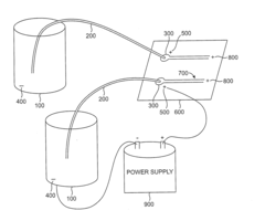
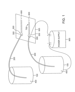
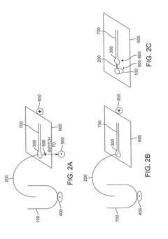
[0131]FIG. 3 illustrates a microfluidic device 600 for detecting the presence of HIV, HBV, and / or HCV antigens (protein analytes) in a sample. The microfluidic device has electrodes 500 and 800 for use, along with LVR electrod 400, in electrophoresis. A biological sample suspected of containing HIV, HBV or HCV is introduced into the large volume reservoir (not shown) and mixed with antibodies specific to HIV, HCV, and HBV protein analytes. The antibodies have an ionic moiety attached. Antibody / analyte complexes are allowed to form and, the complexes are electrophoresed over the capture sites (700a, 700b and 700c) in the Analysis area 700 (optionally after copncentration in staging reservoir 300). Reagents and unbound molecules flow into eleuent chamger 1000, and may be removed. The HIV capture site 700a has antibody capture agents specific to an HIV protein analyte. The HBV capture site 700b has antibody capture agents specific to an HBV protein analyte and the HCV ...

example 2

HIV / HBV / HCV test for viral nucleic acid analytes

[0134]To detect viral nucleic acid analytes in patient samples, ionic or nonionic detergent is added to the patient sample to disrupt the viral coating and expose the nucleic acids in the large volume reservoir. If nonionic detergent is used and the samples have a low ion content, electrophoresis can be used to drive the viral nucleic acids directly into the analysis area 700. Alternatively, the viral nucleic acids are driven by electrophoresis to the staging reservoir 300 first. Subsequently, these viral nucleic acids are delivered from the staging reservoir to the analysis area 700 by electrophoresis or other means such as micropumps.

[0135]If ionic detergents are used, further sample preparation is used as follows. Magnetic microparticles conjugated with complementary nucleic acids or analogs (binding agents) are added to the sample containing the viral RNA or DNA analyte. The viral RNA or DNA binds to the binding agents on the micro...

example 3

HIV / HBV / HCV test for antibody analytes

[0137]An example to detect antibodies against HIV, HBV or HCV viral antigens in the patient samples is performed as follows:

[0138]A specific viral antigen as binding agent for each antibody is conjugated to magnetic microparticles via a cleavable bond such as a double stranded DNA with a restriction site Alternatively, anti-human Fc antibodies modified with ionic moieties can be used. The microparticles are mixed with blood samples from human patients in a large volume reservoir to capture any antibodies for specific viral antigens. After wash steps to reduce unwanted impurities, the complex is freed from the microparticles by cleaving with a restriction enzyme.. The complex is then driven by electrophoresis into a staging reservoir. After concentration into the staging reservoir, the complex is driven to analysis sites (700a, 700b, 700c). As an example, to detect the complexes, capture agents with specific affinity to one of the entities in the...


PUM

PropertyMeasurementUnit
volumeaaaaaaaaaa
volumeaaaaaaaaaa
volumeaaaaaaaaaa

Abstract

An apparatus and methods for concentrating samples for application to microfluidic devices are disclosed. The methods involve electrophoresing charged molecules from a high volume sample into a smaller volume. The analyte of interest can be a charged molecule or can be modified to be charged using, for example, one or more ionic moieties.

Description

CROSS-REFERENCE TO RELATED APPLICATIONS[0001]The application claims benefit of U.S. Provisional Application No. 60 / 723715, filed Oct. 4, 2005 and No. 60 / 820566, filed Jul. 27, 2006, the entire contents of which are incorporated herein by reference.FIELD OF THE INVENTION[0002]The invention relates to microfluidics and detection of analytes present at low concentrations in a sample.BACKGROUND OF THE INVENTION[0003]Microfluidic systems have great potential for use in a clinical laboratory setting. However, these devices are limited by the fact that they have capacity for only very small sample volumes, typically on the order of a few microliters or less. When substances to be analyzed are found in a sample at a very low concentration, sensitivity can be limited. One way to overcome this limitation is by using an analyte amplification step to increase the analyte concentration either before or after introduction of the sample to a microfluidic device. For example, very small amounts of ...

Claims


Application Information

Patent Timeline
Patent Type & Authority Applications(United States)
IPC IPC(8): G01N27/26
CPCB01L3/502715B01L2200/027B01L2300/0636G01N27/447G01N1/40G01N33/536G01N2001/4038B01L2400/0421B33Y80/00
Inventor HU, CELINE
Owner HEADWAY TECH INC