Universal ultrasound holder and rotation device

a technology of rotation device and ultrasound holder, which is applied in the field of apparatus for holding and positioning an imaging instrument, can solve the problems of affecting the successful performance of the procedure, affecting the accuracy of the 3-d image, and affecting the accuracy of the image, so as to achieve the effect of little wobbl

Active Publication Date: 2008-09-11
EIGEN HEALTH SERVICES LLC
View PDF13 Cites 19 Cited by
  • Summary
  • Abstract
  • Description
  • Claims
  • Application Information

AI Technical Summary

Benefits of technology

[0008]Provided herein are devices and methods for the use of such devices for mounting variously configured medical imaging probes for imaging applications. Further, systems and methods are provided for acquiring medical images. In one aspect, a device allows for interfacing / holding most conventional ultrasound probes such that the probes may be attached to a positioning device using a common interface. As ultrasound probes come in various sizes and lengths, the device may adjust to different lengths, widths and shapes of different probes. Hence, the device may work in a substantially universal manner while securely holding probes with little wobble or other problems. The device may also be lightweight and compact to allow it to be used efficiently. The device may also allow use of a biopsy needle and / or a histological gun while a probe is held within the device.

Problems solved by technology

Accordingly, movement of a medical imaging device between the acquisition of individual images makes it more difficult to properly align (e.g., register) the different images for purposes of generating accurate 3-D images.
However, in instances where it is desirable to obtain multiple images for 3-D image generation, manual manipulation of the device may result in movement between images.
Relative movement between the imaging device and the tissue area of interest during imaging and / or biopsy / treatment may impede the successful performance of these procedures.
However, original equipment manufactures (OEMs) of ultrasound probes do not have a standardized design.
Further, such positioning assemblies have typically been complicated and mechanically cumbersome.

Method used


Image

  • Universal ultrasound holder and rotation device
  • Universal ultrasound holder and rotation device
  • Universal ultrasound holder and rotation device

Examples


Embodiment Construction

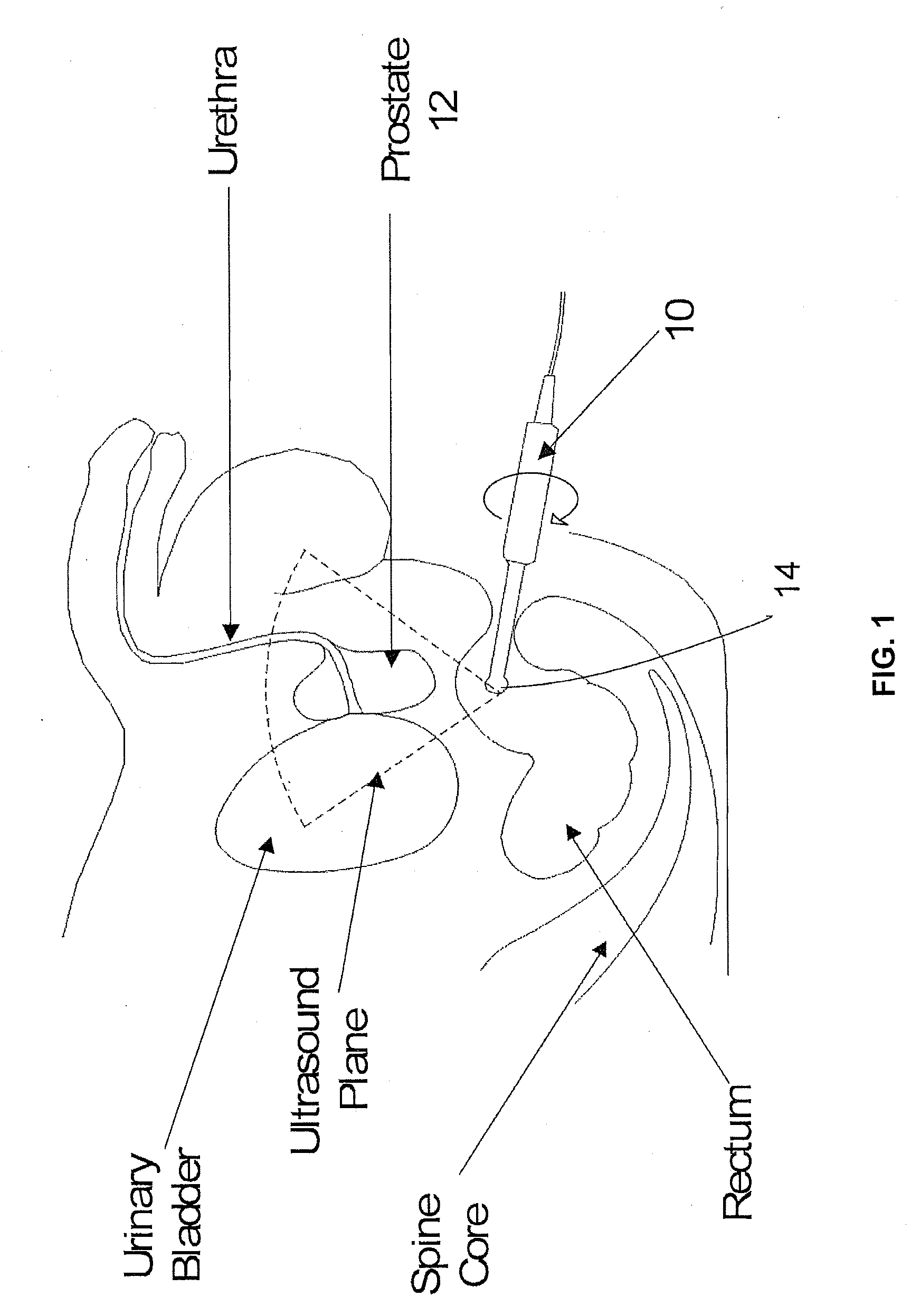
[0035]Reference will now be made to the accompanying drawings, which assist in illustrating the various pertinent features of the present disclosure. Although the present disclosure is described primarily in conjunction with transrectal ultrasound imaging for prostate imaging, it should be expressly understood that aspects of the present invention may be applicable to other medical imaging applications. In this regard, the following description is presented for purposes of illustration and description.

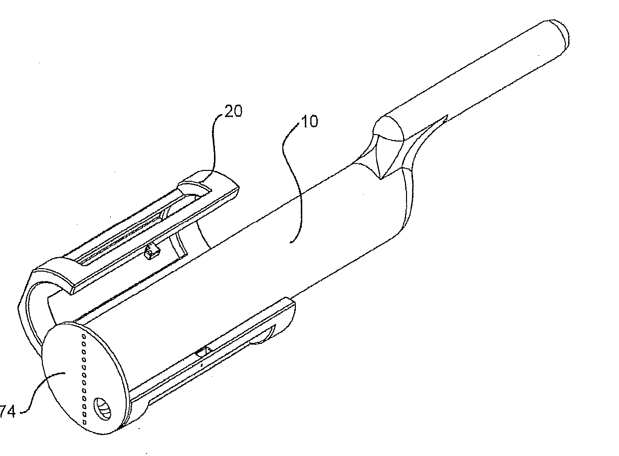
[0036]Disclosed herein are systems and methods that facilitate obtaining medical images and / or performing medical procedures. More specifically, a medical imaging device holder (i.e., holding device or cradle) is provided that is adapted to securely support multiple differently configured ultrasound probes. Further, a simplified rotational mechanism is provided.

[0037]The probe cradle may be interfaced with the rotational mechanism such that a supported probe may be rotated about a fixe...


PUM


Abstract

Provided herein are devices and methods for mounting variously configured medical imaging probes for imaging applications. In one aspect, a holding device allows for interfacing/holding most conventional ultrasound probes such that the probes may be attached to a positioning device using a common interface. As ultrasound probes come in various sizes and lengths, the device may adjust to different lengths, widths and shapes of different probes. Hence, the device may work in a substantially universal manner while securely holding probes with little wobble or other problems.

Description

CROSS-REFERENCE TO RELATED APPLICATION[0001]This application claims priority under 35 U.S.C. 119 to U.S. Provisional Application No. 60 / 893,317, entitled: “UNIVERSAL HOLDER,” filed on Mar. 6, 2007, and No. 60 / 894,602, entitled: “CRADLE WITH MANUAL RATCHET,” filed on Mar. 13, 2007, the contents of which are incorporated herein as if set forth in full.FIELD OF INVENTION[0002]The present invention is directed to an apparatus for holding and positioning a medical imaging instrument. More particularly, the invention relates to an apparatus adapted to hold a plurality of differently configured medical imaging instruments such that those instruments may be secured and / or rotated about at least one fixed axis.BACKGROUND OF THE INVENTION[0003]Medical imaging instruments are often utilized by doctors and other medical professionals to conduct non-invasive examinations. That is, medical imaging instruments, including X-ray, magnetic resonance (MR), computed tomography (CT), ultrasound, and var...

Claims


Application Information

Patent Timeline
Patent Type & Authority Applications(United States)
IPC IPC(8): A61B8/12
CPCA61B8/12A61B8/4218A61B8/4209
Inventor SURI, JASJITSURI, KANWARKHEMKA, ANIMESHKUMAR, DINESH
Owner EIGEN HEALTH SERVICES LLC
Why Eureka
Try Eureka