
Electronic Stool Subtraction in Ct Colonography

a technology of ct colonography and electronic stools, which is applied in the field of ct colonography, can solve the problems of reducing the efficacy of diagnostic procedures, time-consuming and uncomfortable patient preparation processes, and patients may forego diagnostic procedures altogether, and achieves the effect of accurate and efficient method of implementation

Active Publication Date: 2008-11-06
MAYO FOUND FOR MEDICAL EDUCATION & RES
View PDF27 Cites 15 Cited by
  • Summary
  • Abstract
  • Description
  • Claims
  • Application Information

AI Technical Summary

Benefits of technology

[0010]The present invention is an accurate and efficient-to-implement method for electronically removing stool from colonography images. The data processing method effectively estimates the fraction of stool present at each voxel. The stool removal process estimates partial volume and system blur effects for each voxel by attempting to emulate certain aspects of the image acquisition process.

Problems solved by technology

Unfortunately, these colon preparation processes can be time consuming and uncomfortable for the patient.
Patient compliance with the preparation processes is sometimes poor, resulting in reduced efficacy of the diagnostic procedure.
Perhaps even worse, some patients may forego the diagnostic procedure altogether to avoid the inconvenience of the preparation process.
However, accurate stool subtraction is difficult for several reasons.
Stool boundaries are also irregular and unpredictable, and partial volume (blurring) effects between stool and air and between stool and tissue will create pixels with intermediate values that are hard to recognize.
Furthermore, the effects of noise vary between patients, and can alter voxel intensities significantly.
Unfortunately this preparation approach results in some amount of discomfort and inconvenience to the patient and, possibly, reduced compliance.
Accurate stool subtraction on an unprepared colon is typically more difficult.

Method used


Image

  • Electronic Stool Subtraction in Ct Colonography
  • Electronic Stool Subtraction in Ct Colonography
  • Electronic Stool Subtraction in Ct Colonography

Examples


Embodiment Construction

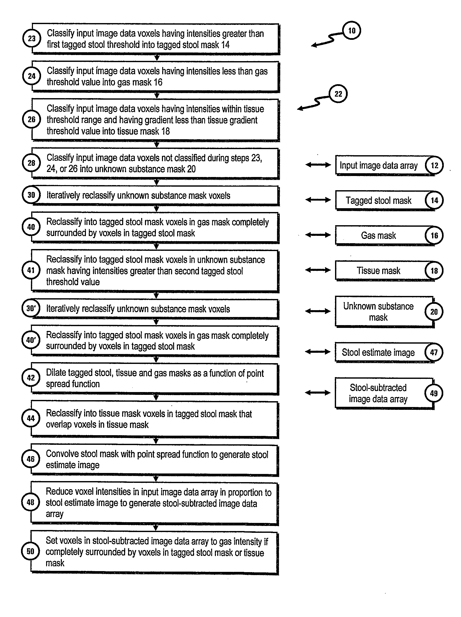
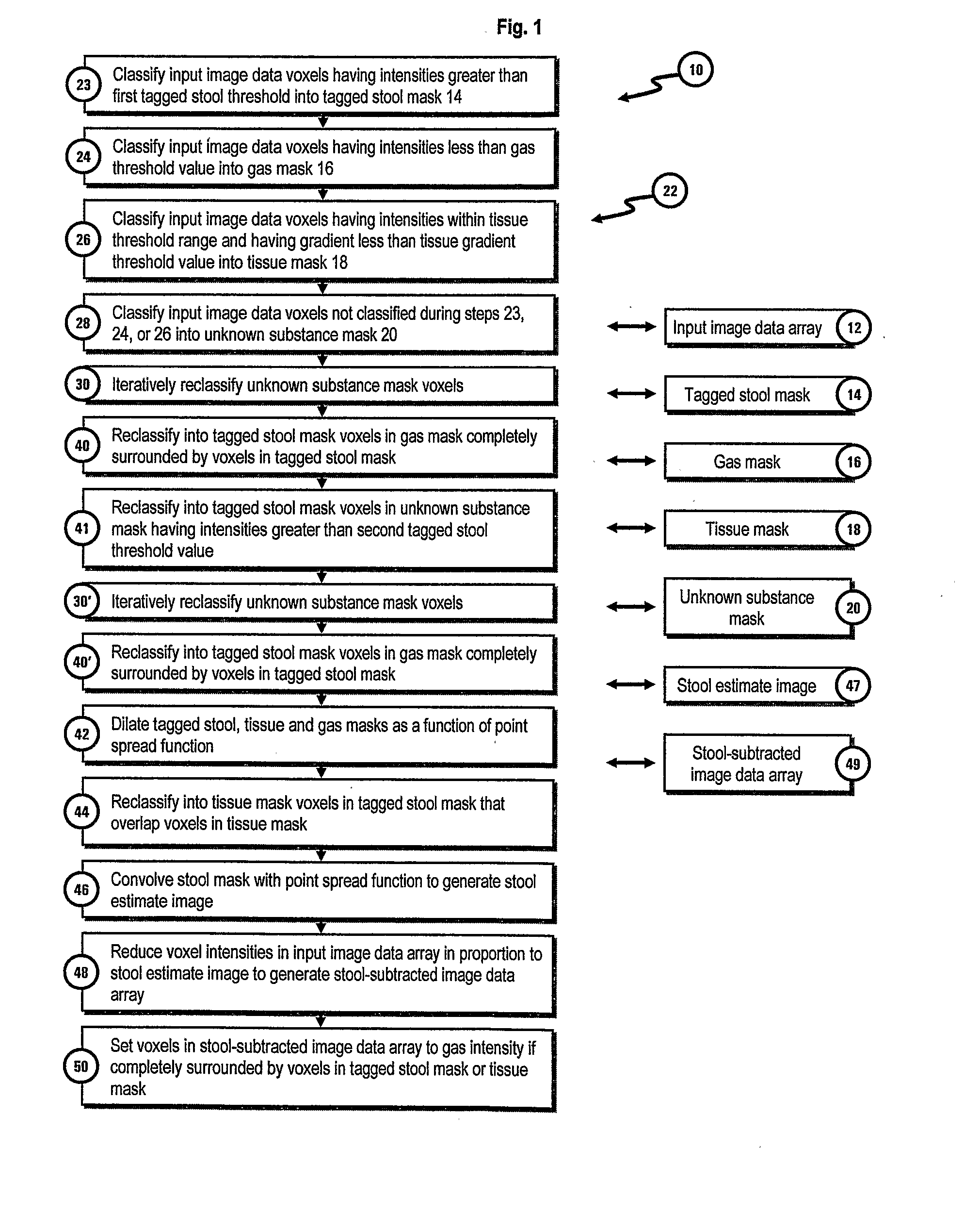
[0018]FIG. 1 is a flow diagram illustrating the electronic stool subtraction method 10 of the present invention. The data processing operations of method 10 can be performed by a conventional computer system (not shown). The method is performed on an array of input image data 12 representative of a 3-dimensional image of a colon. Input image data array 12 includes data representative of the intensity of the image at each location or voxel in the image. In one embodiment of the invention the voxel data uses the Houndsfield scale to represent the range of voxel intensities. On the Houndsfield scale a value of −1000 houndsfield units (HU) is used to represent the density of air and a value of 0 HU is used to represent water. Bone will generally have densities corresponding to between 500 and 1000 HU.

[0019]Conventional low dose (e.g., 50 mAs) CT colonography was performed with 1.25 mm collimation and 1.25 mm reconstruction intervals to generate the image data array 12 used to develop th...


PUM


Abstract

A method for processing CT colonography input image voxel data representative of 3-dimensional images of a colon having gas and stool tagged with stool tagging agent, to remove the stool from the images. The input image voxel data is generated by an imaging instrument having a characteristic point spread function representative of instrument blurring. The point spread function of the instrument can be empirically determined, and the image data processed as a function of the point spread function to accurately identify and remove the tagged stool. In one embodiment of the invention, portions of the image data representative of the tagged stool and colon tissue are dilated as a function of the point spread function. In another embodiment, portions of the image data representative of the tagged stool are convolved with the point spread function to determine the fractional amount of stool present in the image portions, and the tagged stool subtracted by reducing the intensities of the associated portions of the image by an amount proportional to the fractional amount of stool present.

Description

REFERENCE TO RELATED APPLICATION[0001]This application claims the benefit of U.S. Provisional Application Ser. No. 60 / 652,679, filed on Feb. 14, 2005 and entitled Electronic Stool Subtraction Algorithm For CT Colonography Without Bowel Preparation, which is incorporated herein by reference in its entirety.GOVERNMENT LICENSE RIGHTS[0002]This invention was made with funding support from the U.S. government. The U.S. government may have certain rights to this invention.FIELD OF THE INVENTION[0003]This invention relates generally to the field of CT colonography. In particular, the invention is a method for electronically identifying and removing stool from CT colonography images.BACKGROUND OF THE INVENTION[0004]Colonography, the use of electronic imaging technologies such as computed tomography (CT) to generate images of a patient's colon for purposes of colorectal cancer screening, is generally known. Descriptions of this diagnostic methodology can, for example, be found in the followi...

Claims


Application Information

Patent Timeline
IPC IPC(8): G06T7/20
CPCG06T7/0081G06T7/0097G06T2207/20224G06T7/0091G06T5/005G06T5/30G06T2207/10081G06T2207/30028G06T7/11G06T7/155G06T7/174G06T5/77
Inventor MANDUCA, ARMANDOCARSTON, MICHAEL J.WENTZ, ROBERT J.JOHNSON, C. DANIEL
Owner MAYO FOUND FOR MEDICAL EDUCATION & RES
Learn More