Molecular imaging methods for diagnosis and evaluation of ocular and systemic diseases

a technology of ocular and systemic diseases and molecular imaging, which is applied in the direction of echographic/ultrasound-imaging preparations, diagnostic recording/measuring, ultrasonic/sonic/infrasonic diagnostics, etc., can solve problems such as damage to dr

Inactive Publication Date: 2008-12-11
MASSACHUSETTS EYE & EAR INFARY
View PDF3 Cites 21 Cited by
  • Summary
  • Abstract
  • Description
  • Claims
  • Application Information

AI Technical Summary

Benefits of technology

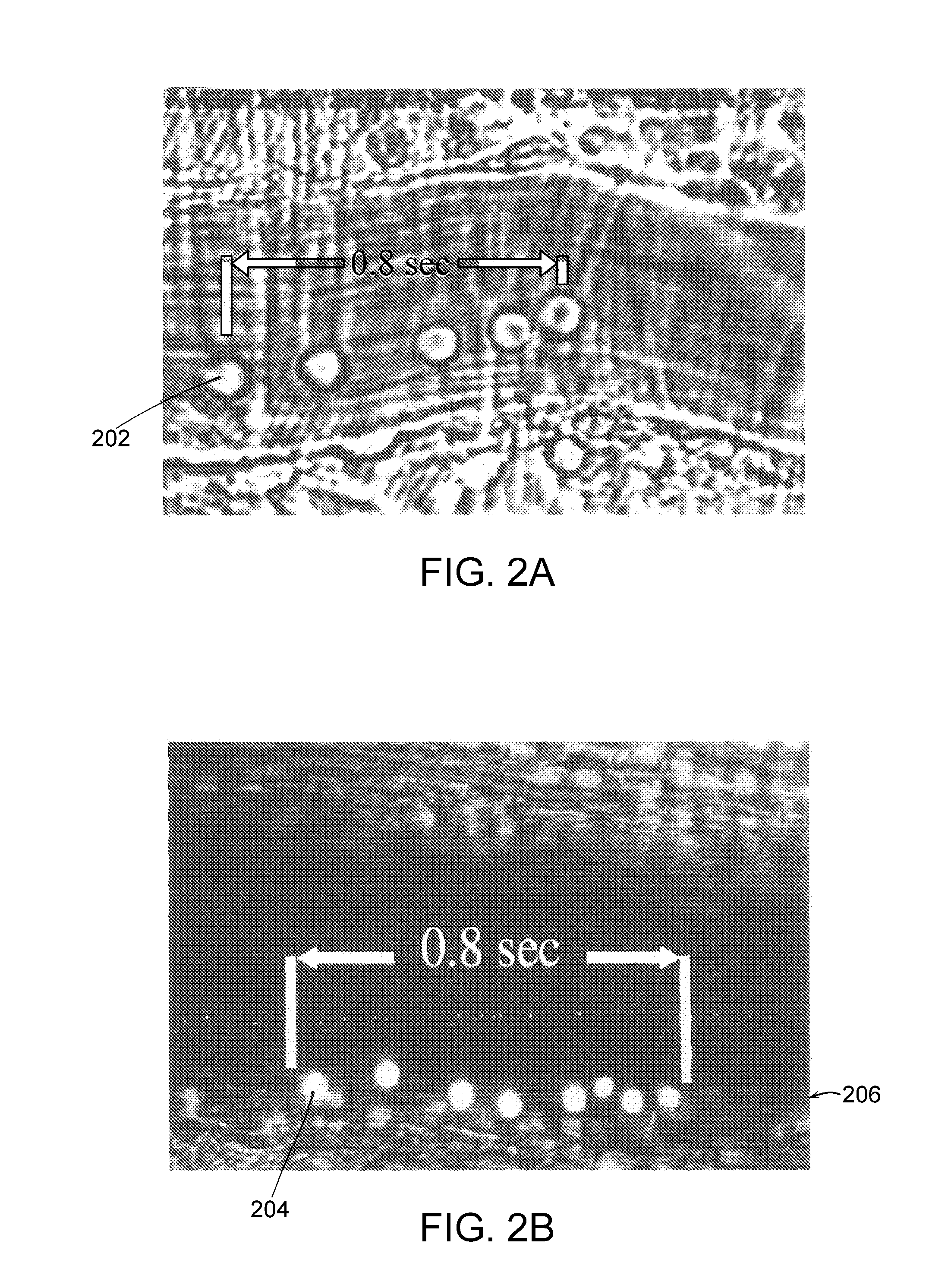
[0032]The non-invasive detection device may capture a sequence of images over time to detect movement of one or more of the microparticles. The sequence of images may be used, for example, to determine a rolling velocity of one or more of the microparticles, thereby providing information about ligands on the intraluminal surface of the blood vessel.

Problems solved by technology

Much of the damage in DR is due to vascular changes, such as endothelial apoptosis and microvascular leakage.

Method used


Image

  • Molecular imaging methods for diagnosis and evaluation of ocular and systemic diseases
  • Molecular imaging methods for diagnosis and evaluation of ocular and systemic diseases
  • Molecular imaging methods for diagnosis and evaluation of ocular and systemic diseases

Examples


Embodiment Construction

[0068]It is contemplated that devices, systems, methods, and processes of the claimed invention encompass variations and adaptations developed using information from the embodiments described herein. Adaptation and / or modification of the devices, systems, methods, and processes described herein may be performed by those of ordinary skill in the relevant art.

[0069]Throughout the description, where apparatus, devices, and systems are described as having, including, or comprising specific components, or where processes and methods are described as having, including, or comprising specific steps, it is contemplated that, additionally, there are apparatus, devices, and systems of the present invention that consist essentially of, or consist of, the recited components, and that there are processes and methods according to the present invention that consist essentially of, or consist of, the recited processing steps.

[0070]It should be understood that the order of steps or order for perform...


PUM


Abstract

This invention relates generally to minimally-invasive, in vivo methods of detecting one or more ligands on an intraluminal surface of a blood vessel using microparticles coated with one or more ligand binding partners. More particularly, in certain embodiments, the invention relates to minimally-invasive, in vivo methods of detecting endothelial and leukocyte antigens that are predictive of diabetic retinopathy (DR) and/or other conditions using protein-conjugated microparticles detectable by a non-invasive detection system, for example, a scanning laser opthalmoscope. In other embodiments, the invention relates to targeted delivery of drugs or other substances to specific regions of an intraluminal surface of a blood vessel using drug-containing microparticles coated with one or more ligand binding partners.

Description

RELATED APPLICATION[0001]This application claims priority to and the benefit of U.S. provisional patent application Ser. No. 60 / 902,004, filed Feb. 16, 2007, the disclosure of which is incorporated herein by reference in its entirety.GOVERNMENT RIGHTS[0002]The invention was supported, in part, by grant AI050775 from the National Institute of Health (NIH). The Government has certain rights in the invention.FIELD OF THE INVENTION[0003]This invention relates generally to minimally-invasive methods for evaluating and treating ocular and systemic diseases using microparticles. More particularly, in certain embodiments, the invention relates to in vivo methods of detecting one or more ligands on an intraluminal surface of a blood vessel by administering microparticles coated with one or more ligand binding partners and detecting the microparticles with a minimally-invasive detection system, for example, a scanning laser opthalmoscope.BACKGROUND OF THE INVENTION[0004]Diabetic retinopathy (...

Claims


Application Information

Patent Timeline
Patent Type & Authority Applications(United States)
IPC IPC(8): A61K49/06A61K49/00A61K49/22A61K9/14A61P27/02A61P43/00
CPCA61B3/1025A61P27/02A61P43/00A61B3/1233A61B3/1241A61B5/14546
Inventor HAFEZI-MOGHADAM, ALI
Owner MASSACHUSETTS EYE & EAR INFARY
Why Eureka
Try Eureka