Multimetric Biosensors and Methods of Using Same
a biosensor and multimeric technology, applied in the direction of fusion polypeptides, dna/rna fragmentation, fungi, etc., can solve the problem that none of these studies has designed and employed single-chain biosensors with multimeric or dimeric moieties
- Summary
- Abstract
- Description
- Claims
- Application Information
AI Technical Summary
Benefits of technology
Problems solved by technology
Method used
Image

Examples
example 1
A Ligand-Binding Scaffold for L-Tryptophan
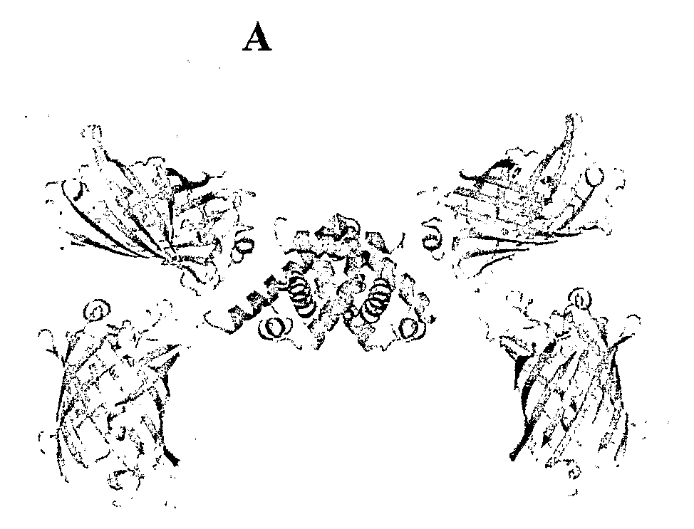
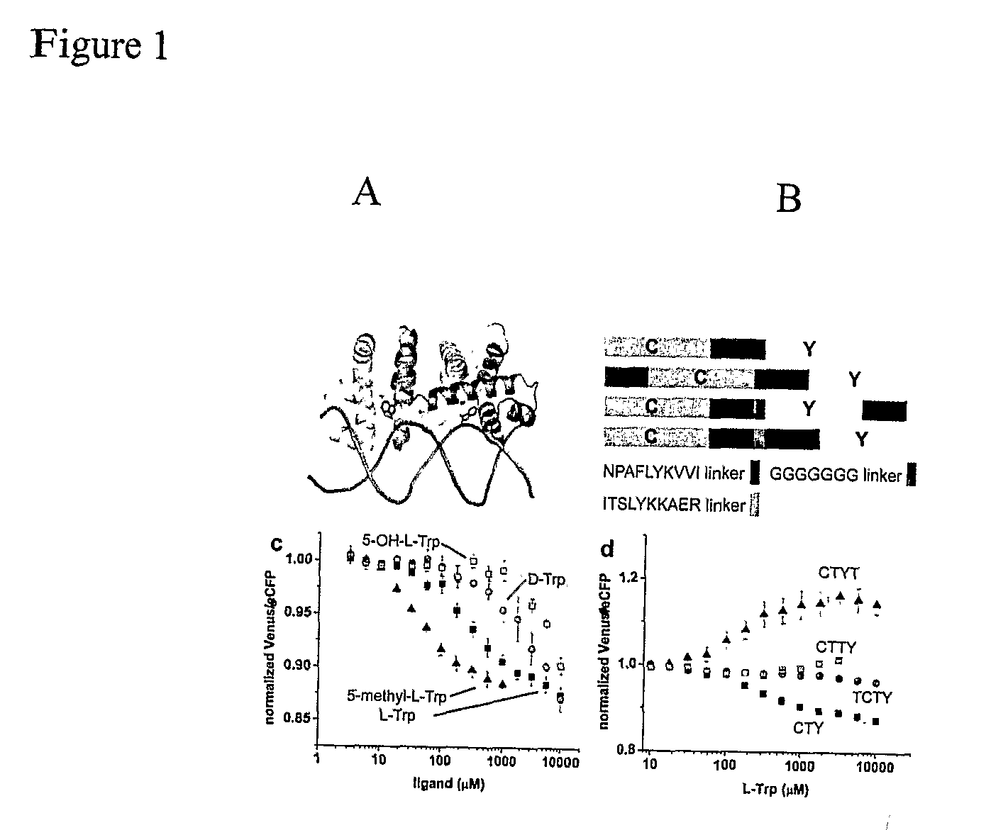
[0099]In this Example, the tryptophan operon repressor TrpR is the ligand binding protein moiety used to generate biosensors. The E. coli tryptophan operon repressor TrpR is an all-helical protein of 108 amino acids organized into 6 α-helices that selectively binds L-tryptophan with micromolar affinity (FIG. 1A) (Marmorstein et al. 1987). The active conformation of TrpR is a dimer in which 5 of the 6 helices are involved in intermolecular contacts (Schevitz et al. 1985). With both chains contributing to the tryptophan-binding sites, two TrpR molecules are necessary to form two functional sites (FIG. 1A). In the absence of tryptophan, a part of TrpR is unfolded (Reedstrom et al. 1995), which likely corresponds to the helix-turn-helix motifs that form the ‘DNA-reading heads’, since they undergo structural rearrangements upon binding of tryptophan in TrpR crystal structures (Zhang et al. 1987) and their flexibility is essential for the recognit...
example 2
Twin-Cassette FLIPW Nanosensor Variants
[0100]As a result of the unique conformational properties of TrpR, the functional FLIPW-CTY sensor consisted of two TrpR molecules and four fluorophores tightly packed together, which potentially could affect the binding affinity due to steric hindrance or result in signal loss due to averaging of the fluorophore signals. It was rationalized that sensors with a single fluorophore pair would have improved sensing characteristics, and therefore the three possible sensors containing two TrpR copies in a single gene product (treating the two fluorophores as equivalent) were constructed (FIG. 1B). In variants FLIPW-TCTY and FLIPW-CTYT the termini of the green fluorescent protein variants span the distance between the N- and C-termini of the respective TrpR molecules in the dimer (˜22 Å, see FIG. 10). One of the fluorophores in these variants should therefore be rotationally restrained by these attachment points, which could improve signal change due...
example 3
Molecular Modeling of FLIPW Sensors
[0101]In this Example, molecular modeling was performed to explain the observed FRET signal changes. The original sensor, FLIPW-CTY, is predicted to dimerize, resulting in two pairs of eCFP-Venus fluorophores close to one another on either side of the TrpR dimer (FIG. 7A). This sensor showed the highest FRET efficiency and a significant FRET decrease upon ligand binding. The FLIPW-CTYT sensor is modeled to form the functional TrpR dimer intra-molecularly, resulting in a single eCFP and a single Venus molecule per sensor (FIG. 7B). This sensor has a lower initial FRET ratio, consistent with the greater distance between the fluorophore dipoles, but the relative FRET change is higher, perhaps due to the rigidification of the Venus molecule by its fusion to both TrpR monomers. The FLIPW-CTTY and FLIPW-TCTY sensors do not show sufficient ligand-dependent ratio changes to be useful as sensors. Molecular modeling explains the surprising disparity between ...
PUM
| Property | Measurement | Unit |
|---|---|---|
| Molar density | aaaaa | aaaaa |
| Molar density | aaaaa | aaaaa |
| Molar density | aaaaa | aaaaa |
Abstract
Description
Claims
Application Information