Medical image processing apparatus and medical image diagnosis apparatus

Active Publication Date: 2009-01-08
TOSHIBA MEDICAL SYST CORP
View PDF5 Cites 62 Cited by
  • Summary
  • Abstract
  • Description
  • Claims
  • Application Information

AI Technical Summary

Benefits of technology

[0028]In the sixth aspect, the apparatus receives the morphological image of the heart from outside, forms the functional image showing the state of wall motion of the heart by detecting the data indicating the function or the morphology of the heart, and displays the synthetic image based on the morphological image and the functional image. Furthermore, in the sixth aspect, the apparatus specifies the culprit coronary arte

Problems solved by technology

However, it is known that the correlation between blood vessels having developed arteriosclerosis or the like (culprit coronary artery) and the constriction rate is, in general, not particularly high.
Therefore, it is not easy to diagnose or treat with high accuracy by referring to only the constriction rate.
However, it is not possible to accurately specify the position of a culprit coronary artery from only functional images.
Therefore, diagnosis and treatment with accuracy is difficult.
However, in the conventional techniques mentioned above, it is difficult to diagnose or treat with favorable accuracy for the following reasons.
First, in the case of referring to only morphological images or only functional images, there is a problem of the accuracy of the diagnosis as mentioned above.
In particular, in the case of referring to morphological images, there is a risk of overl

Method used


Image

  • Medical image processing apparatus and medical image diagnosis apparatus
  • Medical image processing apparatus and medical image diagnosis apparatus
  • Medical image processing apparatus and medical image diagnosis apparatus

Examples


Example

First Embodiment

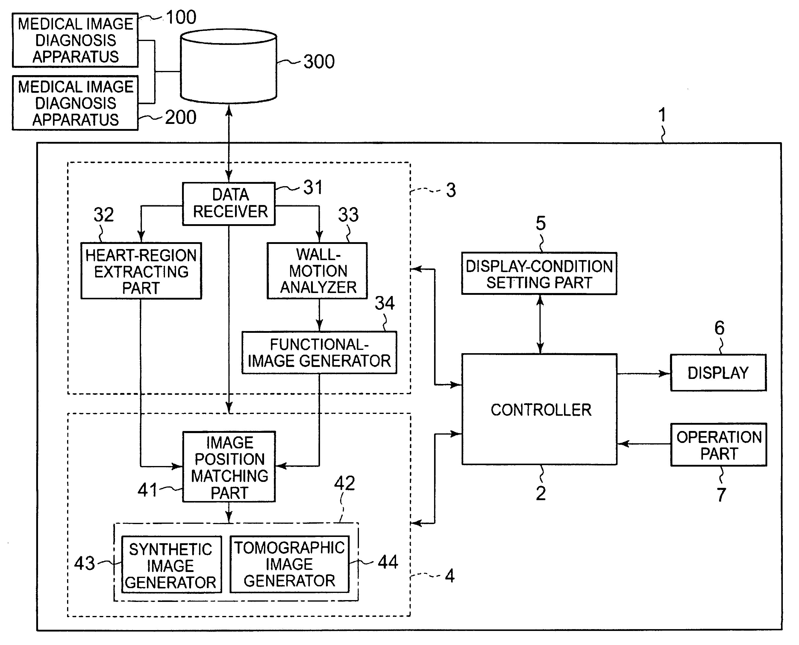
[0047]The medical image processing apparatus according to the first embodiment realizes highly accurate diagnosis and treatment by acquiring a morphological image showing the morphology of a heart and a functional image showing the state of wall motion of the heart to display a synthetic image thereof.

[0048]FIG. 1 shows an example of the configuration of the medical image processing apparatus according to the present embodiment. The medical image processing apparatus 1 shown in FIG. 1 is connected to a medical image database 300 via a communication line such as a LAN (Local Area Network).

[0049]The medical image database 300 stores and manages medical information such as medical images acquired by various types of medical image diagnosis apparatuses. The medical image database 300 includes a large-capacity storage device such as a hard disk drive and a computer program for managing the medical information stored in the storage device.

[0050]The medical image database ...

Example

Second Embodiment

[0157]A medical image processing apparatus according to a second embodiment of the present invention acquires a morphological image showing the morphology of a heart and a functional image showing the state of wall motion of the heart, extracts a coronary artery region from the morphological image, and displays a synthetic image of the coronary artery region and the functional image.

[0158]An example of the configuration of the medical image processing apparatus according to the present embodiment is shown in FIG. 4. A medical image processing apparatus 10 comprises a coronary-artery region extracting part 35 included in the data-acquiring part 3, in addition to the configuration of the medical image processing apparatus 1 of the first embodiment (refer to FIG. 1).

[0159]The coronary-artery region extracting part 35 extracts a coronary-artery region from the heart region extracted by the heart-region extracting part 32 from the morphological image. The coronary-arter...

Example

Third Embodiment

[0195]A medical image processing apparatus according to a third embodiment of the present invention acquires a morphological image showing the morphology of a heart and a functional image showing the state of wall motion of the heart, extracts a heart region from the morphological image, and displays a synthetic image of the heart region and the functional image. Furthermore, the medical image processing apparatus according to the present embodiment makes it possible to display a tomographic image at a desired cross-sectional position of a heart based on the functional image.

[0196]A configuration example of the medical image processing apparatus according to the present embodiment is shown in FIG. 7. The medical image processing apparatus 20 comprises a landmark-site extracting part 36 included in the data-acquiring part 3, in addition to the configuration of the medical image processing apparatus 1 in the first embodiment (refer to FIG. 1). Furthermore, the data pr...


PUM


Abstract

A medical image processing apparatus comprises: an acquiring part configured to acquire a morphological image that is formed by a first apparatus and shows the morphology of an organ of an object, and a functional image that is formed by a second apparatus different from the first apparatus and shows the state of the organ; a display; and a processor configured to cause the display to display a synthetic image based on the morphological image and the functional image.

Description

BACKGROUND OF THE INVENTION[0001]1. Field of the Invention[0002]The present invention relates to a medical image processing apparatus and a medical image diagnosis apparatus, and specifically relates to a technology of displaying by synthesizing a morphological image and a functional image of an object.[0003]2. Description of the Related Art[0004]In recent years, the propagation of medical image diagnosis apparatuses for acquiring images representing the morphology or the function of an object has been promoted. Medical image diagnosis apparatuses include the X-ray diagnosis apparatus, X-ray CT (Computed Tomography) apparatus, MRI (Magnetic Resonance Imaging) apparatus, ultrasound diagnosis apparatus, and nuclear medical diagnosis apparatus.[0005]Ischemic heart disease is one of diseases in which the effectiveness of a medical image diagnosis apparatus is demonstrated. Ischemic heart disease is a disease in which constriction is caused in a coronary artery due to the effects of arte...

Claims


Application Information

Patent Timeline
IPC IPC(8): A61B5/00A61B5/055A61B6/03A61B8/08G01T1/161
CPCA61B2019/5289G06F19/321G06T2207/30048G06T2207/10072G06T7/0028A61B2090/364G06T7/33G16H30/20G16H30/40
Inventor WAKAI, SATOSHIYAMAGATA, HITOSHI
Owner TOSHIBA MEDICAL SYST CORP
Why Eureka
Try Eureka