
Method and apparatus for metal artifact reduction in computed tomography
a computed tomography and metal artifact technology, applied in image enhancement, instruments, applications, etc., can solve the problem of no automatic and robust algorithms for metal artifact reduction, and achieve the effect of reducing the importan
- Summary
- Abstract
- Description
- Claims
- Application Information
AI Technical Summary
Benefits of technology
Problems solved by technology
Method used
Image

Examples
Embodiment Construction
Method and Materials
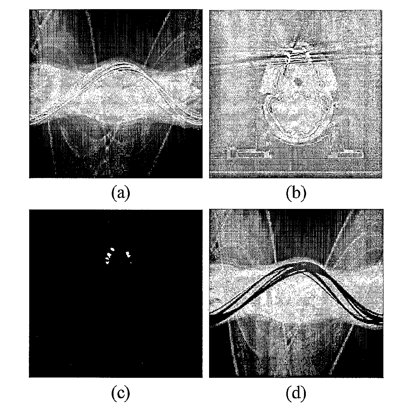
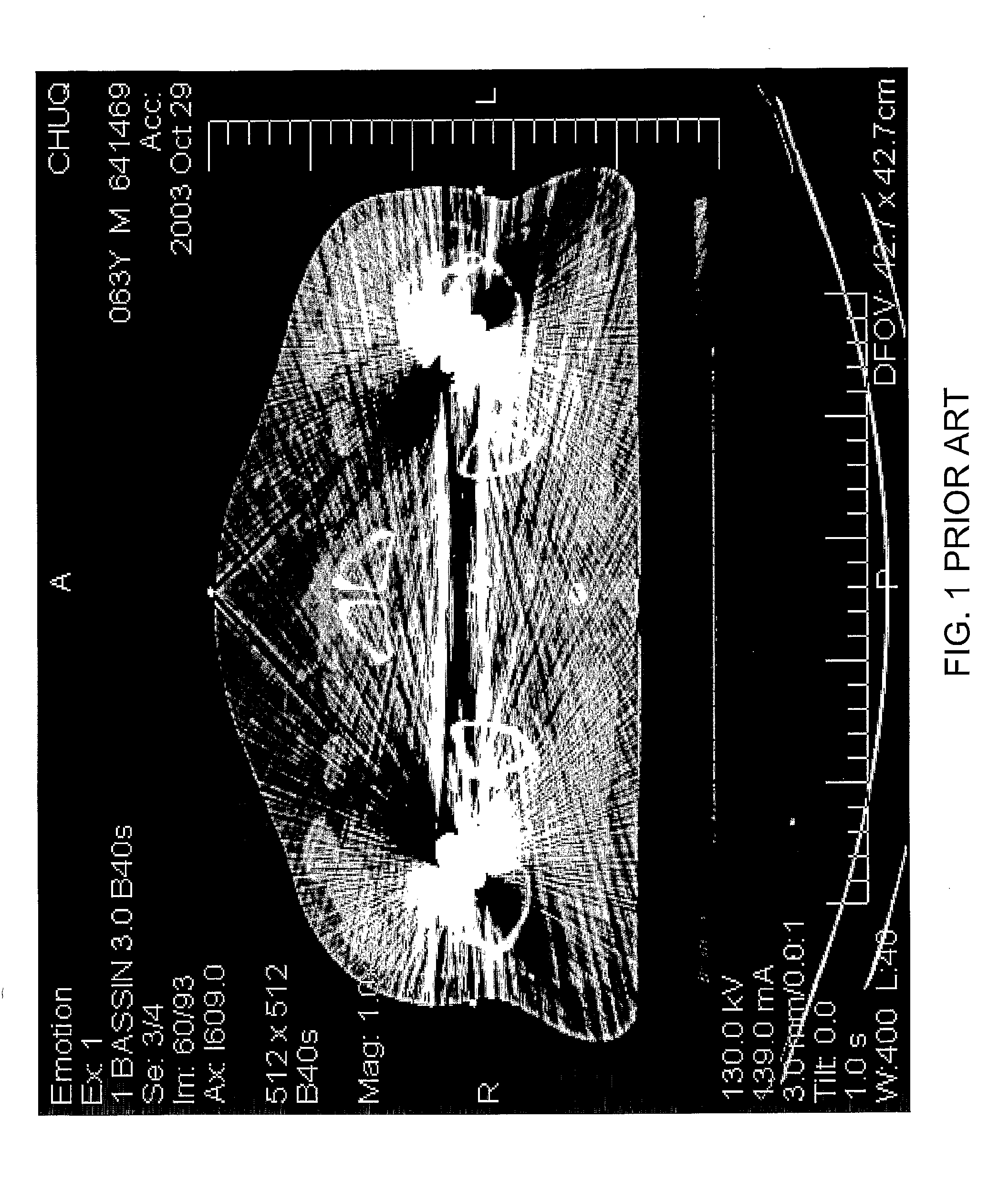
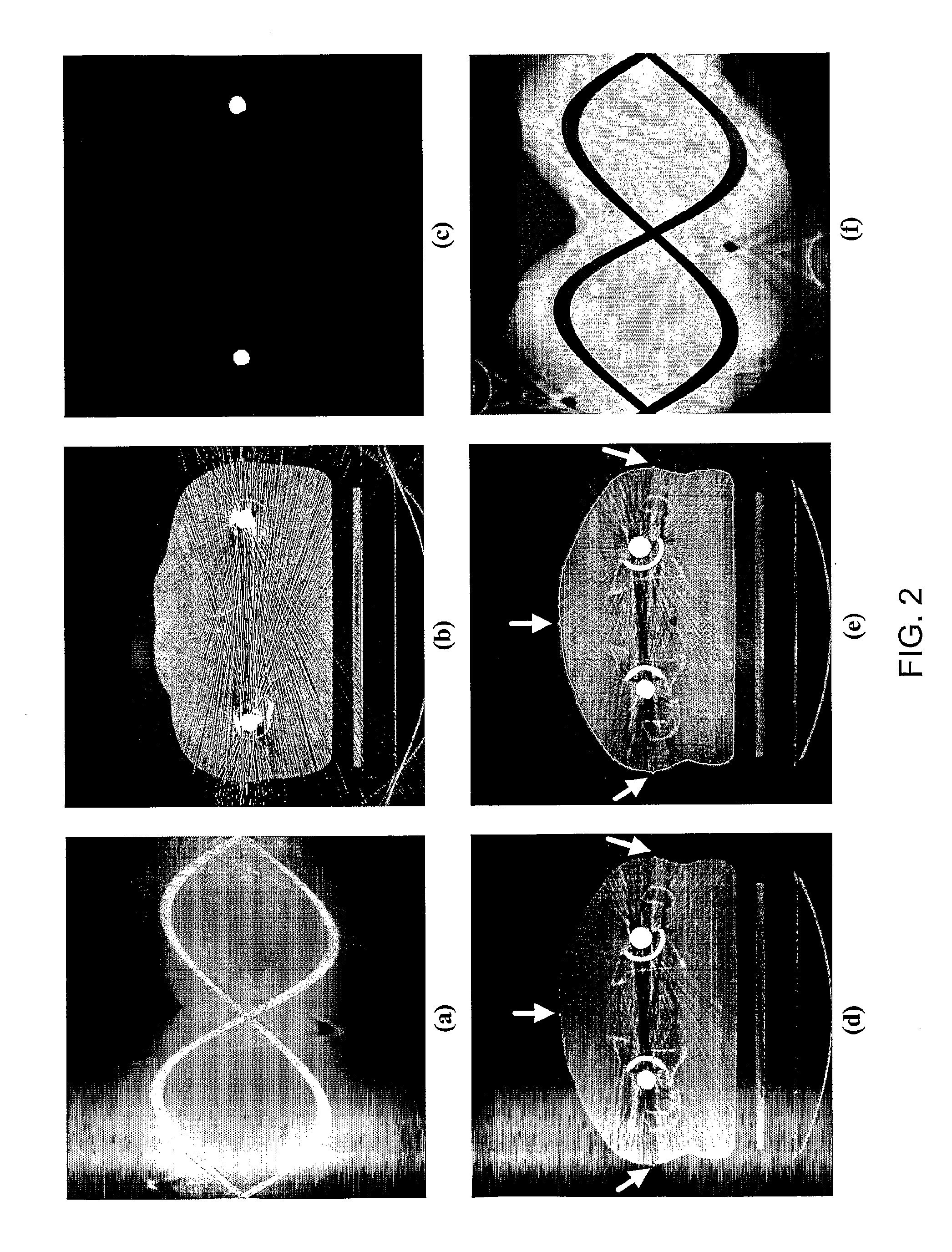
[0042]In a first example, the algorithm is based on the interpolation of missing projections in raw projection data. The modified projection data is used to generate slice images by scanner standard reconstruction algorithm. No further modification in the employed operators is required for this reconstruction. The resulting tomographies are still subject to minor artifact in the area near to the boundary of metal implants, but there are significant gains in image quality for regions of interest such as prostate.
[0043]Three extensions are introduced: the first step is to detect the projections affected by metal implants. Some authors proposed to isolate the correspondence of the metal implants directly from the projection, but have difficulties to fix the appropriate thresholds because of the complex structure of the projection data. Others are identifying the sinusoidal curves resulting from metal implant in the projection data. Although these approaches are inte...
PUM
 Abstract
Description
Claims
Application Information