Device and Method for ReShaping Mitral Valve Annulus

a technology of mitral valve and annulus, which is applied in the field of medical devices and methods, can solve the problems of affecting and many patients cannot withstand the trauma of open-heart surgery, so as to improve the coaption of mitral valve leaflets and reduce the distance

Active Publication Date: 2009-03-19
EDWARDS LIFESCIENCES CORP
View PDF17 Cites 163 Cited by
  • Summary
  • Abstract
  • Description
  • Claims
  • Application Information

AI Technical Summary

Benefits of technology

[0008]In one preferred embodiment, an implantable body is configured for deployment in the right atrium. The body is shaped to apply a lateral force along the atrial septum at a location adjacent to the mitral valve. The force causes the atrial septum to deform, thereby affecting the anatomy on the left side of the heart. More particularly, by pressing on the atrial septum, the anterior leaflet of the mitral valve is pushed toward the posterior leaflet. The amount of force can be selected such that the anterior leaflet is pushed a sufficient amount for closing the gap in the mitral valve and reducing or eliminating mitral valve regurgitation.
[0014]In another aspect, a method for repairing a mitral valve involves providing a repair device having a deployment mechanism for independently applying first and second fastener elements to first and second regions of a mitral valve annulus. The repair device is used to grasp the first region of tissue with a vacuum force and then deploy a first fastener element into the first region of tissue. The first region of tissue is then disengaged from the repair device while leaving the first fastener element deployed therein. The repair device is then used to grasp the second region of tissue with a vacuum force and then deploy the second fastener element into the second region of tissue. The second region of tissue is then disengaged. The first and second fastener elements are then pulled together for reducing the distance between the first and second regions of tissue, thereby improving coaption of the mitral valve leaflets.

Problems solved by technology

These geometric alterations result in incomplete leaflet coaptation during ventricular systole, thereby producing regurgitation.
Although surgical techniques are typically effective for treating heart valve regurgitation, due to age or health considerations, many patients cannot withstand the trauma associated with an open-heart surgical procedure.
Although implants configured for use in the coronary sinus have shown promising results, it has been found that this treatment may not be effective for all patients.
For example, in certain cases, the coronary sinus may be too weakened or fragile to support the implant.
In other cases, due to variations in heart anatomy, the location of the coronary sinus may not be well-situated for treating the mitral valve.
For example, the coronary sinus may be above or below the mitral valve annulus, thereby diminishing the effectiveness of the implant.

Method used


Image

  • Device and Method for ReShaping Mitral Valve Annulus
  • Device and Method for ReShaping Mitral Valve Annulus
  • Device and Method for ReShaping Mitral Valve Annulus

Examples


Embodiment Construction

[0031]Various embodiments of the present invention depict medical implants and methods of use that are well-suited for treating mitral valve regurgitation. It should be appreciated that the principles and aspects of the embodiments disclosed and discussed herein are also applicable to other devices having different structures and functionalities. For example, certain structures and methods disclosed herein may also be applicable to the treatment of other heart valves or other body organs. Furthermore, certain embodiments may also be used in conjunction with other medical devices or other procedures not explicitly disclosed. However, the manner of adapting the embodiments described herein to various other devices and functionalities will become apparent to those of skill in the art in view of the description that follows.

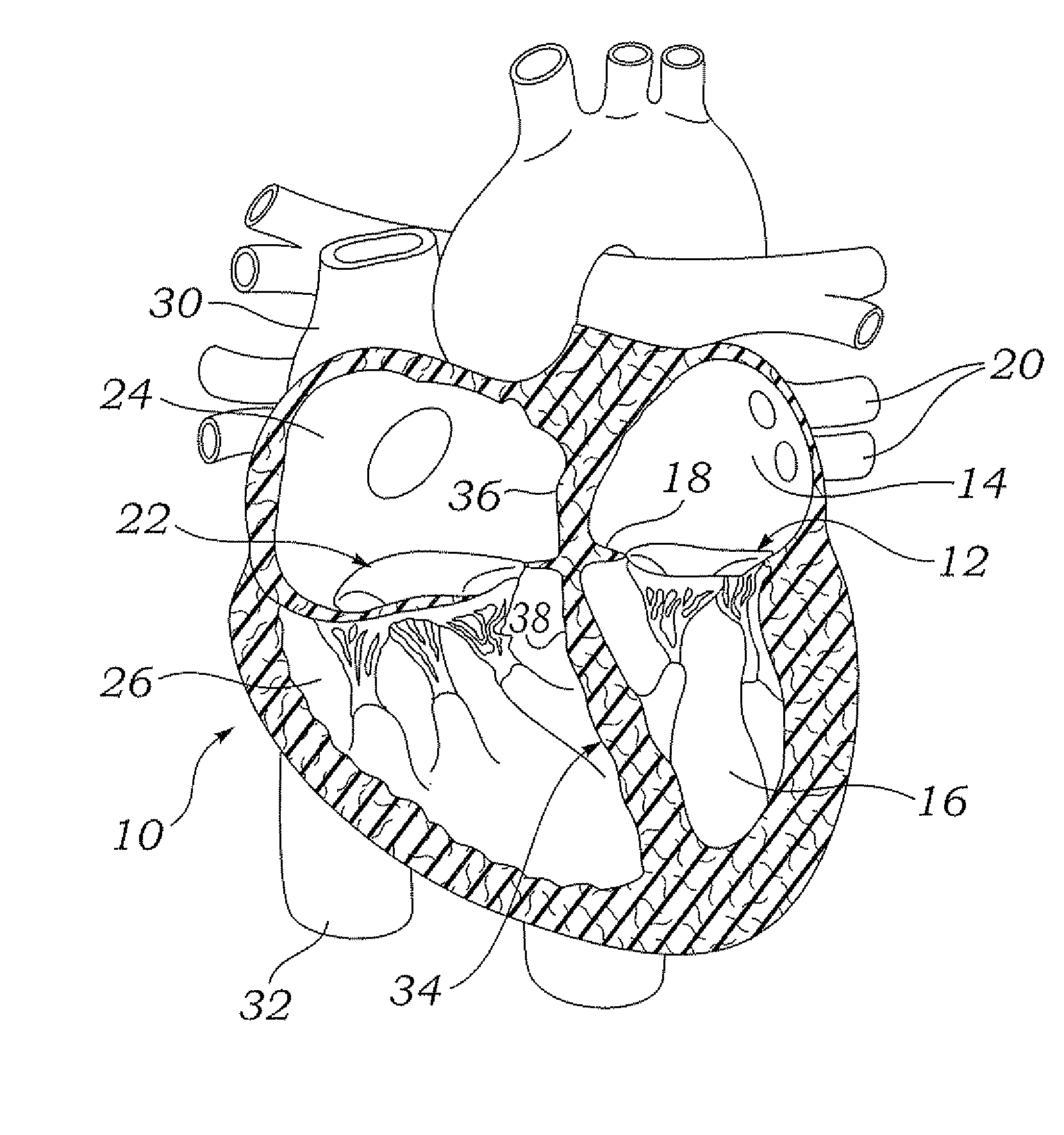
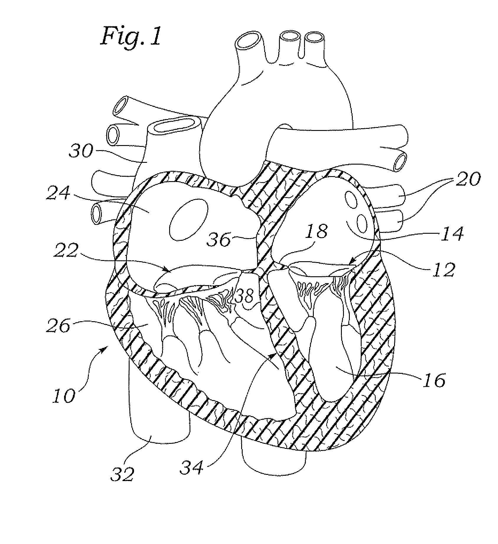
[0032]With reference now to FIG. 1, a four-chambered heart 10 is illustrated for background purposes. On the left side of the heart, the mitral valve 12 is located b...


PUM

PropertyMeasurementUnit
lateral forceaaaaaaaaaa
compressive forceaaaaaaaaaa
forceaaaaaaaaaa

Abstract

Devices and methods for reshaping a mitral valve annulus are provided. One preferred device is configured for deployment in the right atrium and is shaped to apply a force along the atrial septum. The device causes the atrial septum to deform and push the anterior leaflet of the mitral valve in a posterior direction for reducing mitral valve regurgitation. Another preferred device is deployed in the left ventricular outflow tract at a location adjacent the aortic valve. The device is expandable for urging the anterior leaflet toward the posterior leaflet. Another preferred device comprises a tether configured to be attached to opposing regions of the mitral valve annulus.

Description

RELATED APPLICATIONS[0001]The present application is a continuation of application Ser. No. 11 / 519,645, filed Sep. 11, 2006, which claims priority to Provisional Application No. 60 / 716,012, filed on Sep. 9, 2005.FIELD OF THE INVENTION[0002]The present invention relates to medical devices and methods and, more particularly, to medical devices and methods for repairing a defective mitral valve in a human heart.BACKGROUND[0003]Heart valve regurgitation, or leakage from the outflow to the inflow side of a heart valve, occurs when a heart valve fails to close properly. Regurgitation often occurs in the mitral valve, located between the left atrium and left ventricle, or in the tricuspid valve, located between the right atrium and right ventricle. Regurgitation through the mitral valve is typically caused by changes in the geometric configurations of the left ventricle, papillary muscles and mitral valve annulus. Similarly, regurgitation through the tricuspid valve is typically caused by ...

Claims


Application Information

Patent Timeline
Patent Type & Authority Applications(United States)
IPC IPC(8): A61F2/06A61B19/00A61F2/82
CPCA61F2/2445A61F2/82A61F2/2454A61F2/2442A61F2/246A61F2/2478A61F2/2487A61F2002/8483A61F2210/0004A61F2210/0014A61F2220/0016A61F2230/0004
Inventor HAUSER, DAVID L.BOBO, JR., DONALD E.SOLEM, JAN OTTO
Owner EDWARDS LIFESCIENCES CORP
Why Eureka
Try Eureka