Capsule endoscope system and method of processing image data thereof

a technology of endoscope and endoscope, applied in the field of endoscope system, can solve the problems of patients and medical staff suffering from various inconveniences, time waste, pain and unpleasantness, etc., and achieve the effect of accurate diagnosis

Inactive Publication Date: 2009-05-21
INTROMEDIC
View PDF14 Cites 33 Cited by
  • Summary
  • Abstract
  • Description
  • Claims
  • Application Information

AI Technical Summary

Benefits of technology

[0021]Another object of the present invention is to provide a capsule endoscope system and method of processing image data thereof, by which internal organs in numerous image data received from a capsule endoscope are easily discriminated from each other.
[0028]In particular, if the attribute information indicates a diagnostic result by a doctor, when the image data is stored, doctor's comments inputted by the doctor are interconnected to the stored image data. Hence, the present invention facilitates overall diagnostic result of patient to be confirmed and analyzed.
[0030]Therefore, the present invention facilitates a doctor to make an accurate diagnosis from numerous image data received from capsule endoscope.

Problems solved by technology

Unfortunately, the endoscope causes pain and unpleasantness accompanying with the examination to a patient.
Specifically, although endoscopes have been used for various diagnoses and checkups, patients and medical staffs have suffered from various inconveniences due to sizes and rigidity of conduit.
Nonetheless, the doctor has to keep watching the entire images displayed on the monitor.
As the doctor diagnoses a patient's case by checking the whole images displayed on the monitor, time is uselessly wasted and it is a time-consuming job.
Since the related art fails to support attribute information containing diagnostic details on the still images displayed on the separate display area at all, it is difficult to effectively obtain a diagnosis on a disease-possible part.
Yet, the related art enables the received image to be displayed on the monitor only but fails to provide any logical tools that facilitate a doctor to discriminate internal organs from each other.
Since the doctor should keep an eye on the images to recognize changes of the internal organs while the whole images pass in accordance with a flow of time, the doctor has difficulty in diagnosing a precise disease-possible part.

Method used


Image

  • Capsule endoscope system and method of processing image data thereof
  • Capsule endoscope system and method of processing image data thereof
  • Capsule endoscope system and method of processing image data thereof

Examples


Embodiment Construction

[0041]Reference will now be made in detail to the preferred embodiments of the present invention, examples of which are illustrated in the accompanying drawings. Wherever possible, the same reference numbers will be used throughout the drawings to refer to the same or like parts.

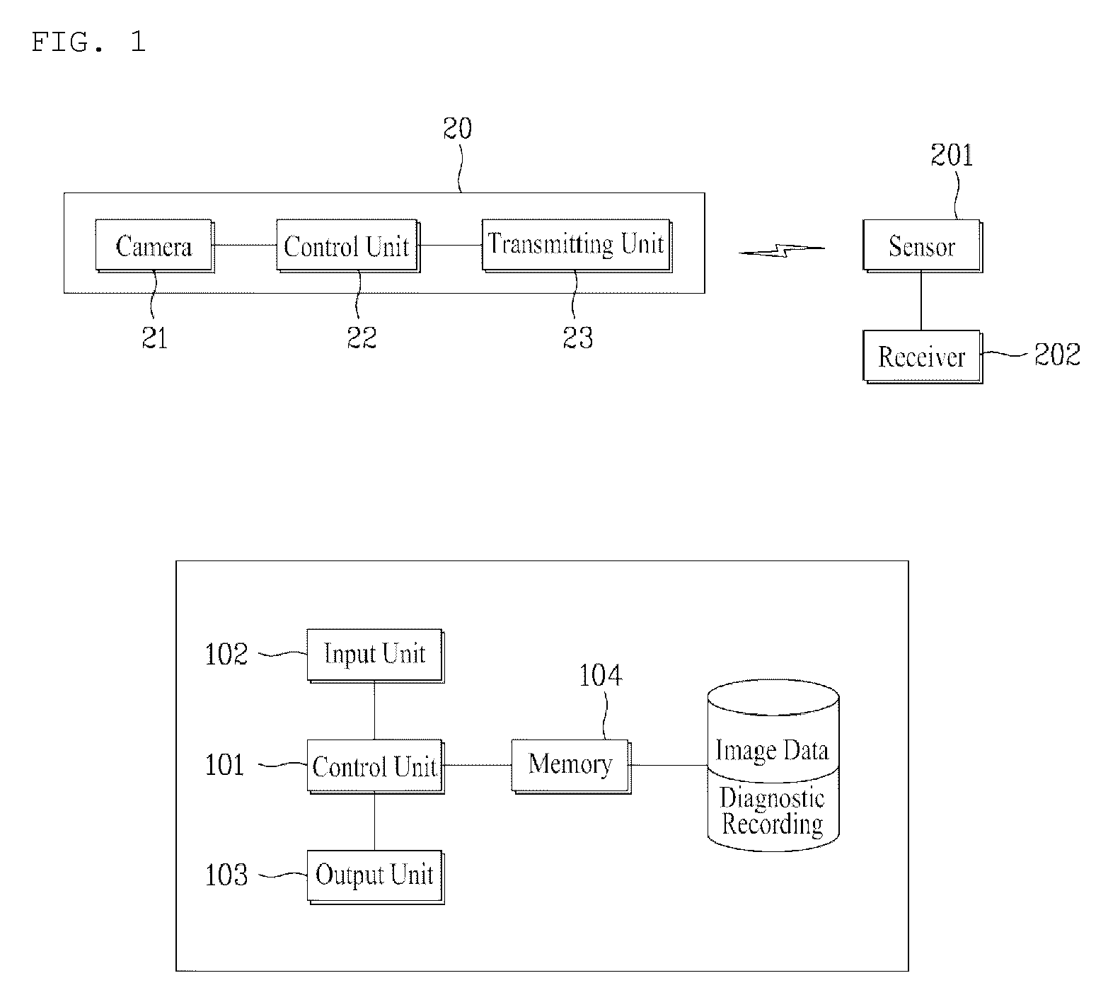
[0042]FIG. 1 is a block diagram of a capsule endoscope system according to one embodiment of the present invention, in which a configuration for displaying image data is shown.

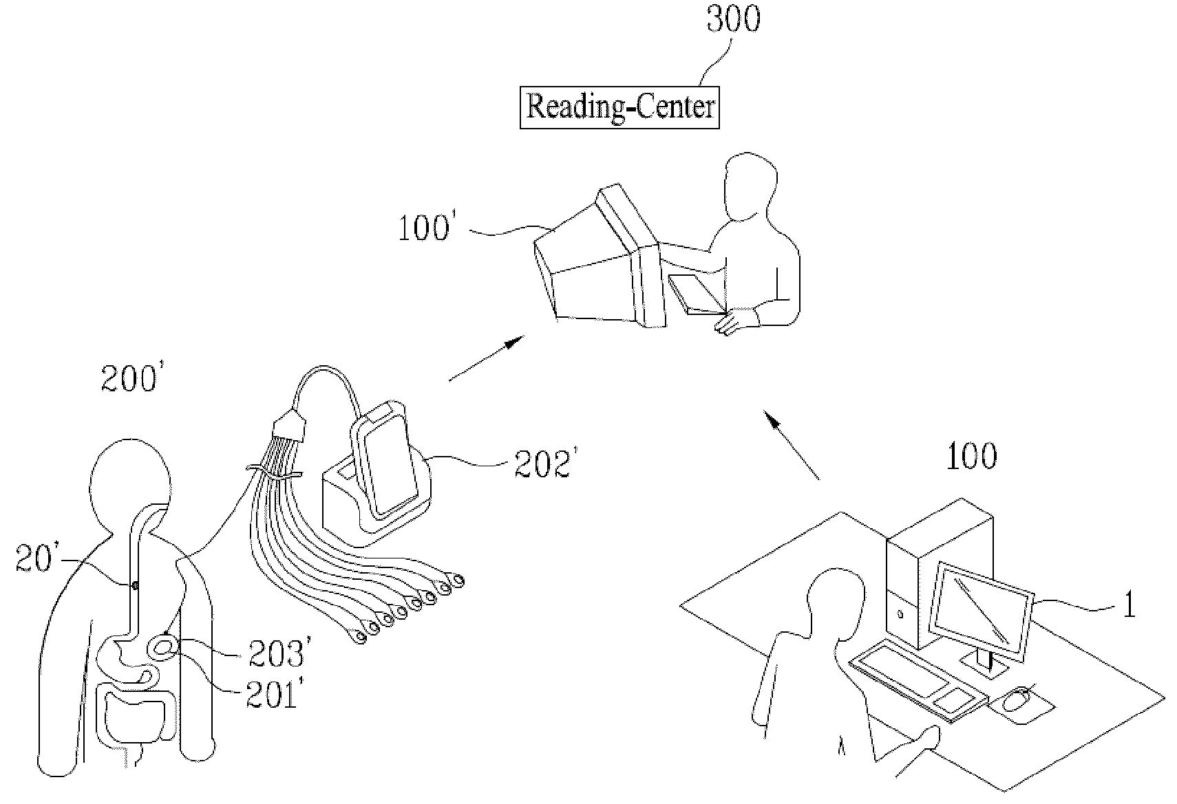
[0043]Referring to FIG. 1, a system according to the present invention includes a capsule endoscope 20, a receiver 202, and a workstation 100.

[0044]The capsule endoscope 20 has a capsule shape and approximately has a radius 11 mmm and a length 24 mm to capture internal organ images of a human body. Once a patient swallows the capsule endoscope 20, the capsule endoscope 20 migrates to the anus from the gullet along a digestive organ for about 10 hours. In the course of the migration, a camera 21 provided within the capsule endoscope 20 ...


PUM


Abstract

The present invention relates to a capsule endoscope system and method of processing image data thereof and is suitable for capturing the inner parts of human body using a capsule endoscope having an internally-usable capsule size and receiving to display image data generated from the capturing. By the present invention, a possible-disease part of a patient is selected and observed through a number of image data received from a capsule endoscope, attribute information containing diagnostic details are supported to facilitate an overall diagnostic result to be interpreted, and internal organs in numerous image data received from a capsule endoscope are easily discriminated from each other.

Description

[0001]This application claims the benefit of the Korean Patent Application No. 10-2007-0116702, filed on Nov. 15, 2007, which is hereby incorporated by reference as if fully set forth herein.BACKGROUND OF THE INVENTION[0002]1. Field of the Invention[0003]The present invention relates to a capsule endoscope system, and more particularly, to a capsule endoscope system and method of processing image data thereof. Although the present invention is suitable for a wide scope of applications, it is particularly suitable for capturing the inner parts of human body using a capsule endoscope having an internally-usable capsule size and receiving to display image data generated from the capturing.[0004]2. Discussion of the Related Art[0005]Generally, an endoscope is an insertion type medical instrument devised to observe internal organs for medical examination of diseases without surgery or autopsy.[0006]Unfortunately, the endoscope causes pain and unpleasantness accompanying with the examinat...

Claims


Application Information

Patent Timeline
Patent Type & Authority Applications(United States)
IPC IPC(8): A61B1/00G06K9/00G16H30/20G16H30/40
CPCA61B1/00045G06F19/321A61B1/041G16H30/20G16H30/40A61B1/04
Inventor SEO, YOUNG DAESHIM, HAN BOLEE, DONG HA
Owner INTROMEDIC
Why Eureka
Try Eureka