
Virtual non-invasive blood analysis device workstation and associated methods

a workstation and non-invasive technology, applied in the field of blood sample collection and analysis, can solve the problems of pain to the individual, injury to the individual's skin, and many laboratories do not carry out venous cutdown procedures, so as to improve the effect of non-invasive blood analysis

Inactive Publication Date: 2009-06-25
SEIKO EPSON CORP
View PDF6 Cites 11 Cited by
  • Summary
  • Abstract
  • Description
  • Claims
  • Application Information

AI Technical Summary

Benefits of technology

[0016]In view of the foregoing background, it is therefore an o...

Problems solved by technology

Many laboratories do not carry out venous cutdown procedures as a routine method of collecting blood.
They are time consuming, slow, can be painful to the individual whose blood is required for testing, screening and analysis.
There are risks of injury to the individual whose skin and vein must be punctured or pierced with a needle, or cut, in the venous cutdown procedure, to direct the flow of blood into the vacuum-sealed test tube.
There is a risk of infection since a foreign body is being introduced into the body.
The venous cutdown procedure is fraught with risks.
In the catheterization process, some degree of risk of interruption of prescribed medicines exists, including infection and even blood loss.
There are risks of the blood samples being destroyed at any point between the site where it is collected and the laboratory where it is to be analyzed.
There is also risk of injury to the individual taking the sample.
Personnel have been known to accidentally prick themselves with contaminated needles.

Method used


Image

  • Virtual non-invasive blood analysis device workstation and associated methods
  • Virtual non-invasive blood analysis device workstation and associated methods
  • Virtual non-invasive blood analysis device workstation and associated methods

Examples


Embodiment Construction

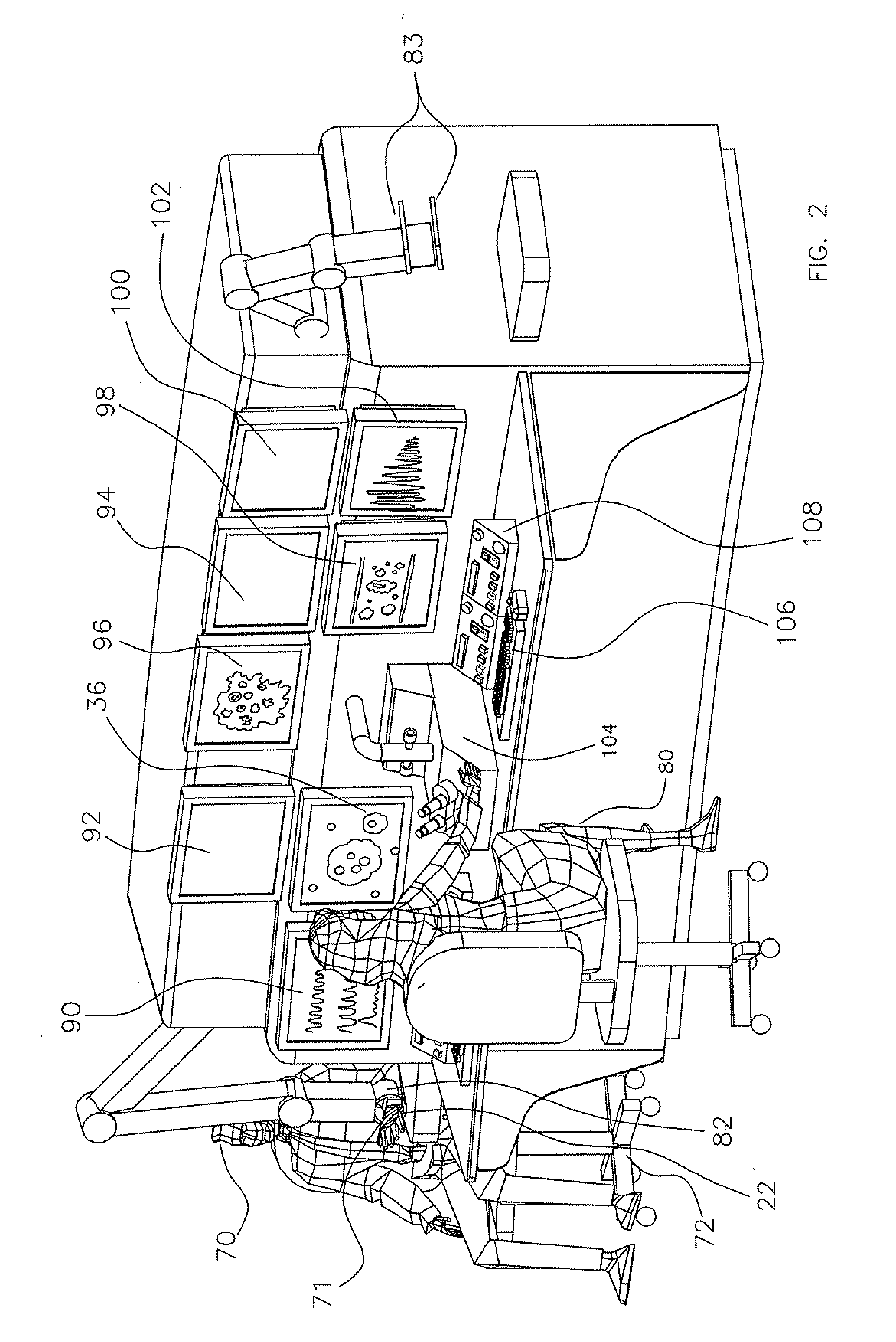
[0041]The present invention will now be described more fully hereinafter with reference to the accompanying drawings, in which preferred embodiments of the invention are shown. This invention may, however, be embodied in many different forms and should not be construed as limited to the embodiments set forth herein. Rather, these embodiments are provided so that this disclosure will be thorough and complete, and will fully convey the scope of the invention to those skilled in the art. Like numbers refer to like elements throughout.

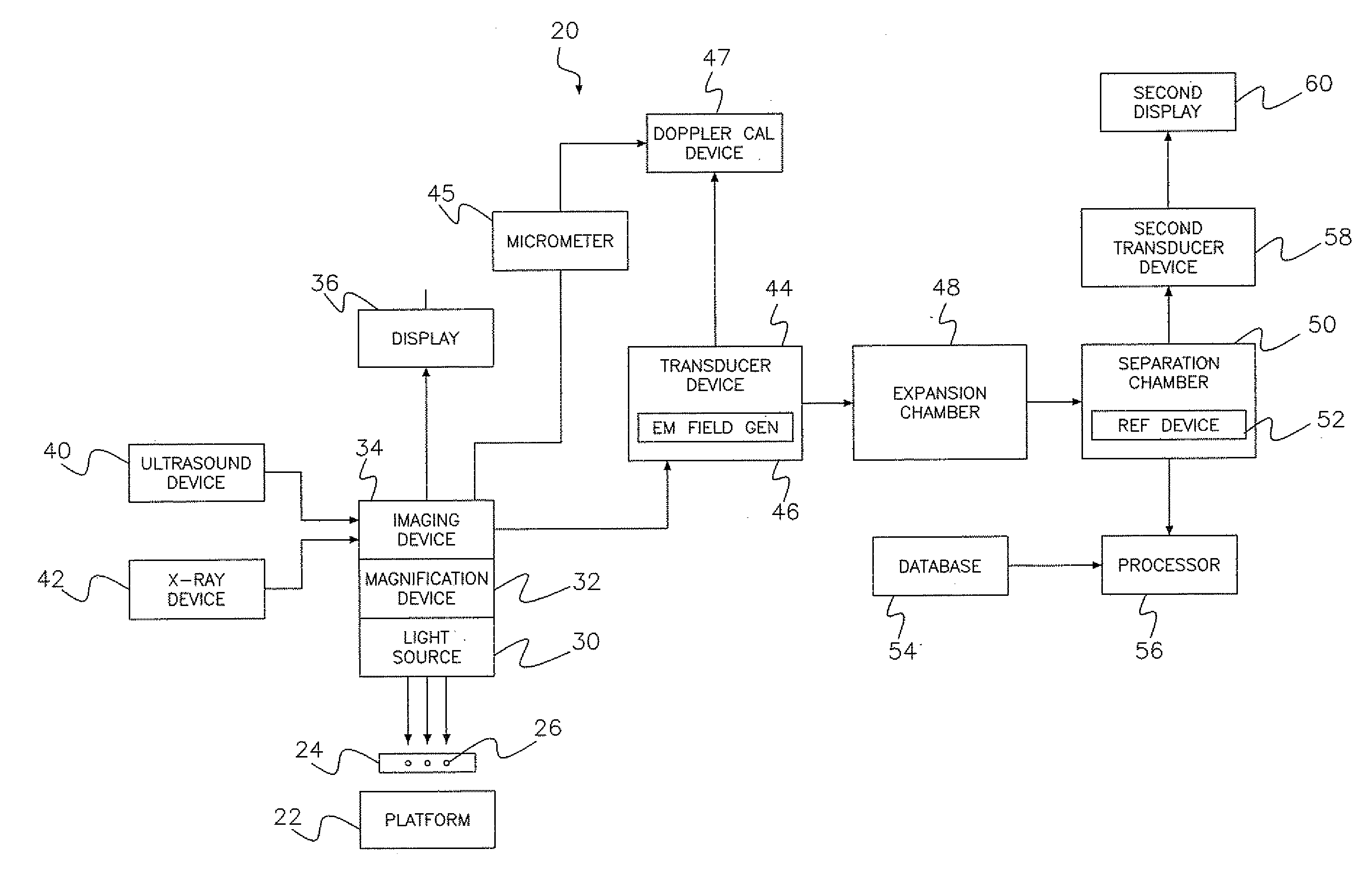
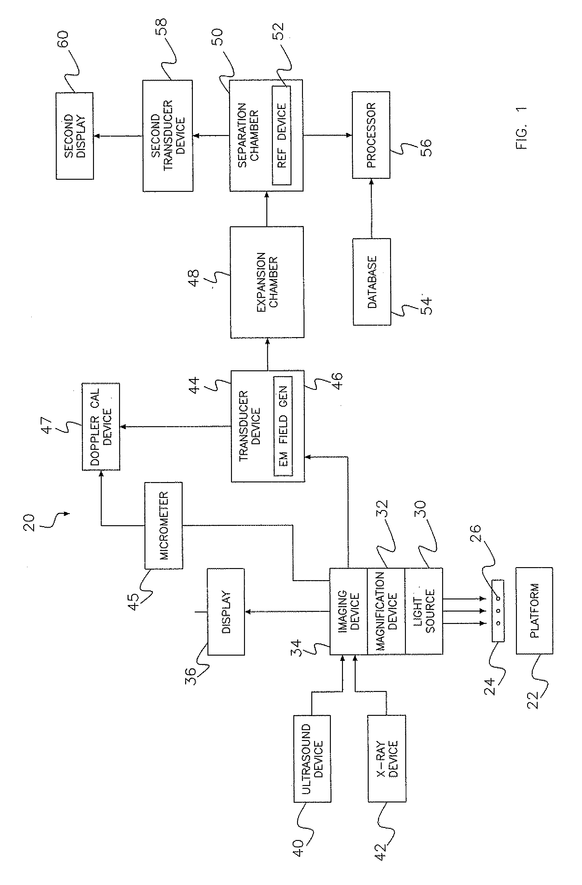
[0042]Referring initially to FIG. 1, a virtual non-invasive blood analysis device workstation 20 includes a support platform or interphase 22 for supporting a body part 24 of an individual or patient. The body part 24 includes blood vessels 26 carrying blood. A light source 30 is adjacent the body part 24 for illuminating a portion of the blood vessels 26. A magnification device 32 magnifies particles of substances in the illuminated portion of the blood v...


PUM


Abstract

A virtual non-invasive blood analysis device workstation includes a light source adjacent the body part of a person for illuminating a portion of a blood vessel therein. A magnification device magnifies particles of substances in the illuminated portion of the blood vessel, and an imaging device captures images of the magnified particles. A transducer device generates electromagnetic waves based on the captured images being exposed to an electromagnetic field, with the electromagnetic waves forming color bands. Each color band corresponds to a respective particle of substance within the blood vessel. A separation chamber separates at least a portion of the color bands within the electromagnetic waves. The separated color bands represent current characteristics of a selected particle of substance within the blood vessel. A processor matches the separated color band according to the selected particle of substance with at least one of the color bands in the database, and compares the current characteristics of the separated color band to the known characteristics of the at least one matched color band.

Description

RELATED APPLICATION [0001]This application claims the benefit of U.S. Provisional Application Ser. No. 61 / 015,247 filed Dec. 20, 2007, the entire contents of which are incorporated herein by reference.FIELD OF THE INVENTION [0002]The present invention relates to the field of blood sample collection and analysis, and more particularly, to a non-invasive blood analysis through an electromagnetic separation process.BACKGROUND OF THE INVENTION [0003]Across the United States and around the world, blood samples are being taken from individuals for analysis. The analysis is intended to identify and quantify substances in the individual's blood. Ideally, this is done at the time the sample is taken. The assumption is that the sample will remain pure from the time it is collected at the collection site until analyzed in a laboratory, which is usually off-site.[0004]Blood is a key organ-tissue in the individual's body, and is an indicator of current health status, which is a predictor of futu...

Claims


Application Information

Patent Timeline
IPC IPC(8): A61B5/1455
CPCA61B5/0008A61B5/0059A61B5/02007A61B5/1072A61B6/4417A61B6/504A61B8/00A61B8/06A61B6/5247A61B5/14546
Inventor NELSON, RILEY H.
Owner SEIKO EPSON CORP
Learn More