
Finger-mounted or robot-mounted transducer device

a transducer and finger-mounted technology, applied in the field of multi-purpose transducers, can solve the problems of insufficient ablation lesions, difficulty in using the drag and burn approach with an endoluminal catheter, and constant movement of the heart, and achieve the effect of convenient manipulation, convenient access, and easy manipulation

Inactive Publication Date: 2009-06-25
ST JUDE MEDICAL ATRIAL FIBRILLATION DIV
View PDF14 Cites 293 Cited by
  • Summary
  • Abstract
  • Description
  • Claims
  • Application Information

AI Technical Summary

Benefits of technology

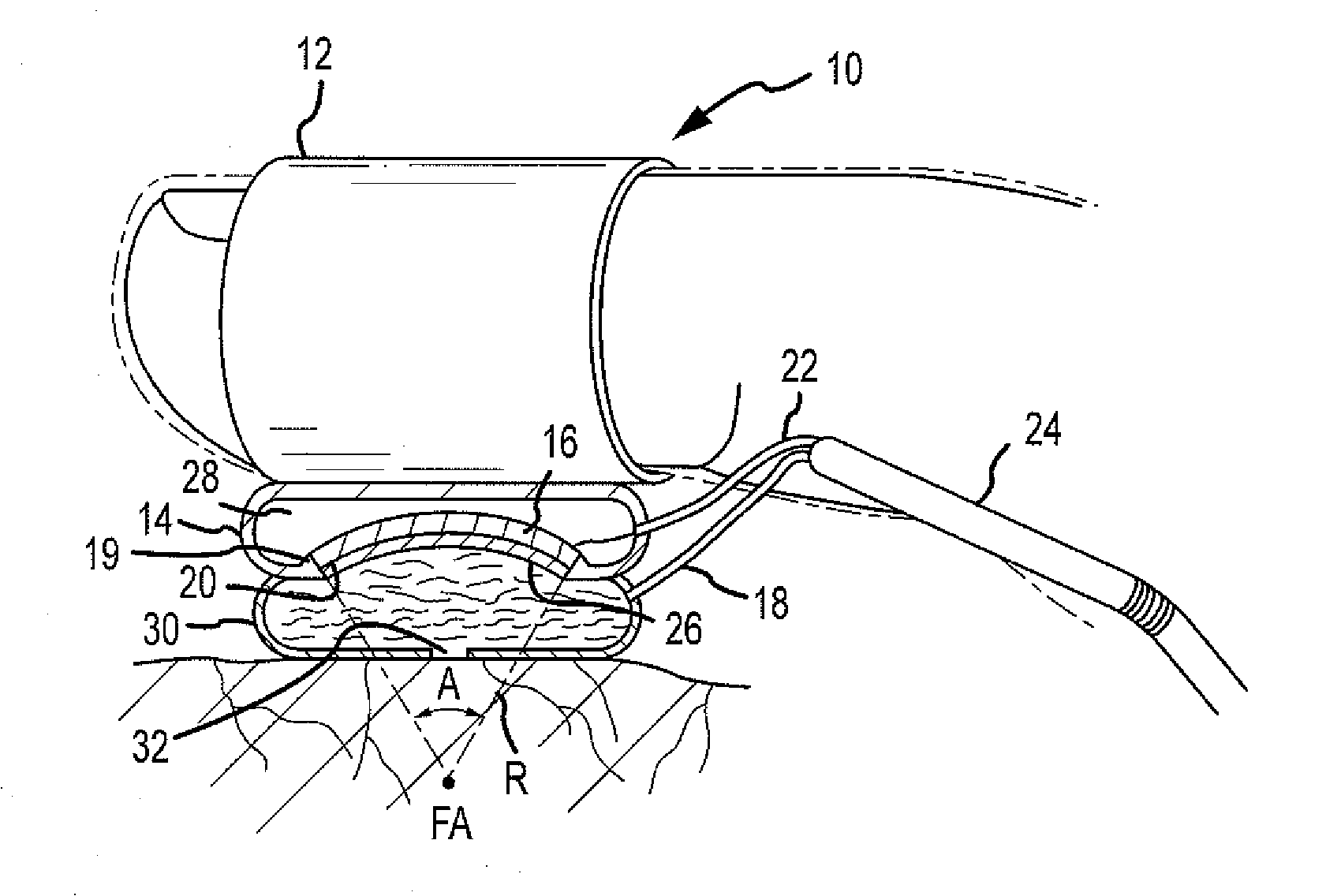
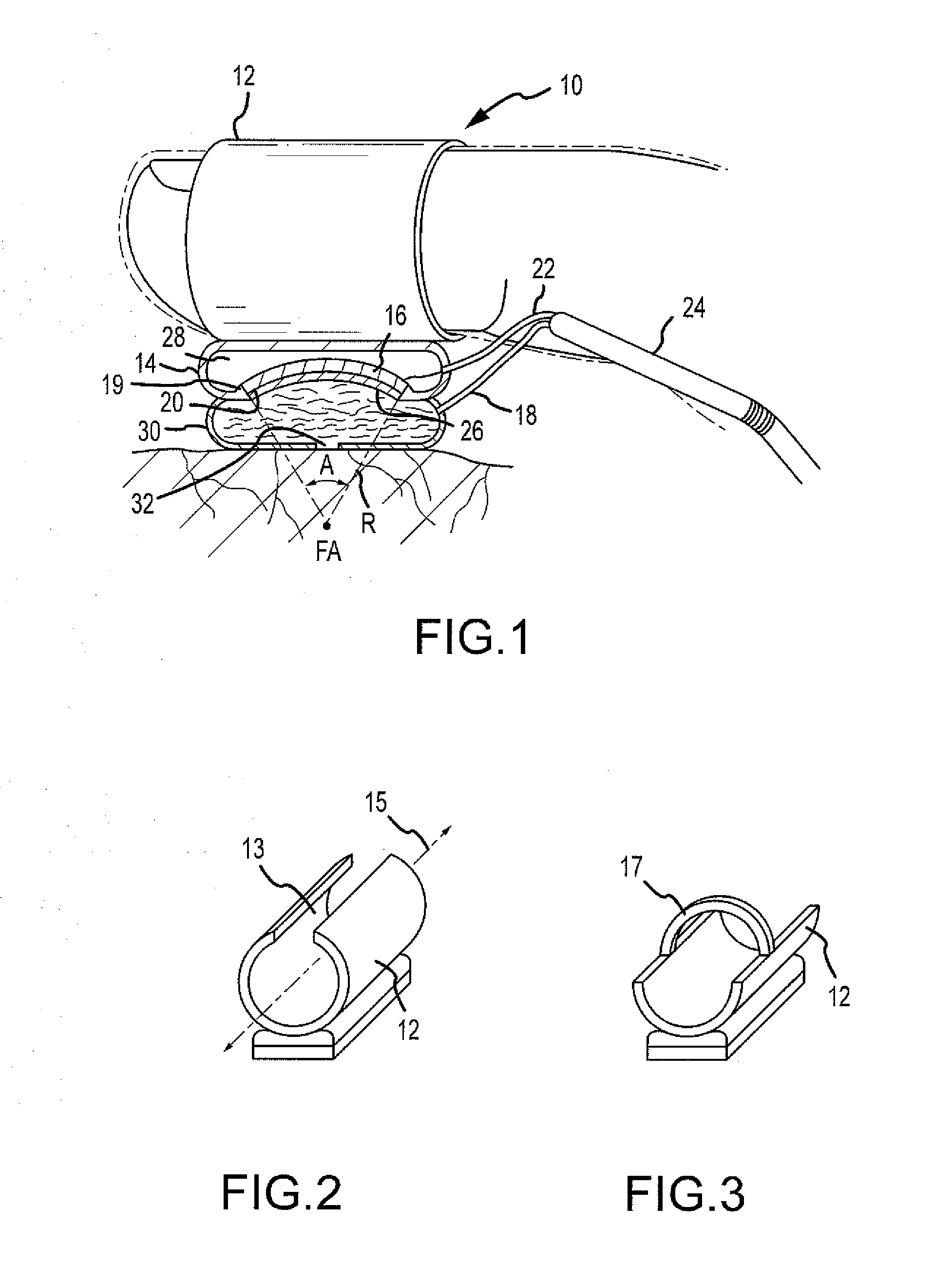
The patent describes a device for procedures and surgeries that can be easily manipulated, such as a finger-mounted tool or a robot's articulator. The device includes a transducer element that can transmit ablating energy to tissue, a gas reservoir to prevent energy from being transmitted away from tissue, and a membrane for cooling and acoustic coupling fluid. The device can be used for therapeutic applications and provides a more efficient and targeted method of applying ultrasound.

Problems solved by technology

Frequently the points are strung together to form elongated blocking lesions; however, this is quite difficult to do using the drag and burn approach with an endoluminal catheter.
One challenge in obtaining an adequate ablation lesion is the constant movement of the heart, especially when there is an erratic or irregular heart beat.
Another difficulty in obtaining an adequate ablation lesion is retaining sufficient and uniform contact with the cardiac tissue across the entire length of the ablation element surface.
Without sufficiently continuous and uniform contact, the associated ablation lesions may not be adequate.
This problem is most severe with catheters.
This location may create difficulties for endocardial ablation devices.
The elongated and flexible catheter-based endovascular ablation devices are difficult to manipulate into the geometries required for forming pulmonary lesions and to maintain in such positions against the wall of a beating heart.
This procedure is time-consuming and may result in lesions which do not completely encircle the pulmonary veins or which contain gaps or discontinuities.
In some cases, procedures may be modified or avoided altogether because of the inability to easily and / or sufficiently access the tissue that has been targeted for treatment.
While a large assortment of tools (e.g., surgical tools) have been designed with pliable or formable shapes in order to try to ameliorate this situation, there remain procedures and surgeries in which the available tools are not sufficient and / or preferred.
In addition, the use of minimally-invasive surgeries (MIS) is increasing, which means that smaller incisions provide for even less ability to navigate tools and instruments.

Method used


Image

  • Finger-mounted or robot-mounted transducer device
  • Finger-mounted or robot-mounted transducer device

Examples


Embodiment Construction

[0017]FIG. 1 illustrates a partial sectional view of a finger-mounted transducer device 10 in accordance with an embodiment of the invention. Device 10 may be configured for use in therapeutic applications. Device 10 may be mounted to a gloved finger as generally illustrated in the depicted embodiment. A surgical glove is illustrated in the figure in phantom. However, in other embodiments, device 10 may instead, for example, be mounted to a finger and then utilized under a glove with the ablative energy then passing through the glove in that case. While the device is described and illustrated as configured for connection (e.g., mounting) to a finger (e.g., a surgeon's finger), the device may also be configured for connection (e.g., mounting) to a robot finger-like appendage. The use of robots to perform procedures and / or surgeries is increasing, and the device can be configured to be utilized for various applications. The finger-mounted transducer device 10 may include a mounting bo...


PUM


Abstract

A transducer device for therapeutic applications is disclosed. The transducer device may include a mounting body configured for mounting the device to a finger of an operator of the device. The transducer device may also include a transducer housing connected to the mounting body that defines a receiving portion. The transducer device may further include a transducer element disposed in the receiving portion that is configured for connection to an energy supply and configured to transmit energy from an emitting surface. The transducer device may further include a gas reservoir disposed between the transducer element and the mounting body that is configured to prevent transmission of energy. The transducer device may further include a membrane connected to the transducer housing and disposed adjacent the emitting surface of the transducer element, and a cooling lumen for providing fluid to the membrane. A method of applying therapeutic ultrasound is also disclosed.

Description

BACKGROUND OF THE INVENTION[0001]a. Field of the Invention[0002]The instant invention is directed toward a multipurpose transducer device, including a finger-mounted or robot-mounted transducer device for therapeutic applications.[0003]b. Background Art[0004]In a normal heart, contraction and relaxation of the heart muscle (myocardium) takes place in an organized fashion as electrochemical signals pass sequentially through the myocardium from the sinoatrial (SA) node located in the right atrium to the atrialventricular (AV) node and then along a well defined route which includes the His-Purkinje system into the left and right ventricles. Atrial fibrillation results from disorganized electrical activity in the heart muscle, or myocardium. The surgical maze procedure has been developed for treating atrial fibrillation and involves the creation of a series of surgical incisions through the atrial myocardium in a preselected pattern so as to create conductive corridors of viable tissue ...

Claims


Application Information

Patent Timeline
Patent Type & Authority Applications(United States)
IPC IPC(8): A61B8/00
CPCA61B19/2203A61B2018/00023A61N7/02A61B2019/261A61B2019/2242A61B8/4272A61B8/4227A61B90/53A61B34/71A61B34/30
Inventor SLIWA, JOHN W.
Owner ST JUDE MEDICAL ATRIAL FIBRILLATION DIV
Learn More