
Wearable photoactivator for ocular therapeutic applications and uses thereof

a technology of ocular therapy and photoactivator, which is applied in the field of wearable photoactivator for ocular therapy applications, can solve the problems of poor visual rehabilitation prospects and virtually useless systemic administration of anti-infective agents, and achieve the effect of convenient exchang

Inactive Publication Date: 2009-07-30
THE JOHN HOPKINS UNIV SCHOOL OF MEDICINE +1
View PDF5 Cites 104 Cited by
  • Summary
  • Abstract
  • Description
  • Claims
  • Application Information

AI Technical Summary

Benefits of technology

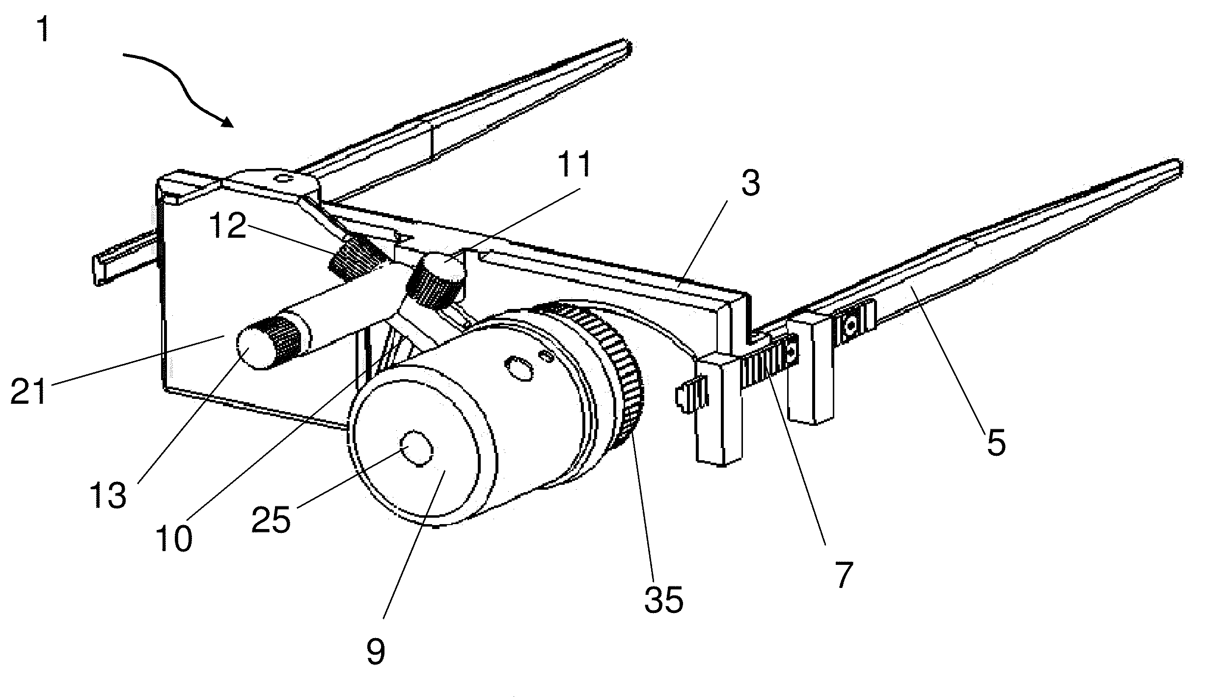
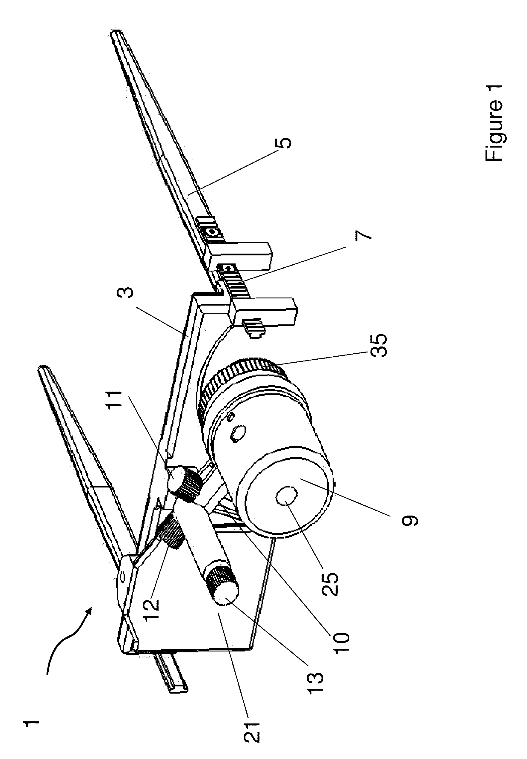
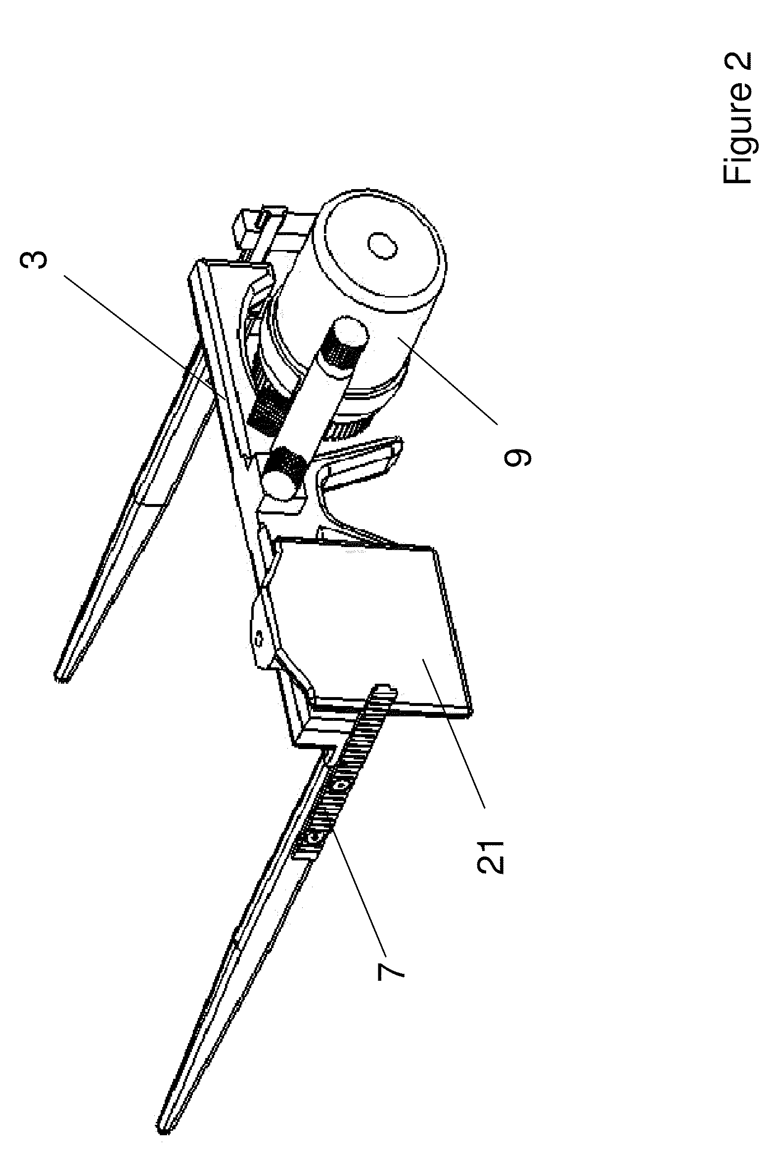
[0008]The invention provides a light source housing having one or more lenses. In some embodiments, the housing includes lenses of more than one size. Such lens housings can be rotated to allow for the alignment of the desired lens(es) with the light source(s). In alternative embodiments, the lens housing attached to the proximal end of the housing can be easily exchanged to provide lenses of the desired size. In an embodiment, the angle of the lenses can be adjusted in the lens housing. The housing can also include an opening that can serve one or more purposes. The opening can be used by an ophthalmologist to align the housing with the appropriate portion of the eye of the subject. The ophthalmologist can introduce the photoactive therapeutic agent through the opening in the housing, using an automated dropper device, or manually using, for example, a medicine dropper. The opening can also be used by the subject to allow the subject to watch television or other form of visual entertainment during the treatment. The eye of the subject not being treated can be covered with an occluder that optionally includes an opening for use by the ophthalmologist, the subject, or both.

Problems solved by technology

Depending on the pathogen responsible of the infection, the prognosis for visual rehabilitation may be poor.
Systemic administration of anti-infective agents is virtually useless, as therapeutic levels are not reached in the area where the infection develops.
Therefore, the cornea constitutes an ideal tissue to harbor living microorganisms, as it is bathed by the tear film with nutrients, and lacks of vessels to allow the protective systems of the body react against pathogenic microorganisms.

Method used


Image

  • Wearable photoactivator for ocular therapeutic applications and uses thereof
  • Wearable photoactivator for ocular therapeutic applications and uses thereof
  • Wearable photoactivator for ocular therapeutic applications and uses thereof

Examples


example 1

Treatment of Corneal Infection Using UV-A Photoactive Therapeutic Agent and a Wearable Photoactivator

[0093]A patient presents with acanthamoebic keratitis in one eye. A lid speculum is used to hold the patient's eye open. The recumbent patient is fitted with a wearable photoactivator of the invention having a UV-A light source. The housing of the light source is adjusted to provide light over 3 to 10 mm spot size on the eye, depending on the area to be exposed, based on the extent of the infection. The fluence of the light is such that it warrants its absorption in the layers of the cornea before penetrating into other ocular structures, thereby reducing the exposure of other structures to the light. A dropper is inserted through an opening in the housing to apply riboflavin to the eye in the form of drops and the riboflavin solution concentration is in the range of about 0.1% to 5% to completely bathe the eye in riboflavin.

[0094]The riboflavin is instilled in the eye every 5 minute...

example 2

Treatment of Keratoconus Using UV-A Photoactive Therapeutic Agent and a Wearable Photoactivator

[0095]A patient presents with keratoconus in one eye. A lid speculum is used to hold the patient's eye open. The recumbent patient is fitted with a wearable photoactivator of the invention having a UV-A light source. The optical system for the light source is adjusted to provide light of 8-9 mm spot size on the eye. The fluence of the light is such that it warrants its absorption in the layers of the cornea before penetrating into other ocular structures. A dropper is inserted through an opening in the housing to apply riboflavin to the eye in the form of drops and the riboflavin solution concentration is in the range of 0.1% to 5% to completely bathe the eye in riboflavin.

[0096]The riboflavin is instilled in the eye every 5 minutes for 15 minutes in the form of eyedrops or soaked in a filter paper disc placed on the surface of the cornea to impregnate the sroma with the photochemical subs...


PUM


Abstract

The invention provides a wearable device for delivery of light of a desired wavelength and power to the cornea of a subject. The device includes a frame for attachment of a light source housing which includes a light source and a lens positioned in the housing to allow light to be directed to the eye of the subject, and the light source is operably linked to a power source. The invention provides method for the prevention and treatment of ocular disease including infection, neoplasia, and corneal dystrophies. The device of the invention can be used in conjunction with photoactive therapeutic agents.

Description

RELATED APPLICATIONS[0001]This application claims priority to U.S. Provisional Patent Application Ser. No. 60 / 994,979 filed on Sep. 24, 2007, which is incorporated by reference herein in its entirety.STATEMENT OF RIGHTS TO INVENTIONS MADE UNDER FEDERALLY SPONSORED RESEARCH[0002]This work was supported in part by the National Eye Institute grant numbers 1R43 EY 015055. The government has certain rights in the invention.BACKGROUND OF THE INVENTION[0003]Infectious keratitis is probably one of the most feared diseases of the cornea. Depending on the pathogen responsible of the infection, the prognosis for visual rehabilitation may be poor. Topical antibiotics and anti-infectives are prone to resistance development, since microorganisms have mechanisms to transform and avoid the effects of these medications. Systemic administration of anti-infective agents is virtually useless, as therapeutic levels are not reached in the area where the infection develops. Therefore, the cornea constitut...

Claims


Application Information

Patent Timeline
IPC IPC(8): A61F9/00A61M37/00
CPCA61F9/0079A61F9/008A61F2009/00872A61N2005/0652A61N5/062A61N5/0624A61N5/0613
Inventor SOLTZ, ROBERTSOLTZ, BARBARA ANNBEHRENS, ASHLEY
Owner THE JOHN HOPKINS UNIV SCHOOL OF MEDICINE
Learn More