Microscopy system having automatic and interactive modes for forming a magnified mosaic image and associated method

a microscopy system and mosaic image technology, applied in the field of microscopy systems having automatic and interactive modes, can solve the problems of unsuitable quantitative evaluation, impractically large scanning time, etc., and achieve the effect of avoiding blurred images and growing faster

Inactive Publication Date: 2009-08-06
TRIPATH IMAGING INC
View PDF22 Cites 24 Cited by
  • Summary
  • Abstract
  • Description
  • Claims
  • Application Information

AI Technical Summary

Benefits of technology

[0026]In view of the deficiencies of the existing image scan devices and methods, the present invention provides a robust, error-tolerant, cost-efficient, high-speed system and method for collecting and assembling contiguous image tiles to form large overview images (virtual slides) of excellent quality with a minimum need for system alignment, calibration, and / or special hardware, while at the same time keeping the potential of being operated in an interactive mode in a user-friendly way, or being operated as a completely automatic unsupervised rare event detection system. Due to these features, the device is suited to be applied to and operated in routine environments of small and mid-sized laboratories as a multi purpose system for accommodating small slide volumes of different natures, while at the same time also meeting the needs of large laboratories as a high-speed, high-throughput system focused on high volume applications and virtual slide scanning for biological slides. The system comprises a microscope device (see FIGS. 1 and 2) with built-in automation functionality, a motorized stage, a fast autofocus device, and an RGB progressive area scan camera. For automatic high volume routine slide processing the system includes an automatic handling device for a plurality of slides such as 50 slides or optionally 200 slides. The motorized microscope stage and slide handler are connected via a controller to a PC or other computer device. The PC is preferably a top end model with good processing power and well equipped with system memory. The camera is linked to the same PC via a frame grabber. A bar code reader facilitates the automatic data management and work flow.
[0028]Recently, progressive area scan cameras with excellent color quality have become commercially available. These cameras acquire a full image at a time so that the introduction of an image jitter for moving objects is eliminated (see FIG. 4). Such cameras generally come with an integrated shutter function, which allows one to electronically adjust the exposure times within a wide range. This further allows one to optically freeze the movement of a passing object without expensive strobe illumination. As these cameras typically can acquire 60 full images per second, low cost continuous motion scanning is feasible.
[0030]Interactive operation: The system can be used like a regular routine microscope with additional quantification capabilities. The operator is able to move the motorized stage via a bicoaxial digipot (FIG. 5) in an interactive manner, just as any other manual microscope stage. The bicoaxial digipot simulates the typical way and feeling of operating a manual microscope stage by using a motorized stage equipped with angle encoders which keep track of the slide location coordinates as the stage is moved. Contrary to a line scan device, the progressive area scan camera allows one to capture and evaluate individual frames. As the microscope is equipped with a regular halogen or LED microscope illumination source, and not a strobing device, the operator can watch and select the objects of interest underneath the microscope and store, on demand, individual FOVs for quantitative evaluation. This is especially useful for fast quantitative assessment of marker expression in histological sections, e.g. for the breast cancer panel, where the pathologist quickly selects a small number of areas of interest underneath the microscope, grabs an image of each field of view, and then the system automatically creates a quantitative assessment of the marker expression and generates the final report. The whole evaluation of a slide done in this way takes, for example, less than a minute.

Problems solved by technology

This introduces a jitter in the resulting assembled full image, which renders the image unsuitable for quantitative evaluation.
However, such a solution leads to impractically large scanning times, on the order of hours (scan for a full slide with a 20× objective) for higher resolution scans.

Method used


Image

  • Microscopy system having automatic and interactive modes for forming a magnified mosaic image and associated method
  • Microscopy system having automatic and interactive modes for forming a magnified mosaic image and associated method
  • Microscopy system having automatic and interactive modes for forming a magnified mosaic image and associated method

Examples


Embodiment Construction

[0055]The present inventions now will be described more fully hereinafter with reference to the accompanying drawings, in which some, but not all embodiments of the inventions are shown. Indeed, these inventions may be embodied in many different forms and should not be construed as limited to the embodiments set forth herein; rather, these embodiments are provided so that this disclosure will satisfy applicable legal requirements. Like numbers refer to like elements throughout.

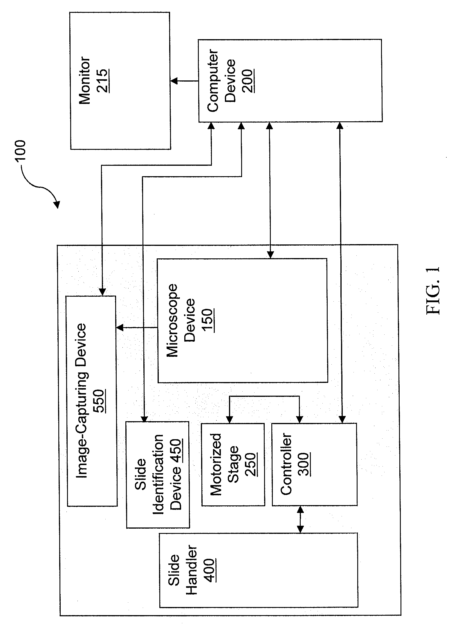
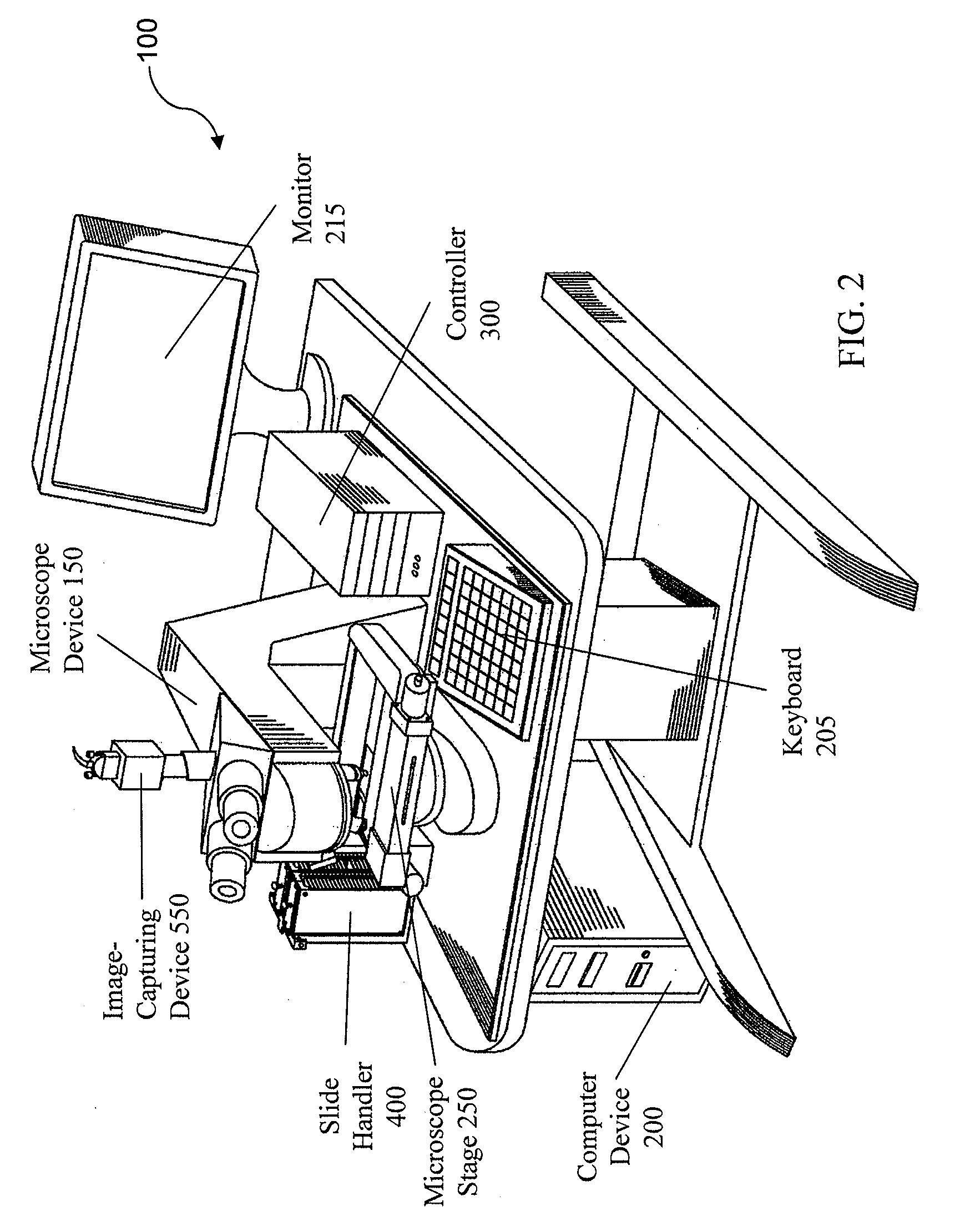
[0056]FIG. 6 shows a block diagram of one embodiment of an automatic-interactive hybrid microscopy system 100 according to the present invention. The system 100 includes a bright field microscope device 150 with automatic Köhler illumination. This means that the microscope illumination settings, such as light intensity and opening diameters of the condenser and field stops can be initially set and stored for each objective mounted on the microscope device 150 to provide optimal Köhler illumination when switchi...


PUM


Abstract

A scanning device for biological slides is provided, which can be operated in an interactive routine mode as well as in an unsupervised high speed automatic mode. In the first case, typical components, which the pathologist is used to operating manually, such as the microscope, the stage and the focus, and which have to be motorized for the automatic unsupervised system mode, are configured to simulate manual use, operation, and response. A non-interlaced area scan camera supports the interactive selection and acquisition of individual images in the manual mode, as well as the continuous high-speed scan motion for the rare event detection and virtual slide scan applications of the system. Due to the particular requirements to accommodate both operational modes, methods are described for constructing the virtual slide out of image tiles with varying overlap areas in the x- and y-directions.

Description

CROSS-REFERENCE TO RELATED APPLICATIONS[0001]This application is a divisional application of U.S. application Ser. No. 11 / 204,954, filed Aug. 16, 2005, which claims the benefit of U.S. Provisional Application No. 60 / 602,463, filed Aug. 18, 2004, which are incorporated by reference herein in their entirety.BACKGROUND OF THE INVENTION[0002]1. Field of the Invention[0003]The present invention relates generally to the acquisition and analysis of digital images of objects and areas of interest located on a microscopic slide, and more particularly, to a microscopy system having automatic and interactive modes, and associated method, for acquiring, storing, displaying and analyzing digital images of areas of interest on a microscopic slide which can be much larger than a field of view (FOV) of the microscope.[0004]2. Description of Related Art[0005]Microscopic analysis is a widely used tool for research and routine evaluations specifically in the field of cellular biology, cytology and pat...

Claims


Application Information

Patent Timeline
Patent Type & Authority Applications(United States)
IPC IPC(8): G06K9/64G06K9/36
CPCG01N15/042G01N15/05G01N2035/00039G06T2200/32G02B21/26G02B21/367G06K9/00127G02B21/0016G06V20/69
Inventor SCHMID, JOACHIM HELMUTGAHM, THOMASKOOY, KEVIN RUSSELLDORRER, RAINERKRIEF, BRUNOCHEESEMAN, JOHN ROYAL
Owner TRIPATH IMAGING INC
Why Eureka
Try Eureka