

Detection and Localized Imaging of Cancer Using X-Ray Fluorescent Nanoparticle/Preferential Locator Conjugates
a fluorescent nanoparticle and localized imaging technology, applied in the field of cancer, can solve the problems of other labels, difficult to accept the procedure, and the inability of surgery to remove tumors
- Summary
- Abstract
- Description
- Claims
- Application Information
AI Technical Summary
Problems solved by technology
Method used
Image

Examples
Embodiment Construction
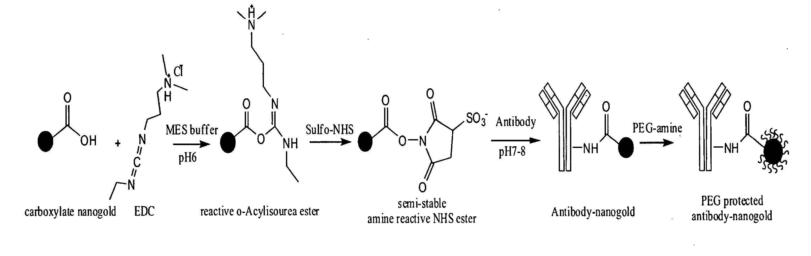
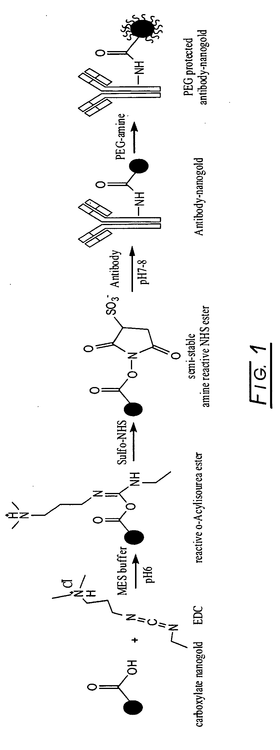
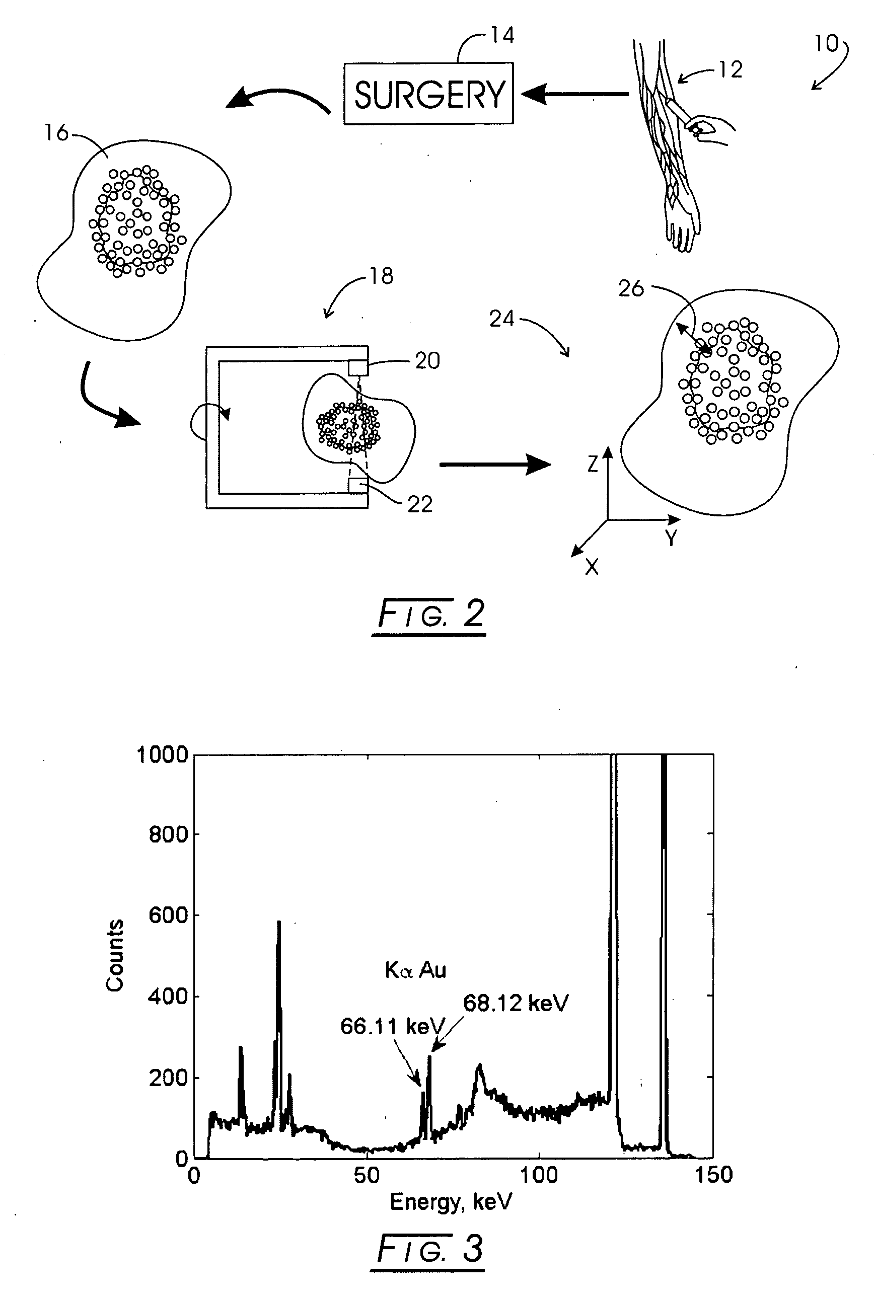
[0022]The fundamental idea is to perform cancer diagnosis and detection with neoplastic tissue preferential locators, such as, for example, cancer-specific antibodies, that have been labeled with a detectable non-radioactive atom that has a relatively high atomic number. In diagnostic practice, for example, the antibodies, preferably monoclonal antibodies (MAbs) are injected into the blood stream of the patient and, after a period of time, preferentially accumulate in malignant sites. Exciting K-shell x-ray emission from the labels and detecting such excitation, then, identify the sites. The excitation is produced by a directed beam of gamma photons that have a sufficiently high energy to remove electrons from the K-shell of the label. The location of the malignant site (neoplastic tissue or cancer, for example) that emits the K-shell x-rays, then, can be determined with a suitable probe or x-ray imaging device.
[0023]This technique has the advantage compared to, for example, mammogr...
PUM
 Abstract
Description
Claims
Application Information