
Method of suppressing obscuring features in an image

a technology of obscuring features and images, applied in image data processing, character and pattern recognition, instruments, etc., can solve the problems of difficult isolation and identification of nodules, difficult to repeat tests, and blunders in the resultant radiography images

Active Publication Date: 2009-08-27
SIEMENS COMP AIDED DIAGNOSIS
View PDF2 Cites 56 Cited by
  • Summary
  • Abstract
  • Description
  • Claims
  • Application Information

AI Technical Summary

Benefits of technology

[0037]c(viii) correlating the smoothed difference profiles of the next and previous columns by applying an affine transformation to the smoothed-difference-profile of the next column so as to minimize the correlation-difference between the previous transformed smoothed-difference-profile and the next transformed smoothed-difference-profile, and
[0044](III) the affine transformation of step c(vi) comprising applying a linear transformation of type x′→ax+b where a defines a degree of stretch and b defines an offset, thereby minimizing correlation-difference between the first smoothed-difference-profile and the transformed second smoothed-difference-profile;

Problems solved by technology

However, in practice patients may inadvertently move or inhale or exhale during the exposure to x-rays, blurring the resultant radiography images somewhat.
Furthermore, since x-ray radiation may itself cause malignancies, radiologists are wary of repeatedly exposing patients to x-rays, particularly with pregnant women, the young, the elderly and / or the ill.
Sometimes, it may be impossible to repeat tests once a problem is determined, as the patient may no longer be available.
The size of a nodule may be smaller, similar or larger than the rib cross-over, and thus isolation and identification of nodules is not easy.
‘False negatives’ where actual nodules or tumors are not identified as such or discarded as ‘noise’ or some artifact of the system are unacceptable since late diagnosis may lead to complications and such mistakes may be fatal.
Despite the importance, in practice the elimination of false positives whilst avoiding false negatives is difficult since tumors may have similar dimensions to the width of ribs or the diameter of blood vessels within the lung, and x-ray images typically have poor contrast.
Explicit rib segmentation is one approached that has been tried in the past and has met with some success however there is often an imprecision in defining the ribs and it will be appreciated that segmentation of the ribs as a precursor to suppression thereof is a computer intensive procedure that expends large amounts of processing power on defining and resolving features to be discarded which is a somewhat inefficient approach.
Additionally, rib suppression based on segmentation which makes use of the varying contrast (grey levels) in the vicinity of the ribs, often results in distortions of the lung tissue which may introduce artifacts, i.e. noise from the processing.
Such distortions may conceal elements of interest as well.

Method used


Image

  • Method of suppressing obscuring features in an image
  • Method of suppressing obscuring features in an image
  • Method of suppressing obscuring features in an image

Examples


Embodiment Construction

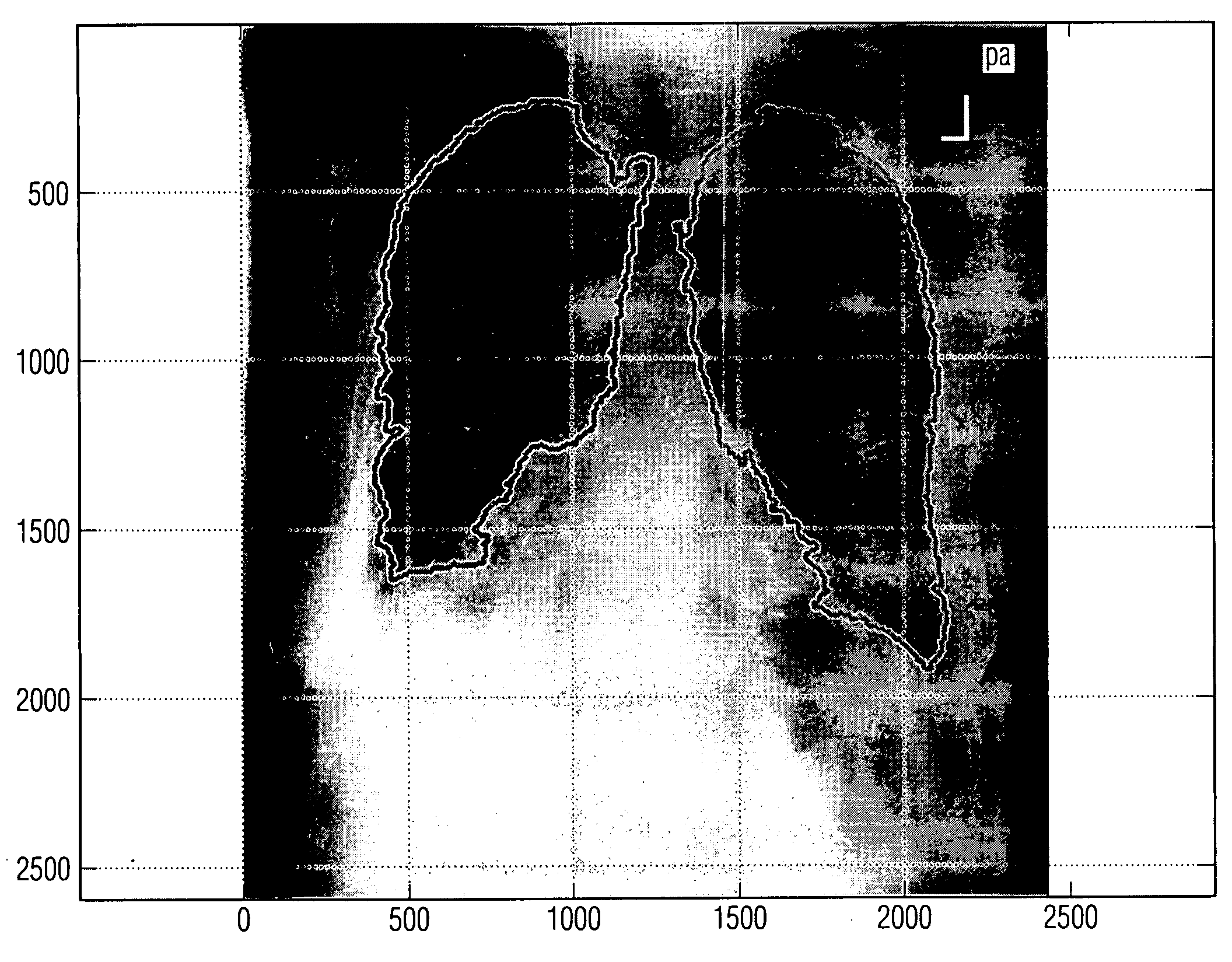
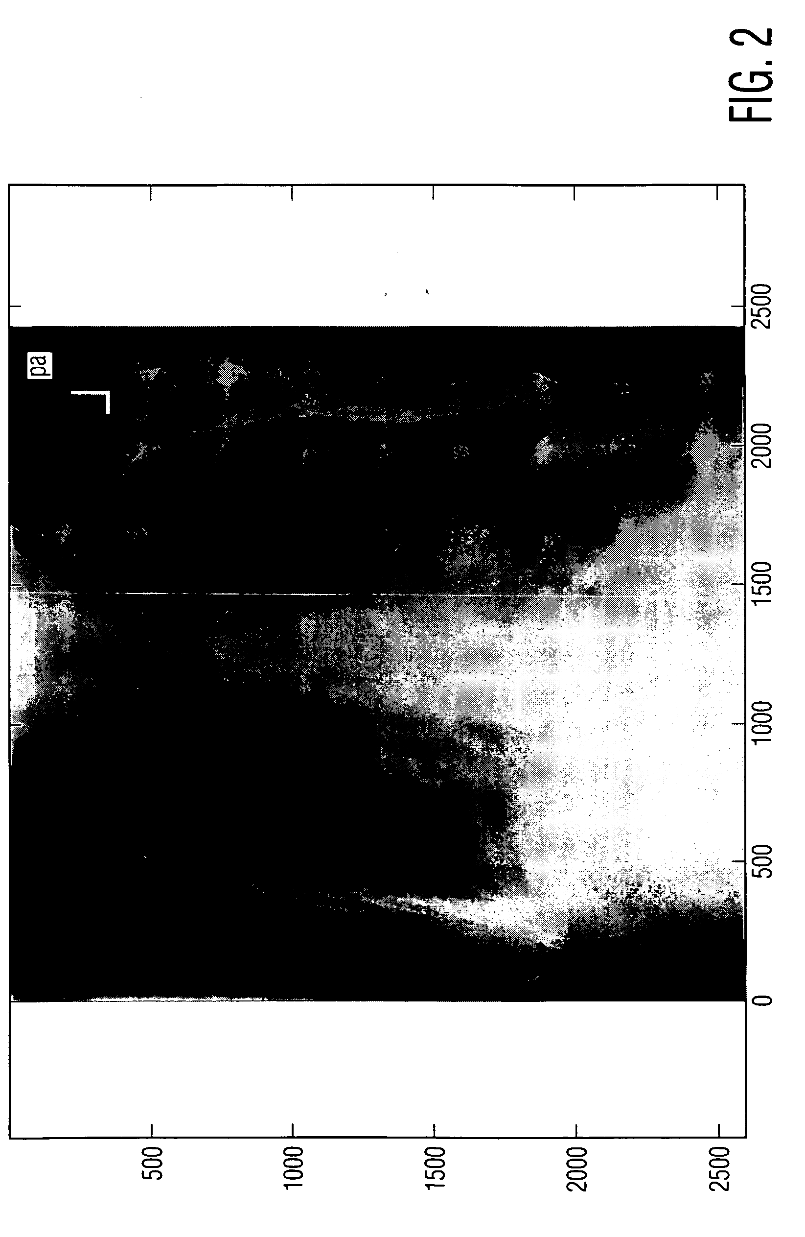
[0080]Chest images are mostly used to diagnose or monitor symptoms of disease and illnesses that manifest themselves by a densification of the soft tissues. The densification leads to a whiter area, confusingly referred to as a shadow, appearing in the x-ray radiograph as conventionally viewed. The lighter shadows of ribs in posterior anterior x-ray radiography images of the chest obscure the features of the soft tissue, particularly nodules in lung tissue, making them difficult to identify. Thus to examine the soft tissue, whether manually or by a Computer Aided Diagnosis CAD system, it is advantageous to suppress or attenuate the rib shadows appearing in x-ray images.

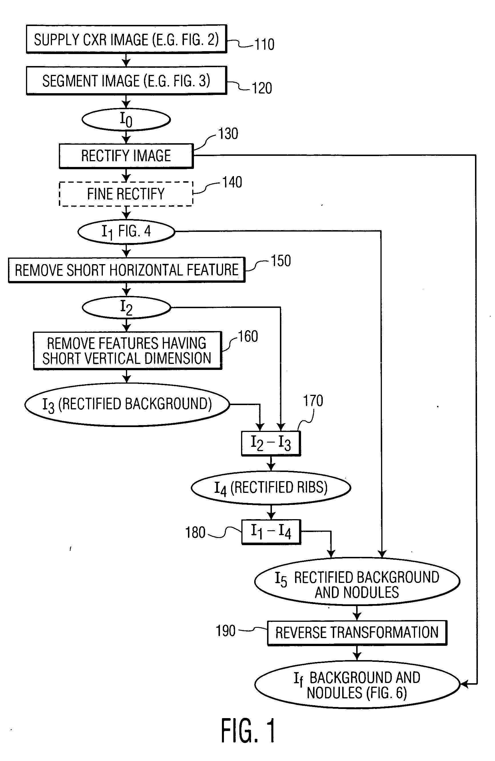
[0081]Specific embodiments of the present invention are directed to identifying ribs showing up in x-ray radiographs to enable their suppression, allowing nodules and other irregularities of interest to be detected. Of note, in preferred embodiments, the ribs are not segmented, and thus a calculation-intensive and thu...


PUM


Abstract

A method of removing obscuring features of relatively standard size and shape from an image comprising the obscuring features, features of interest and background, comprising the steps of applying a geometric transformation to generate a transformed image with the obscuring features aligned with an axis of image; applying an attenuation algorithm to the transformed image to suppress features having a dimension in direction of axis of range appropriate to dimension of the obscuring features in similar alignment, and applying a reverse geometrical transformation to produce a modified image with obscuring features attenuated.

Description

CROSS REFERENCE TO RELATED UNITED STATES APPLICATIONS[0001]This application claims priority from “Straightening The Ribs to Attenuate Them in Chest Images”, Provisional Application No. 61 / 031,783 of Nicolas J. Merlet, filed Feb. 27, 2008, the contents of which are herein incorporated by reference in their entireties.FIELD OF THE INVENTION[0002]The present invention relates to methods of suppressing obscuring features in an image to enable viewing of features of interest. A particular application is for suppressing rib shadows in x-ray radiographs of the thoracic cavity for lung examination purposes, thereby enhancing and improving the detection of lesions.BACKGROUND TO THE INVENTION[0003]The chest x-ray is the most commonly performed diagnostic x-ray examination. In chest x-ray imaging, the heart, lungs, airway, blood vessels and the bones of the spine and chest are imaged in a painless medical test that helps in the diagnosis of medical conditions.[0004]The chest x-ray is typically...

Claims


Application Information

Patent Timeline
Patent Type & Authority Applications(United States)
IPC IPC(8): G06K9/40G06K9/00
CPCG06T7/0081G06T2207/10116G06T5/30G06T5/005G06T2207/30061G06T7/11G06T5/77
Inventor MERLET, NICOLAS J.
Owner SIEMENS COMP AIDED DIAGNOSIS
Learn More