Automatic Recovery of the Left Ventricular Blood Pool in Cardiac Cine Magnetic Resonance Images

a left ventricular blood pool and cine magnetic resonance technology, applied in the field of medical imaging of the heart, can solve the problems of not providing enough information to estimate the peak ejection rate or filling rate, and many physicians can only manually outline the lv

Active Publication Date: 2009-09-17
SIEMENS HEALTHCARE GMBH
View PDF14 Cites 15 Cited by
  • Summary
  • Abstract
  • Description
  • Claims
  • Application Information

AI Technical Summary

Benefits of technology

[0013]The clustering of the 2D+T connected components into the groups based on the first and second measures may include: generating a confidence value for each vertex from the first measures, defining an edge weight for each edge based on the confidence value and the second measures, and clustering the 2D connected components into the groups based on the edge weights.

Problems solved by technology

Manual outlining of the LV is very cumbersome however, and many physicians can only manually outline the end-diastolic (ED) and end-systolic (ES) phases.
While the ejection fraction and cardiac output can be computed from the resulting outlined LVs, they do not provide enough information to estimate peak ejection rate or filling rate.
However, it can be difficult to localize the LV blood pool because MR intensities are not consistent across acquisitions and blood pixels cannot easily be identified in the images.
However, both of these methods are too slow for clinical practice.
Further, models have difficulty capturing variability outside their training sets.
However, many of the datasets tested using the statistical modeling method exhibit breathing artifacts and through-plane motion.
Further, the statistical modeling method assumes that the heart is located in the center of an image, which is rarely a valid assumption.
However, this assumption is not reasonable since physicians tend to increase the coverage of the image stack to correct for potential motion after the acquisition of the localized images to make sure that the LV is visible during all phases.
Further, when the top slice extends into the atrium, the method can not separate the LV from the right ventricle (RV) without user intervention at the mitral valve.

Method used


Image

  • Automatic Recovery of the Left Ventricular Blood Pool in Cardiac Cine Magnetic Resonance Images
  • Automatic Recovery of the Left Ventricular Blood Pool in Cardiac Cine Magnetic Resonance Images
  • Automatic Recovery of the Left Ventricular Blood Pool in Cardiac Cine Magnetic Resonance Images

Examples


Embodiment Construction

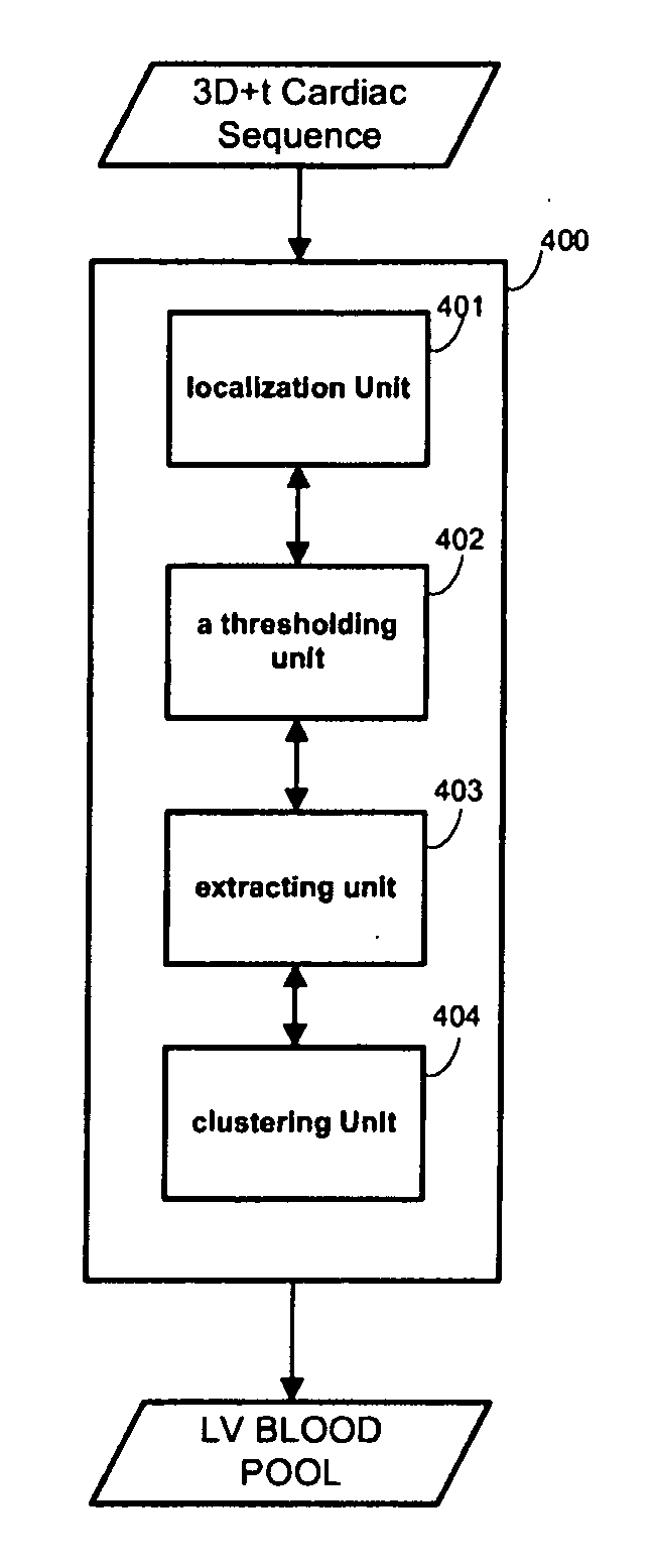
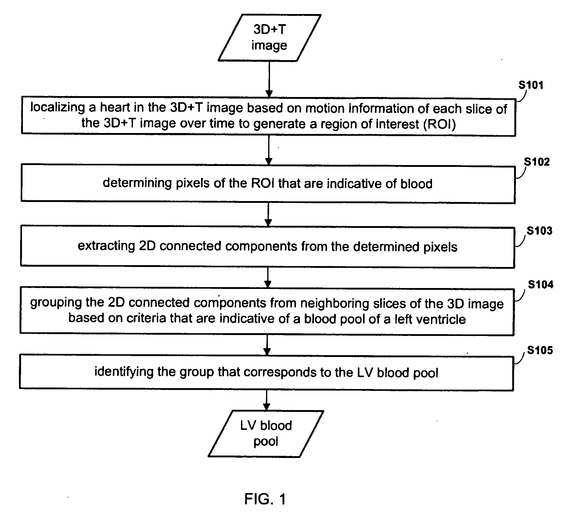
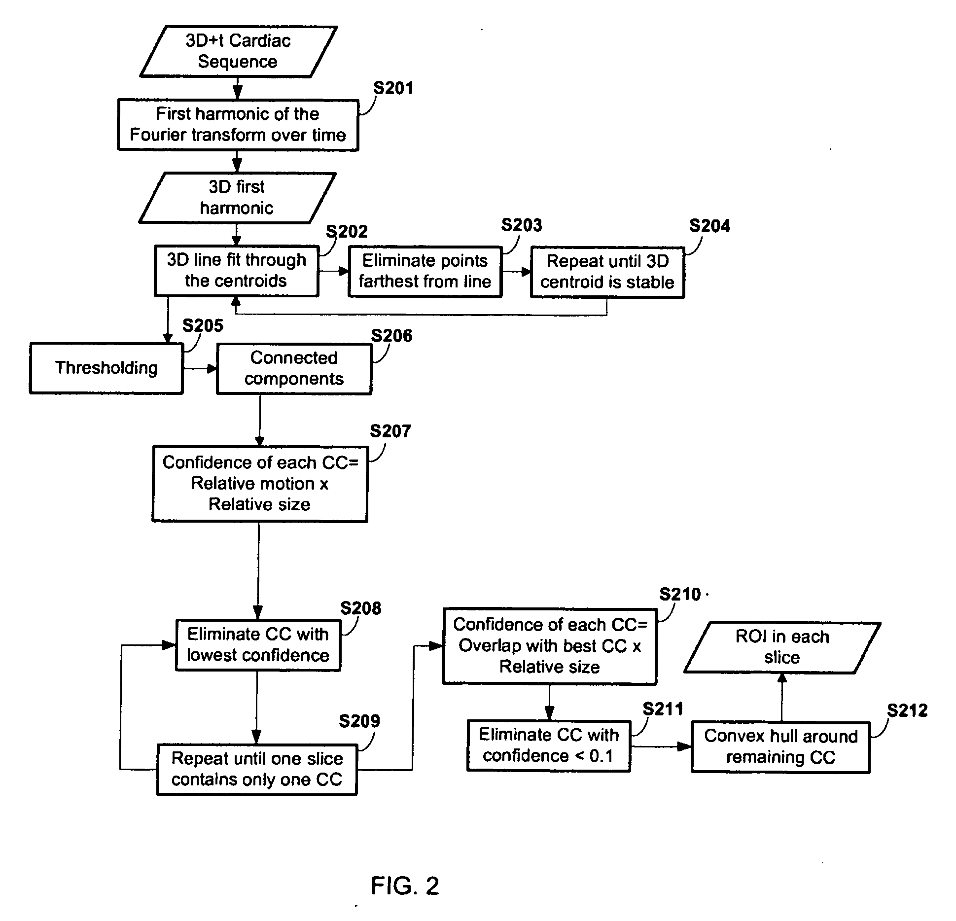
[0028]In general, exemplary embodiments for systems and methods of detecting a blood pool of a left ventricle from a plurality of images will now be discussed in further detail with reference to FIGS. 1-5. This invention may, however, be embodied in different forms and should not be construed as limited to the embodiments set forth herein. Rather, these embodiments are provided so that this disclosure will be thorough and complete, and will fully convey the scope of the invention to those skilled in the art.

[0029]It is to be understood that the systems and methods described herein may be implemented in various forms of hardware, software, firmware, special purpose processors, or a combination thereof. In particular, at least a portion of the present invention may be implemented as an application comprising program instructions that are tangibly embodied on one or more program storage devices (e.g., hard disk, magnetic floppy disk, RAM, ROM, CD ROM, etc.) and executable by any device...


PUM


Abstract

An exemplary embodiment of the present invention includes a method of detecting a left ventricle blood pool. The method includes: localizing a region of interest (ROI) in a three-dimensional temporal (3D+T) image based on motion information of each slice of the 3D image over time, thresholding the ROI to determine pixels of the ROI that correspond to blood, extracting connected components from the determined pixels, clustering the extracted connected components into groups based on criteria that are indicative of a blood pool of a left ventricle, and selecting one of the groups as the left ventricle blood pool.

Description

CROSS-REFERENCE TO RELATED APPLICATIONS[0001]This application claims priority to U.S. Provisional Application No. 61 / 035,887, filed on Mar. 12, 2008, the disclosure of which is incorporated by reference herein.BACKGROUND OF THE INVENTION[0002]1. Technical Field[0003]The present disclosure relates to medical imaging of the heart, and more particularly, to detection of the left ventricular blood pool in the heart.[0004]2. Discussion of Related Art[0005]Cardiovascular disease has become the largest cause of death in the modern world and is an important health concern. Imaging technologies such as magnetic resonance (MR) imaging allow physicians to non-invasively observe the behavior of the heart. Physicians are particularly interested in the left ventricle (LV) because it pumps oxygenated blood out to the rest of the body. To diagnose medical conditions, it is useful to be able to quantify the volume of blood pool of the LV over time and estimate its ejection fraction, cardiac output, ...

Claims


Application Information

Patent Timeline
Patent Type & Authority Applications(United States)
IPC IPC(8): G06K9/00A61B5/055
CPCG06K9/342G06K9/4638G06K2209/05G06T2207/30101G06T7/0093G06T2207/10076G06T2207/30048G06T7/0083G06T7/12G06T7/162G06V10/267G06V10/457G06V2201/03
Inventor JOLLY, MARIE-PIERRE
Owner SIEMENS HEALTHCARE GMBH
Why Eureka
Try Eureka