
Mammography method and apparatus with image data obtained at different degrees of breast compression

a breast compression and image data technology, applied in mammography, medical science, diagnostics, etc., can solve the problems of significant cost, method disadvantage, patient stress, etc., and achieve the effect of higher degree of specificity

Inactive Publication Date: 2009-10-29
SIEMENS HEALTHCARE GMBH
View PDF7 Cites 13 Cited by
  • Summary
  • Abstract
  • Description
  • Claims
  • Application Information

AI Technical Summary

Benefits of technology

[0005]An object of the present invention is to provide a method for the generation of a mammography image that enables a higher degree of specificity relative to a simple mammography image acquisition.
[0008]Elastography is based on the fact that the elastic response of different tissue structures is different. Soft, healthy tissue moves differently than hard, pathological tissue given exertion of a pressure on the tissue. The method according to the invention is based on the recognition that a benign lesion behaves differently (viewed elastographically)—consequently moves differently in space given a pressure application—than a malignant lesion (thus a cancerous tissue). As long as the breast thus represents an essentially homogenous, plastic tissue, the deformation essentially ensues uniformly, and existing tissue parts, glands, lipomas, etc. essentially shift uniformly and homogenously in space. By contrast, malignant lesions are significantly less deformable, such that they shift and deform in an abnormal manner in the homogenous tissue surrounding them. This difference resulting from the different elasticity is utilized in the method according to the invention by the two images or image data sets being acquired at different breast compressions and being computationally combined with one another in order to generate the diagnostic image to be evaluated and output. In this image, image information resulting from the different elasticity behavior (thus from elastography) can be extracted and diagnostically evaluated due to the computational link, meaning that a possible malignant lesion can be detected significantly more precisely than using only a single mammography radioscopy acquisition.
[0009]The method according to the invention thus combines the high sensitivity of x-ray radioscopic mammography with the advantages of an elastography, and the specificity of the method strikingly improves because not only mammographic information but also elastographic information influence the computationally generated final image to be output.
[0012]For example, this computational linking can ensue by forming a difference image data set from the two image data sets and determining a difference image from the difference image data set. This means that the transformed second image or the transformed second image data set is subtracted from the first image or first image data set. Insofar as the tissue is uniformly homogenous, in the ideal case the difference image is contourless to the greatest possible extent because the healthy tissue shifts homogenously and, as a result of the retrograde calculation, ultimately the second image or the second image data set shows or contains the same as the first image or the first image data set. However, if one or more malignant lesions are present that have shifted differently in the deformation due to compression, these are clearly emphasized in a difference operation since, for those malignant lesions, the assumption of a homogenously elastic behavior that forms the basis of the retrograde transformation does not apply, and consequently an actual signal difference within the appertaining pixels occurs in the region of this image section. The subtraction in the framework of the difference image data set generation is only an example; many different calculation formulas or weighting formulas are possible for use in the linking. It is thus possible to unequally weight the two image data sets in the linking in order to ensure that the breast from one image (advantageously the untransformed image) still remains slightly visible in the computationally determined final image to be output, which facilitates orientation for the physician when he or she evaluates the image. The possibility to set parameters for the computational linking (for example by greyscale value windowing or other image processing tools) that accounts for or weights the image contents of the individual image data sets to be linked differently can also be provided to the operator. This means that the operator can make adjustments as needed as to how the computational linking should ensue so that, in the image processing, the processing parameters can be set so that a diagnostically evaluable final image (for example a difference image) is generated.
[0013]As already described, the possibility exists to produce the link (for example by different weighting of the image data sets) so that the breast in one image still remains slightly visible. However, it is also possible to link the image data sets such that only the curve of the edge of the mamma shown in one of the two image data sets (in particular in the untransformed image data set) is shown in the image, possibly in a difference image. This also enables the physician to be able to determine the precise location of a possible finding. For example, an edge detection algorithm can be used to determine the edge in the one image data set.

Problems solved by technology

Not only is this a problematic stress for the patient, but also a significant cost is incurred.
However, these methods likewise have disadvantages.
Ultrasound examinations are time-consuming and must be conducted by the physician himself or herself.
Optical radioscopy is less established as a method and not sufficiently specific, while magnetic resonance examinations are very cost-intensive and time-intensive.

Method used


Image

  • Mammography method and apparatus with image data obtained at different degrees of breast compression
  • Mammography method and apparatus with image data obtained at different degrees of breast compression
  • Mammography method and apparatus with image data obtained at different degrees of breast compression

Examples


Embodiment Construction

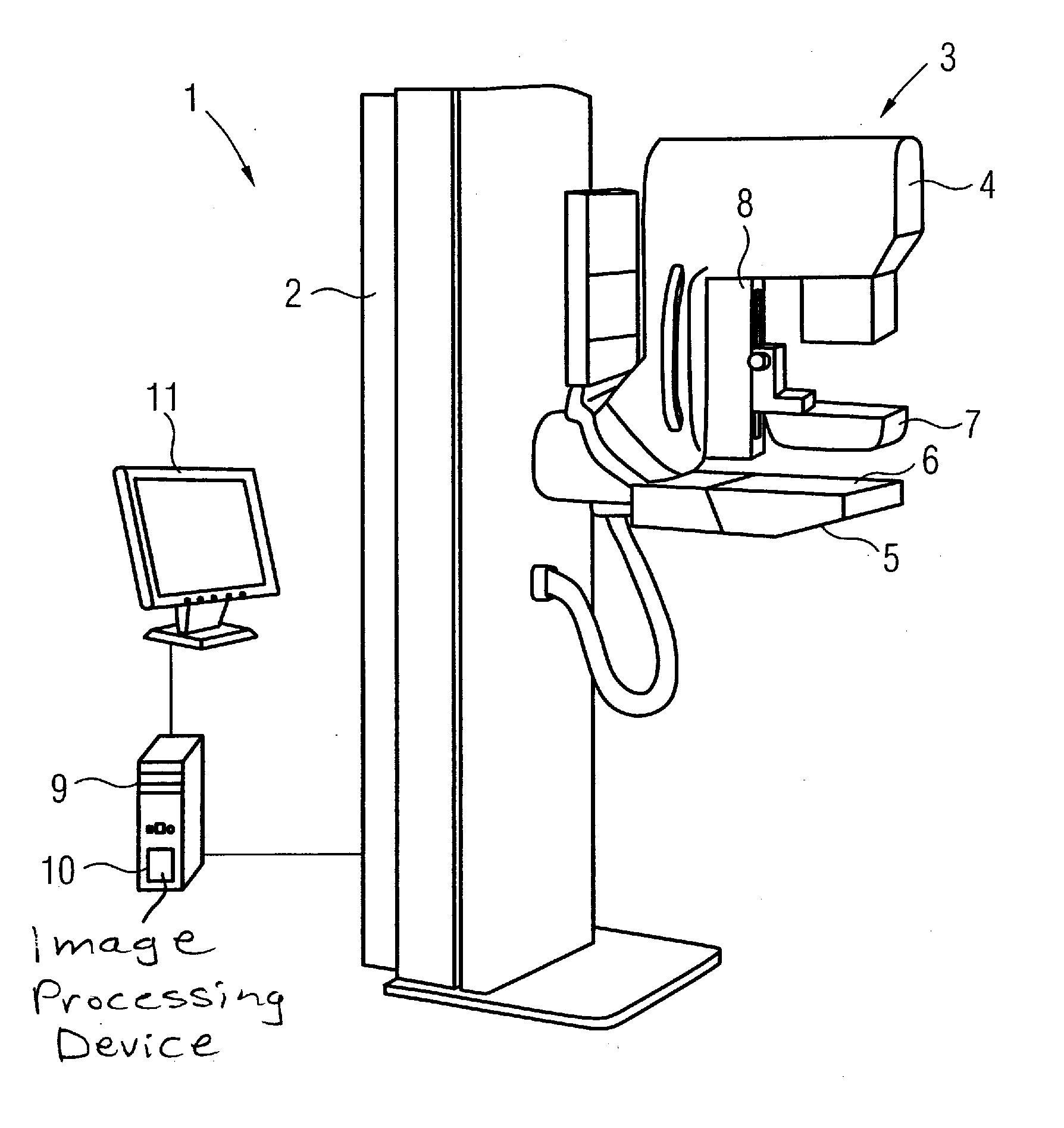
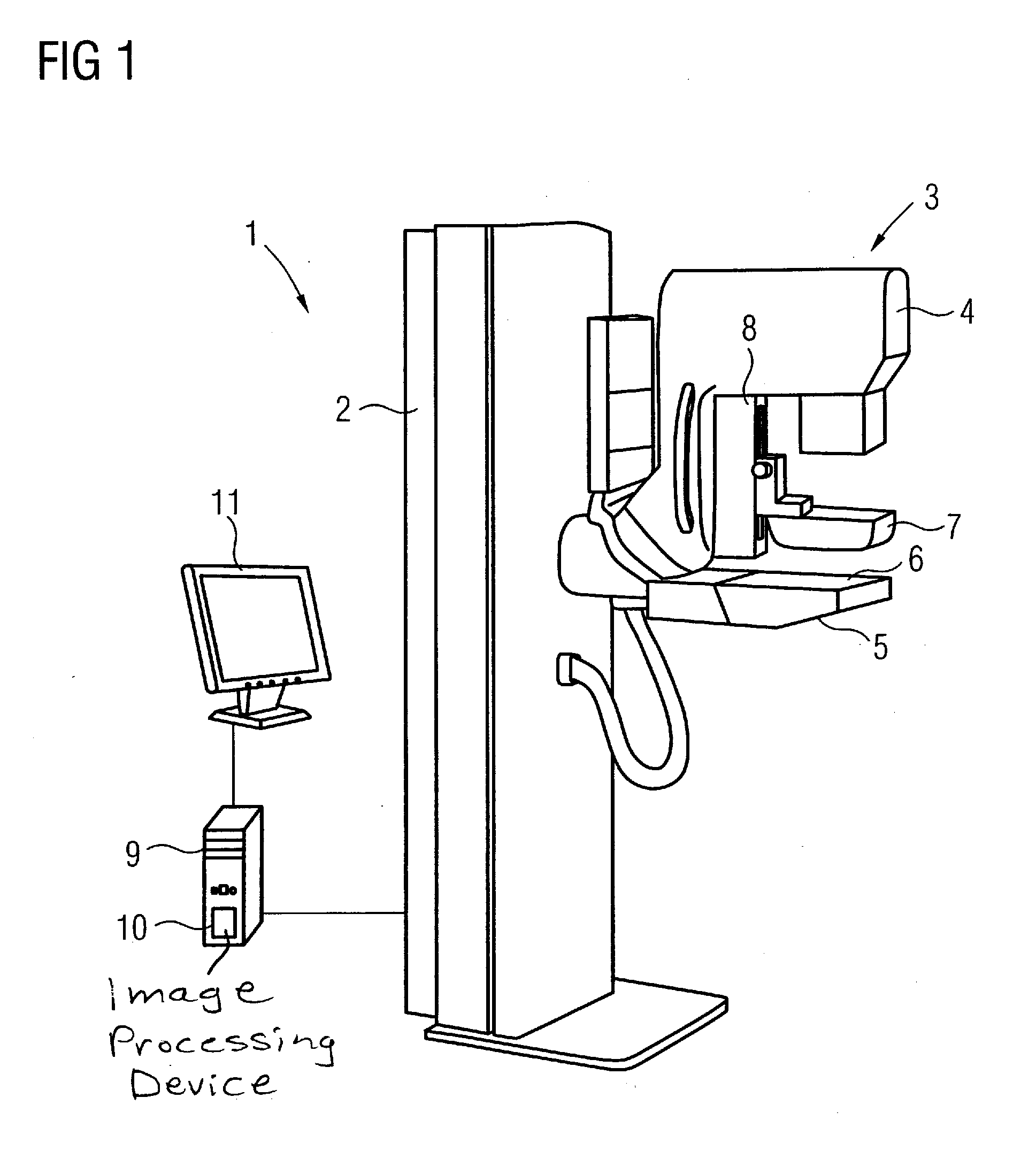
[0018]FIG. 1 shows a known mammography apparatus 1 having a vertical column 2 on which the image acquisition unit 3 is arranged vertically such that it can move. The image acquisition unit 3 has an x-ray source 4 as well as a digital x-ray detector 5 that is arranged below a support plate 6 for the female breast. A compression plate 7 is provided above the support plate 6. The compression plate 7 is likewise vertically driven in an adjustable manner on a vertical support 8. With this compression plate 7 the breast can be compressed in a known manner for image acquisition. A control device 9 is also provided that controls the entire operation of the x-ray apparatus as well as the image acquisition and image evaluation operation, for which a suitable image processing device 10 is provided. Acquired images are output on a monitor 11.

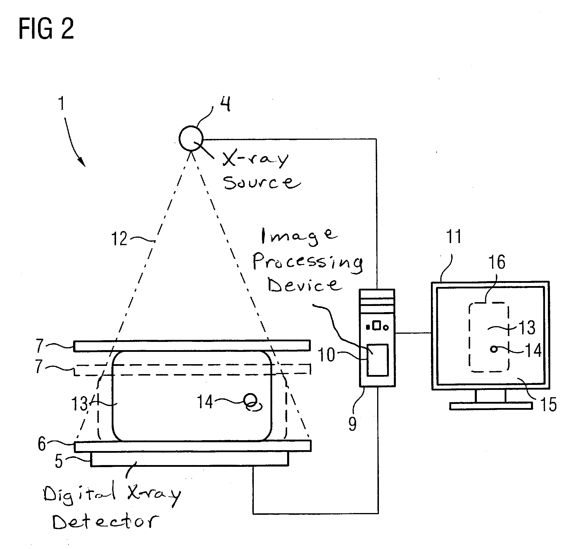
[0019]FIG. 2 shows the mammography apparatus 1 of FIG. 1 in a schematic representation in order to explain the method according to the invention in more de...


PUM


Abstract

In a method and apparatus to generate a mammographic image, the apparatus has a radiation source, a digital radiation detector, and support plate and a compression plate between which the breast is compressed during image acquisition. A first image data set depicting the breast is acquired, with a first degree of compression of the breast. A second degree of compression of the breast is set and a second image data set depicting the breast is acquired. The first and second image data sets are linked for the generation of the diagnostic image.

Description

BACKGROUND OF THE INVENTION[0001]1. Field of the Invention[0002]The present invention concerns a method to generate an image with a mammography apparatus of the type having a radiation source, a digital radiation detector, and a support plate and a compression plate between which the breast is compressed during acquisition of an image.[0003]2. Description of the Prior Art[0004]The most successful method for the examination of the female breast to determine possible suspicious lesions is mammography, either in the framework of a screening independent of suspicion or given an already present suspicion of breast cancer. For this purpose, it is known to use a mammography apparatus having an x-ray source and a digital radiation source as well as a support plate and a compression plate between which the breast is placed while the image is acquired, the breast being compressed by the vertically adjustable compression plate. The breast is located between the radiation source and the digital...

Claims


Application Information

Patent Timeline
IPC IPC(8): A61B6/04
CPCA61B5/0048A61B6/502A61B6/0414A61B5/0053
Inventor HOHEISEL, MARTIN
Owner SIEMENS HEALTHCARE GMBH
Learn More