
Ultrasound imaging apparatus and method for generating ultrasound image

a technology of ultrasound imaging and ultrasound image, applied in tomography, instruments, applications, etc., can solve the problems of difficult simultaneous acquisition of image data representing a treatment target site that requires high spatial resolution, and difficult to acquire three-dimensional image data representing a wide range. achieve the effect of higher temporal resolution

Inactive Publication Date: 2009-12-10
TOSHIBA MEDICAL SYST CORP
View PDF9 Cites 32 Cited by
  • Summary
  • Abstract
  • Description
  • Claims
  • Application Information

AI Technical Summary

Benefits of technology

[0013]An object of the present invention is to provide an ultrasound imaging apparatus that, when a puncture needle is inserted into a patient with observation of image data acquired by three-dimensional scan with ultrasound waves on the patient, can almost simultaneously acquire image data representing a treatment target organ and image data representing the puncture needle and an area surrounding the puncture needle with higher temporal resolution than the former image data, and also provide a method for generating an ultrasound image.
[0015]According to this first aspect, when a puncture needle is inserted into the patient with observation of image data acquired by three-dimensional scan with ultrasound waves, it is possible to almost simultaneously acquire image data representing a treatment target organ and image data representing the puncture needle and an area surrounding the puncture needle with higher temporal resolution than the former image data. Thus, accurate insertion of the puncture needle in a desired position of an examination target organ or a treatment target organ is facilitated. This increases safety and efficiency in examination or treatment using a puncture needle and makes it possible to reduce the burden on the operator and the patient.

Problems solved by technology

However, it is difficult to acquire three-dimensional image data representing a wide range that satisfies both high spatial resolution and high temporal resolution (real-time property).
Therefore, with the method according to the conventional art described above, in the case of acquiring image data representing a relatively wide treatment target site including liver cancer or the like, and image data representing a puncture needle inserted into this treatment target site and an area surrounding the puncture needle, it is difficult to simultaneously acquire image data representing a treatment target site that requires high spatial resolution and image data representing a puncture needle and an area around the puncture needle that requires high temporal resolution and high spatial resolution in order to track the tip of the puncture needle.

Method used


Image

  • Ultrasound imaging apparatus and method for generating ultrasound image
  • Ultrasound imaging apparatus and method for generating ultrasound image
  • Ultrasound imaging apparatus and method for generating ultrasound image

Examples


Embodiment Construction

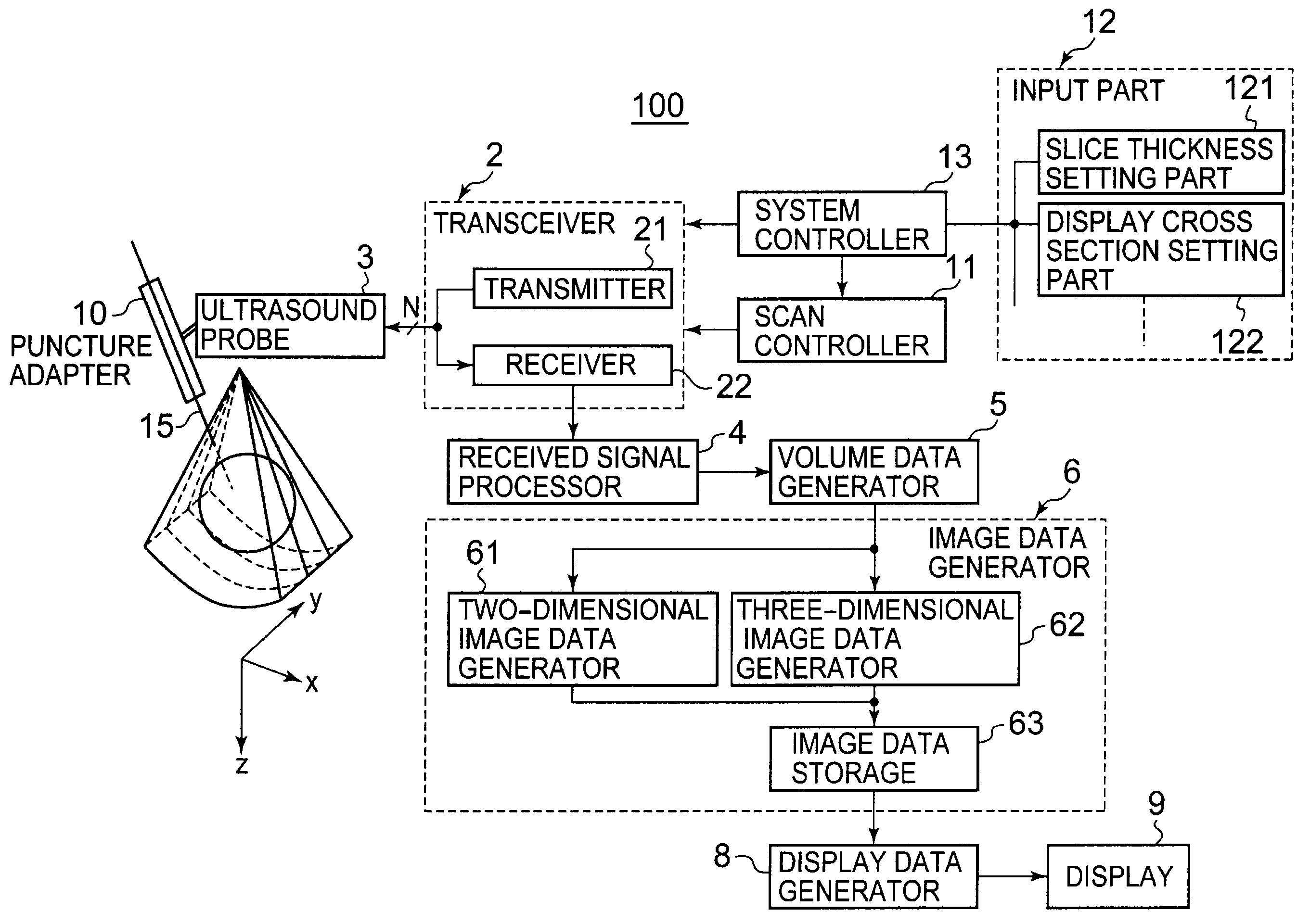
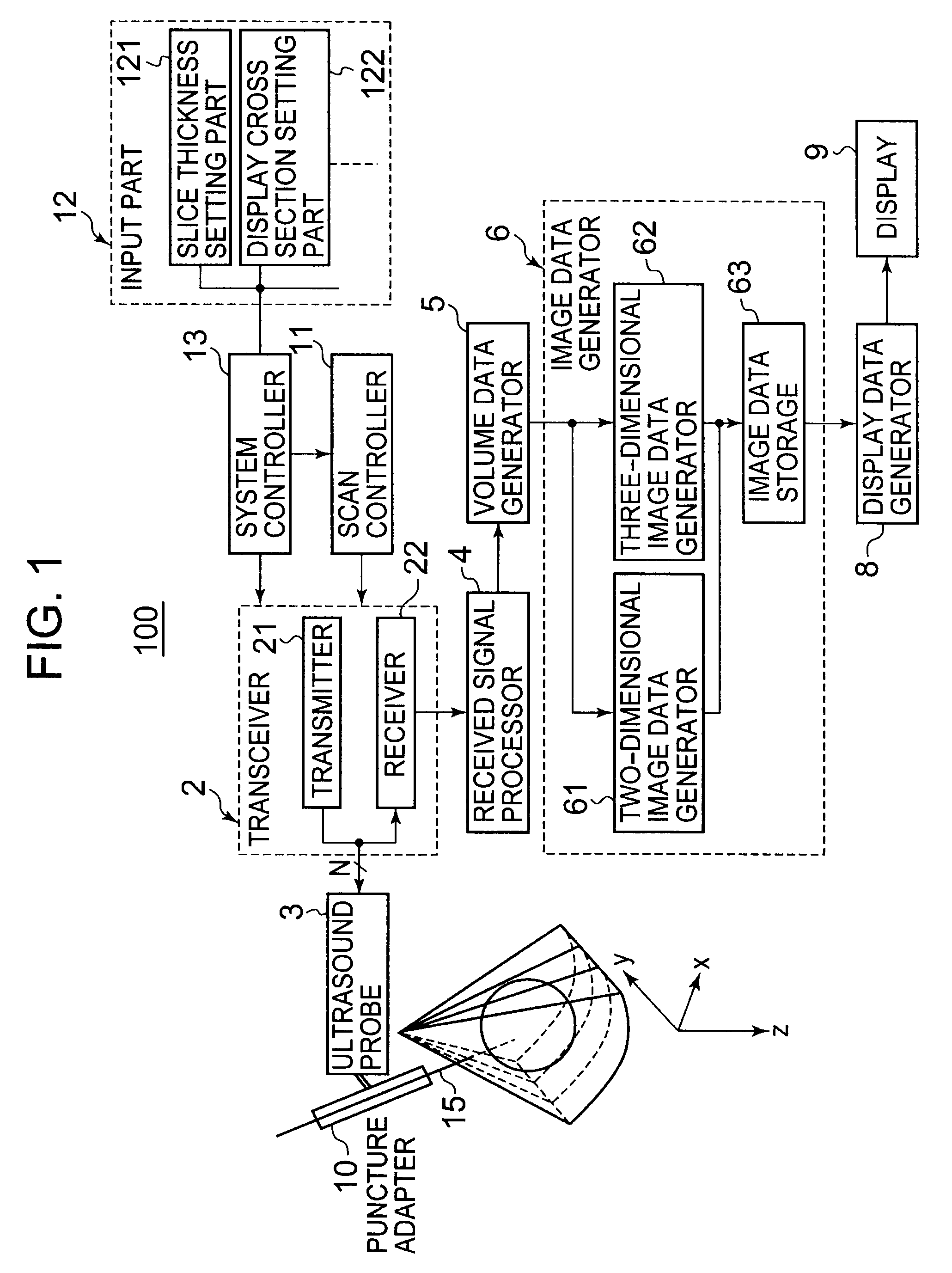
[0036]An ultrasound imaging apparatus according to an embodiment of the present invention will be described below with reference to the drawings.

[0037]In the embodiment of the present invention described below, for a three-dimensional region including a treatment target site of a patient, a puncture needle scanning region having a predetermined slice thickness is firstly set with reference to a cross section (may be referred to as a “puncture cross section” hereinafter) including an insertion direction of a puncture needle inserted along a needle guide of a puncture adapter attached to an ultrasound probe. Subsequently, in the y-direction (normal direction) substantially perpendicular to the puncture cross section, a treatment target scanning region having a predetermined slice thickness adjacent to the puncture needle scanning region is set. Then, based on volume data in the puncture needle scanning region acquired by first three-dimensional scan with ultrasound waves and volume da...


PUM


Abstract

For a three-dimensional region including a treatment target site of a patient, a puncture needle scanning region R1 having a slice thickness d1 is set with reference to a cross section (a puncture cross section) including an insertion direction of a puncture needle. Subsequently, in the y-direction substantially perpendicular to the puncture cross section, two treatment target scanning regions R2 (R21 and R22) each being adjacent to the puncture needle scanning region R1 and having a slice thickness d2 are set. Then, based on volume data in the puncture needle scanning region R1 acquired by first three-dimensional scan with ultrasound waves and volume data in the treatment target scanning regions R2 acquired by second three-dimensional scan performed at a lower volume rate than the first three-dimensional scan, image data is generated for the purpose of supporting puncture.

Description

BACKGROUND OF THE INVENTION[0001]1. Field of the Invention[0002]The present invention relates to an ultrasound imaging apparatus that generates image data for supporting treatment and examination using a puncture needle, and also relates to a method for generating an ultrasound image.[0003]2. Description of the Related Art[0004]The mortality rate for cancer, which is one of the three major diseases, has been increasing yearly, and early diagnosis and treatment of cancer is strongly desired. For example, liver cancer accounts for approximately 10% of all cancers, and the percentage has been increasing. On the other hand, advancement of the technology of medical image diagnosis apparatuses such as an ultrasound imaging apparatus, an MRI apparatus and an X-ray CT system is remarkable, and medical image diagnosis apparatuses has been essential for early detection of various cancers including liver cancer mentioned above.[0005]To be specific, three-dimensional imaging by an X-ray CT syst...

Claims


Application Information

Patent Timeline
IPC IPC(8): A61B8/14
CPCA61B8/06A61B8/0833A61B8/0841A61B8/13A61B8/14A61B8/483G01S15/8993G01S7/52063G01S7/52074G01S7/52085G01S15/8925G01S15/899A61B8/523
Inventor YAMAGATA, HITOSHI
Owner TOSHIBA MEDICAL SYST CORP
Learn More