Method of diagnosis and treatment of tumors using high intensity focused ultrasound

a tumor and ultrasound technology, applied in immunological disorders, antibody medical ingredients, therapy, etc., can solve the problems of invasive surgery removal, invasive biopsy procedure, serious complications, complex and time-consuming,

Inactive Publication Date: 2010-04-15
ALBERT EINSTEIN COLLEGE OF MEDICINE OF YESHIVA UNIV
View PDF60 Cites 55 Cited by
  • Summary
  • Abstract
  • Description
  • Claims
  • Application Information

AI Technical Summary

Problems solved by technology

The biopsy procedure, however, is invasive and involves the removal of a portion of the tissue for analysis.
Surgical removal, however, is invasive and can be quite complex and time consuming.
Surgical treatment can also result in serious complications, such as from anesthesia.

Method used

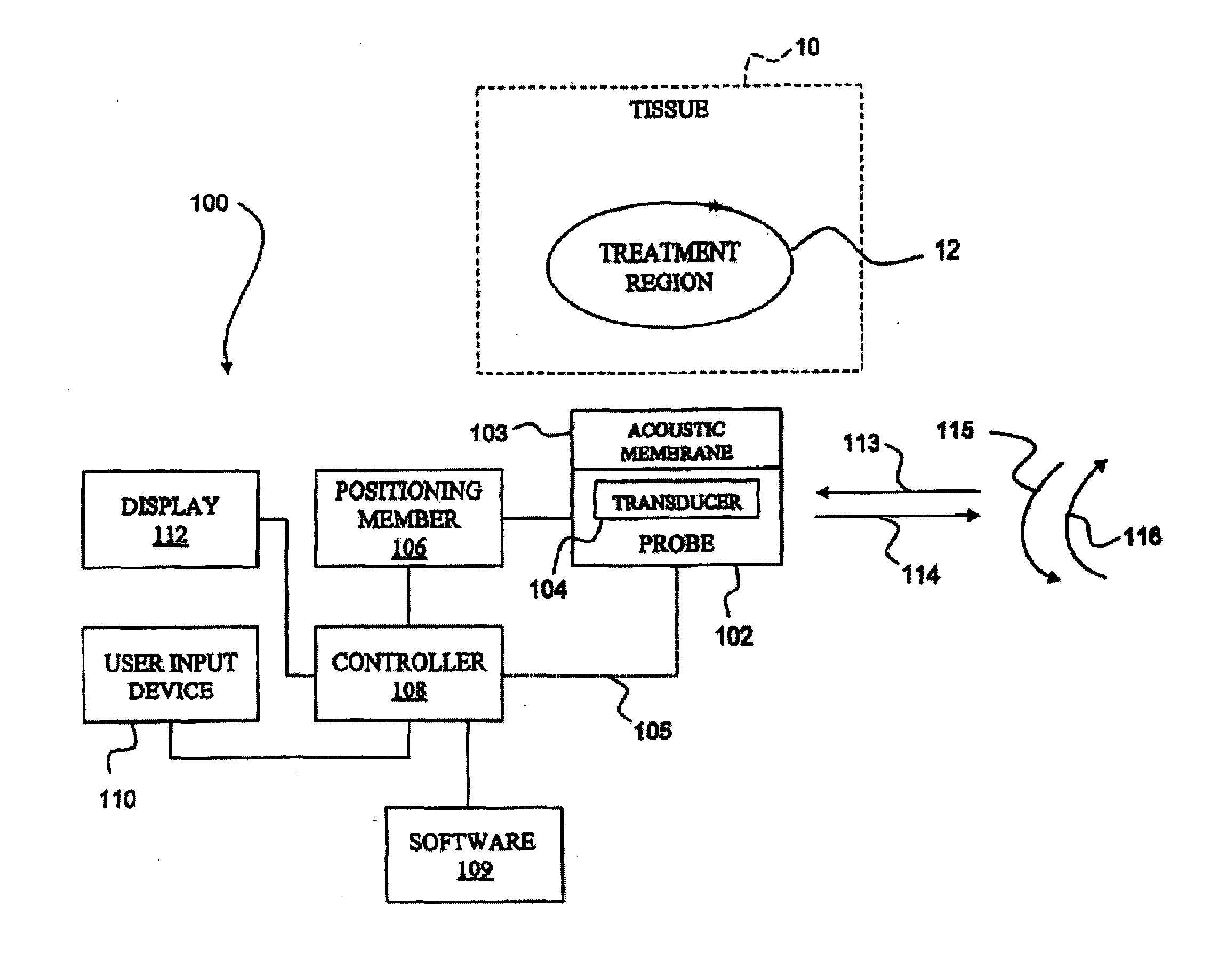
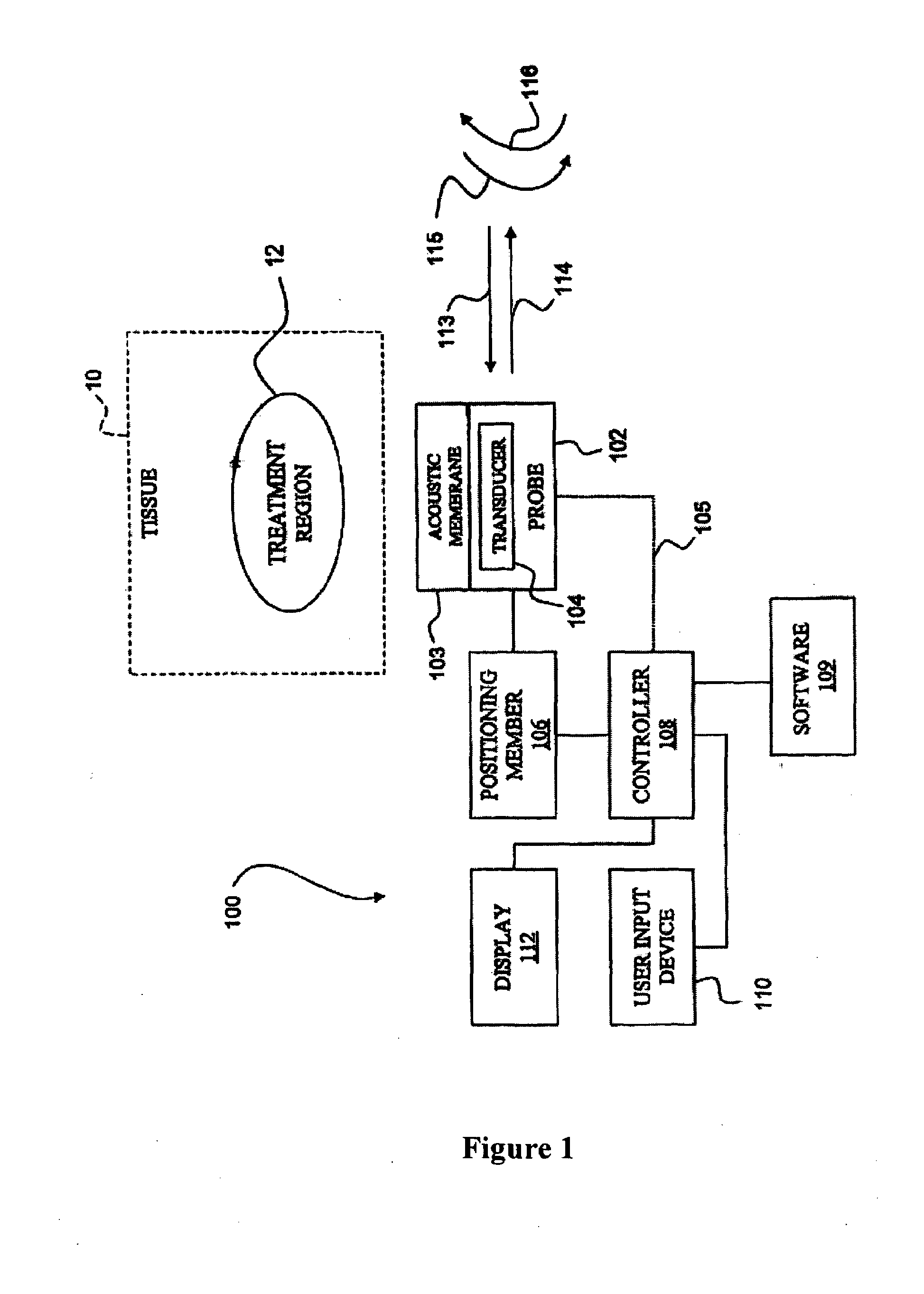
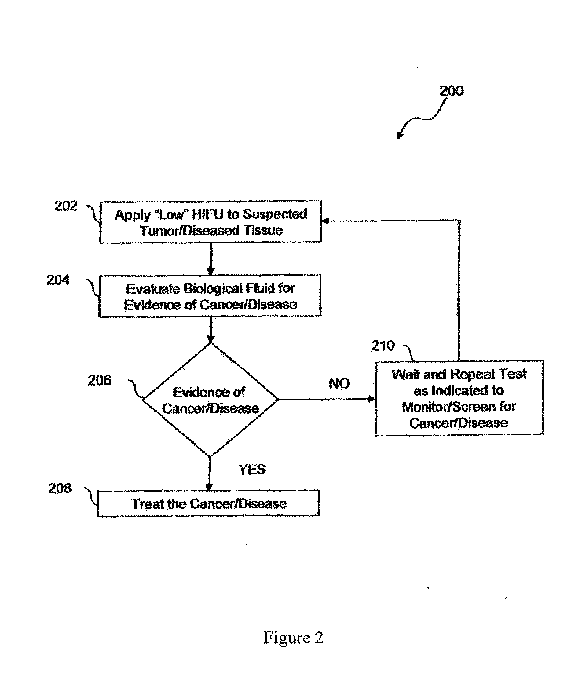

Image

  • Method of diagnosis and treatment of tumors using high intensity focused ultrasound
  • Method of diagnosis and treatment of tumors using high intensity focused ultrasound
  • Method of diagnosis and treatment of tumors using high intensity focused ultrasound

Examples


example 1

[0090]To investigate the complimentary nature of HIFU with immunotherapy, a HIFU system was designed with a probe that is capable of providing the user with control over the acoustic properties of the ultrasound transmission and sized appropriately for use in the preclinical murine model. The immune system response to the sequential application of both LO-HIFU, followed by one-to-two days later, HI-HIFU was addressed in the animal model.

[0091]The modified Sonablate® 500 operates at HI-HIFU with approximate focal spatial peak temporal peak (SPTP) intensities of 1300 to 2000 W / cm2. The HIFU continuous wave of 3 seconds and operating frequency of 4 MHz for the treatment of prostate cancer is used to achieve tissue temperatures in the focal zone of 80° C. to 95° C. The resulting thermal lesions are approximately 3 mm×3 mm×12 mm with a very sharp demarcation with no tissue damage beyond the focal zone.

[0092]For LO-HIFU, the pulse duration is in the micro-second to milli-second range with...

example 2

[0104]The immune system response in patients is amplified as follows. This model involves the sequential application of both LO-HIFU, followed by one-to-two days later, HI-HIFU.

[0105]The Sonablate® 500 operates at HI-HIFU with approximate focal spatial peak temporal peak (SPTP) intensities of 1300 to 2000 W / cm2. The HIFU continuous wave of 3 seconds and operating frequency of 4 MHz for the treatment of prostate cancer is used to achieve tissue temperatures in the focal zone of 80° C. to 95° C. The resulting thermal lesions are approximately 3 mm×3 mm×12 mm with a very sharp demarcation with no tissue damage beyond the focal zone.

[0106]For LO-HIFU, the pulse duration is in the micro-second to milli-second range with approximate focal intensities (SPTP) of 500 W / cm2 and pulse repetition frequencies (PRF) on the order of 1 Hz. Also to limit temperature elevation, a lower center frequency near 1 MHz is generally employed. Thus, cells experience mechanical agitation while remaining viabl...


PUM

PropertyMeasurementUnit
timeaaaaaaaaaa
operating frequencyaaaaaaaaaa

Abstract

A method of diagnosis and treatment of tumors using High Intensity Focused Ultrasound is provided. The method of diagnosing the presence of a tumor in a patient comprises the steps of subjecting a tumor to high intensity focused ultrasound (HIFU) to cause the tumor cells to release cellular material and evaluating the cellular material for a tumor marker. The method of treating a tumor in a patient can also comprise the step of subjecting a tumor to high intensity focused ultrasound (HIFU) to provoke an immune response.

Description

CROSS REFERENCE[0001]The present application claims priority to Provisional Application No. 60 / 989,629, filed Nov. 21, 2007, the entire contents of which are incorporated herein by reference, including any references cited therein.FIELD OF THE INVENTION[0002]The present invention relates to an apparatus and methods for the non-invasive diagnosis and / or treatment of diseased tissue. In particular, the present invention relates to an apparatus and related methods for the non-invasive diagnosis and / or treatment of tumors and metastatic cancer with High Intensity Focused Ultrasound (HIFU).BACKGROUND[0003]Diseased tissue, such as a cancerous tumor, is commonly diagnosed by taking a biopsy of the tissue for pathology. The biopsy procedure, however, is invasive and involves the removal of a portion of the tissue for analysis. Clearly, it is much more desirable to have a non-invasive method for such diagnosis.[0004]One approach to the treatment of diseased tissue, particularly tumors, is su...

Claims


Application Information

Patent Timeline
Patent Type & Authority Applications(United States)
IPC IPC(8): A61M5/00A61N7/00A61M31/00A61K38/19A61K38/20A61K39/395A61K48/00A61K39/00
CPCA61B8/0833A61B10/0051A61B10/007A61B2017/2253A61N2007/0073A61N2007/0082A61B8/08A61B18/04A61N2007/0004A61N7/02A61P35/00A61P37/02A61P43/00
Inventor SANGHVI, NARENDRA T.GUHA, CHANDANFEDEWA, RUSSELL JAMESCARLSON, ROY FRANCISSEIP, RALFCHEN, WO-HSING
Owner ALBERT EINSTEIN COLLEGE OF MEDICINE OF YESHIVA UNIV
Why Eureka
Try Eureka