Method and equipment for image-guided stereotactic radiosurgery of breast cancer

a stereotactic radiosurgery and stereotactic technology, applied in the field of magnetic resonance imaging (mri), stereotactic radiosurgery, breast cancer, can solve the problems of no better than a centimeter of surgery, adversely affecting the quality of life of patients undergoing bct, and complex bct, etc., to achieve the effect of monitoring the safety and operation

Active Publication Date: 2010-04-15
XCISION MEDICAL SYST +1
View PDF9 Cites 23 Cited by
  • Summary
  • Abstract
  • Description
  • Claims
  • Application Information

AI Technical Summary

Benefits of technology

[0012]The present invention provides a method of treating a cancerous region in a breast of a patient. The method comprises (i) imaging the breast in a three-dimensional coordinate system, (ii) stereotactically determining the location of the cancerous region in the breast, (iii) optionally determining the volume of the entire cancerous region or portion thereof to be treated, and (iv) while maintaining the breast in a three-dimensional coordinate system that is identical to or corresponds with the three-dimensional coordinate system used in (i), noninvasively exposing the cancerous region of the breast of the patient to a cancer-treatment effective dose of radiation.
[0013]The present invention further provides equipment for image-guided stereotactic radiosurgery of a cancerous region in a breast. The equipment comprises: (i) means for immobilizing the breast containing the cancerous region; (ii) a couch comprising a channel or right and left openings, wherein the right or left breast of a patient is placed in the channel or the right or left opening, respectively, for treatment when the patient is lying prone on the couch, which optionally comprises a top layer of a self-molding medium; (iii) a stereotactic localization frame beneath the couch for placement of the breast in a coordinate system; (iv) one or two breast shields, which are removably attached to the couch, slidably mounted to the channel, or removably attached to the means for immobilizing the breast; (v) an irradiation unit comprising (i′) a source holder, which comprises a wall having an interior surface, an exterior surface, and channels, which communicate with the interior surface, may or may not communicate with the exterior surface, and each of which can comprise a radiation source, (ii′) a collimator holder, which is adjacent to the interior surface of the source holder and comprises collimators of different sizes in the same arrangement as the channels in the source holder such that relative rotation between the source holder and the collimator holder allows selection of radiation beams of different sizes, resulting in shots of radiation of different sizes, and (iii′) a base housing, which is beneath the exterior surface of the source holder, supports the source holder and the collimator holder, and houses a motor, which rotates the collimator holder and the source holder relative to each other, and another motor, which rotates the collimator holder and the source holder together, when locked; (vi) a curved base support; (vii) three motors, which are operably connected to the couch and each of which moves the couch in a different axis of motion; and (viii) a computerized control system, which controls the movement of the couch and the irradiation unit. The equipment can, and preferably does, further comprise a treatment planning system. The operations of the irradiation unit and the couch are controlled by a computerized control system, which obtains treatment parameters from the treatment planning system, controls the movement of the couch and the irradiation unit according to the treatment plan, monitors the safety and operation of the entire unit, and provides safety interlocks and movement limits when needed.

Problems solved by technology

BCT, however, is a complex, protracted treatment.
Furthermore, mammograms and magnetic resonance images are obtained under different geometric conditions as compared to surgery, lumpectomy is performed without any direct image guidance, and the geometric uncertainty of surgery is no better than about a centimeter.
These drawbacks adversely affect the quality of life of patients undergoing BCT.
Although these ablative methods are meant to be minimally invasive, they have to be performed under general anesthesia.
In addition, it is difficult, if not impossible, to conform precisely the damage generated to the shape of the tumor, such that unnecessary damage to surrounding normal tissue is avoided or minimized.
These techniques have not been demonstrated to be able to replace surgery.
Therefore, these percutaneous image-guided techniques, though less invasive, cannot replace surgery.
In this regard, the inventors are not aware of any device that can deliver a high dose of radiation to a cancerous region of the breast safely and accurately with surgical precision.
Furthermore, unlike the skull, which can be securely fixated into a coordinate system so that there is no geometric difference between imaging and treatment, such immobilization has never been achieved with a breast.

Method used


Image

  • Method and equipment for image-guided stereotactic radiosurgery of breast cancer
  • Method and equipment for image-guided stereotactic radiosurgery of breast cancer
  • Method and equipment for image-guided stereotactic radiosurgery of breast cancer

Examples


Embodiment Construction

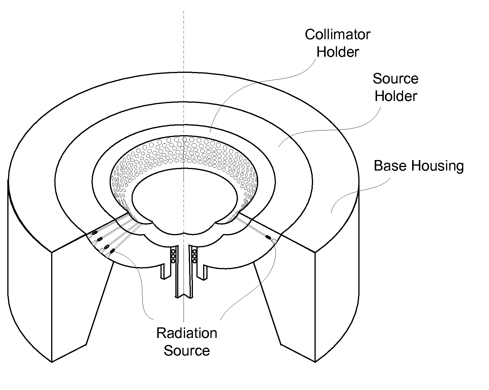
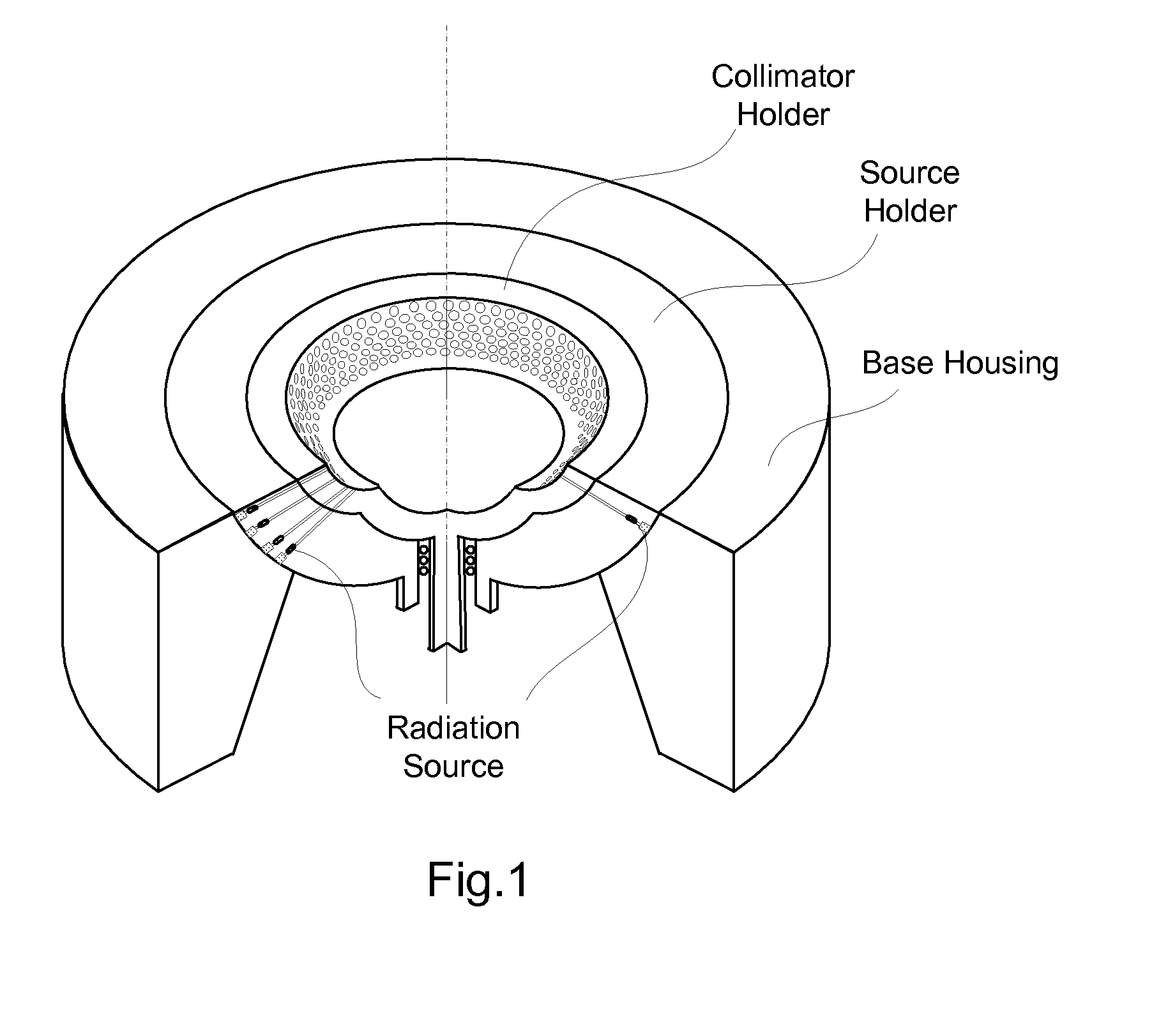
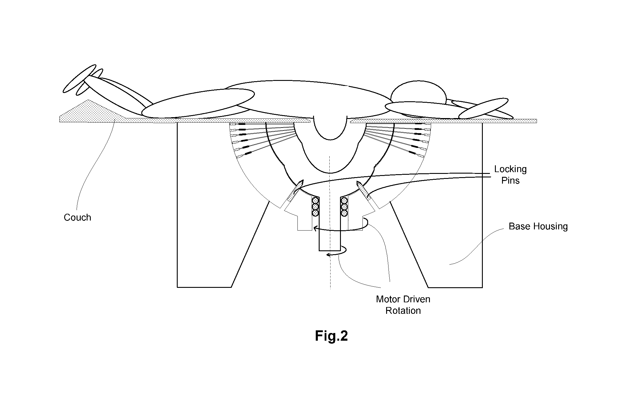
[0019]The present invention provides a method of using stereotactic radiosurgery to treat a cancerous region of a breast. The present invention also provides equipment for use in the method. The method and equipment of the present invention are believed to offer potential advantages over current methods of treatment, including BCT. The potential advantages include, but are not limited to, non-invasive nature, no pain, potential for elimination of radiation treatment for most, if not all, early-stage breast cancers, enhanced quality of life (by shortening the treatment time from 7-10 weeks to hours), absence of scars, reduced radiation to non-cancerous tissue, ease of repetition, and cost-effectiveness, due to the elimination of invasive surgery and subsequent radiation therapy.

[0020]Due to stereotactic localization, it is believed that an accuracy of about 1 mm can be achieved. With breast cancer radiosurgery, a substantial radiation dose is inevitably delivered to the outside of th...


PUM


Abstract

A method of treating a cancerous region in a breast of a patient comprising (i) imaging the breast in a three-dimensional coordinate system, (ii) stereotactically determining the location of the cancerous region in the breast, (iii) optionally determining the volume of the entire cancerous region to be treated, and (iv) while maintaining the breast in a three-dimensional coordinate system that is identical to or corresponds with the three-dimensional coordinate system used in (i), noninvasively exposing the cancerous region of the breast of the patient to a cancer-treatment effective dose of radiation; and equipment for use in such a method.

Description

TECHNICAL FIELD OF THE INVENTION[0001]The present invention pertains to magnetic resonance imaging (MRI), radiation, stereotactic radiosurgery, and breast cancer. Specifically, the present invention pertains to a method and equipment employing γ-radiation in image-guided, stereotactic, radiosurgical ablation of a cancerous region in a breast.BACKGROUND OF THE INVENTION[0002]According to the American Cancer Society, breast cancer is the most prevalent malignancy among women in the United States, with more than 200,000 new cases diagnosed per year (Jemal et al., Cancer J. Clin. 56: 106-130 (2006)). Significant advances have been made in mammography and other imaging modalities over the last few decades. As a result, more patients are diagnosed in the early-stage of the disease, for which breast conservation therapy (BCT) has become the treatment of choice.[0003]Compared with mastectomy, BCT provides a comparable outcome with superior cosmetic results and reduced psychological and emot...

Claims


Application Information

Patent Timeline
Patent Type & Authority Applications(United States)
IPC IPC(8): A61B5/055A61N5/00
CPCA61B5/0555A61B2019/205A61B2019/207A61N5/1084A61N2005/1055A61B6/0435G01R33/58A61B6/00A61B6/02A61B6/022G01R33/4808A61B5/055A61B6/027A61B90/17A61B2090/101A61B5/05
Inventor YU, XINSHENG CEDRICYU, YING SUREGINE, WILLIAM
Owner XCISION MEDICAL SYST
Why Eureka
Try Eureka