Malignancy diagnosis using content - based image retreival of tissue histopathology

a content-based image and histopathology technology, applied in image analysis, image enhancement, instruments, etc., can solve the problems of inability to quantitatively evaluate the system with different feature sets, inability to perform ml methods, and often deadly diagnosis and treatment in the late stages, so as to reduce the dimensionality of extracted feature data and increase magnification

Active Publication Date: 2010-04-22
THE TRUSTEES OF THE UNIV OF PENNSYLVANIA +1
View PDF2 Cites 94 Cited by
  • Summary
  • Abstract
  • Description
  • Claims
  • Application Information

AI Technical Summary

Benefits of technology

[0006]In one embodiment, the invention provides a computer-aided diagnostic method to predict the probability that a histological image contains a malignant region comprising: obtaining a histological image, wherein the histological image is a first of a series of images ordered in increasing magnification; identifying a region or regions of said histological image classified as suspect; extracting one or more image features from at least one of said identified regions; reducing a dimensionality of said extracted feature data; and classifying said extracted region or regions as either benign, malignant, or suspect based on at least one said extracted image feature, whereby if said extracted region or regions are classified as malignant, the histological image has a malignant region, otherwise; if the extracted region or regions are classified as benign, then the histological image does not have a malignant region; or if the extracted region or regions are classified as suspect, the next histological image in the series is obtained and the steps of identifying, extracting, reducing and classifying are repeated.

Problems solved by technology
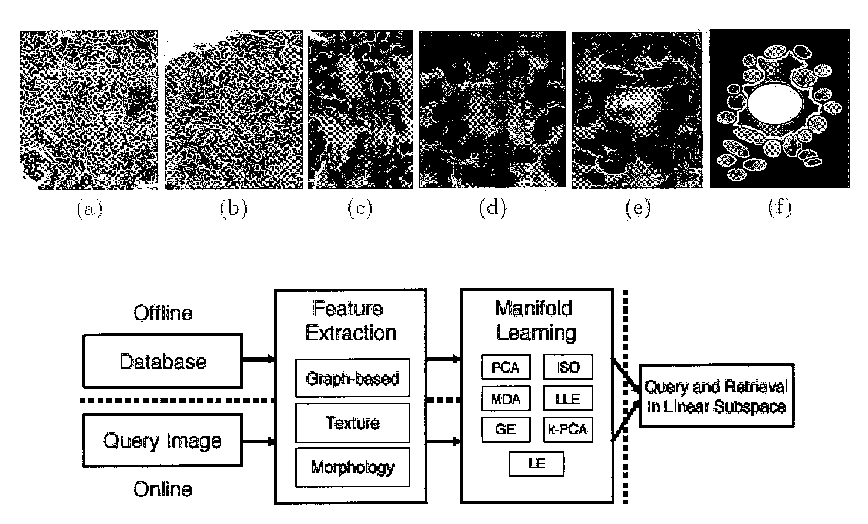
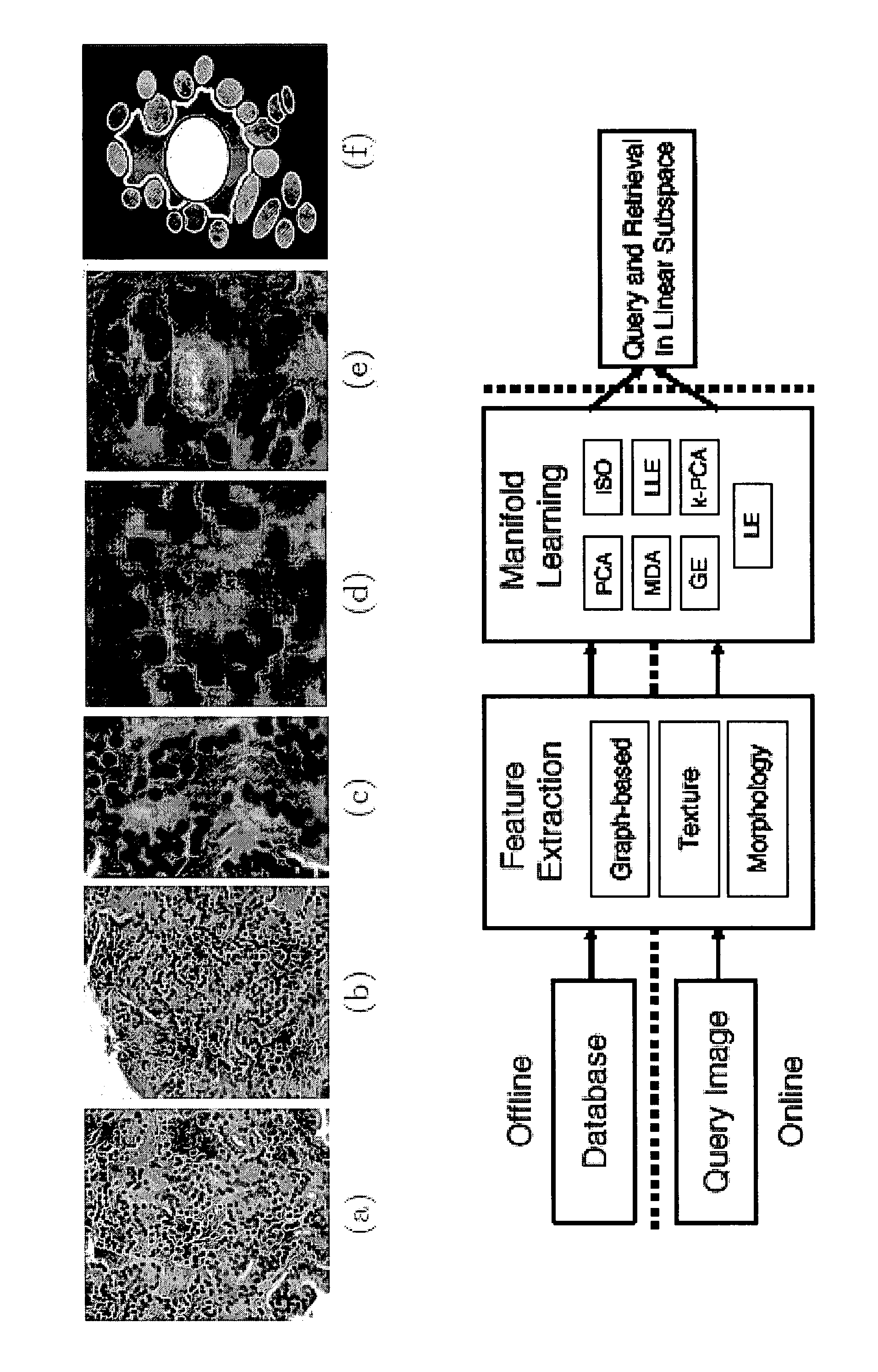
In contrast, diagnosis and treatment in late stages often have deadly results.
However, quantitative evaluation of the system with different feature sets and ML methods was not done.

Method used


Image

  • Malignancy diagnosis using content - based image retreival of tissue histopathology
  • Malignancy diagnosis using content - based image retreival of tissue histopathology
  • Malignancy diagnosis using content - based image retreival of tissue histopathology

Examples


example 1

Using Manifold Learning for Content-Based Image Retrieval of Prostate Histopathology

[0065]Prostate cancer is the most commonly diagnosed cancer among males in the U.S., with 200,000 new cases and 27,000 deaths predicted for 2007 (source: American Cancer Society). Currently manual examination of prostate biopsy samples under a microscope by an expert pathologist is the gold standard of prostate cancer diagnosis and grading. In the U.S., the most common system of numbering or \grading” prostate tissue (assessing degree of malignancy) is the Gleason scale [1], which assigns grades on a scale from 1 (well-differentiated, relatively benign tissue) to 5 (non-differentiated tissue, highly invasive cancer).

[0066]The Gleason paradigm illustrates how cancer grades differ in terms of their architecture (spatial arrangement of nuclei and glands within the tissue with respect to their centers of mass) and morphology (shape and size of glands and nuclei). Glands and nuclei both express architectu...

example 2

Grading of Prostate Cancer Using CBIR

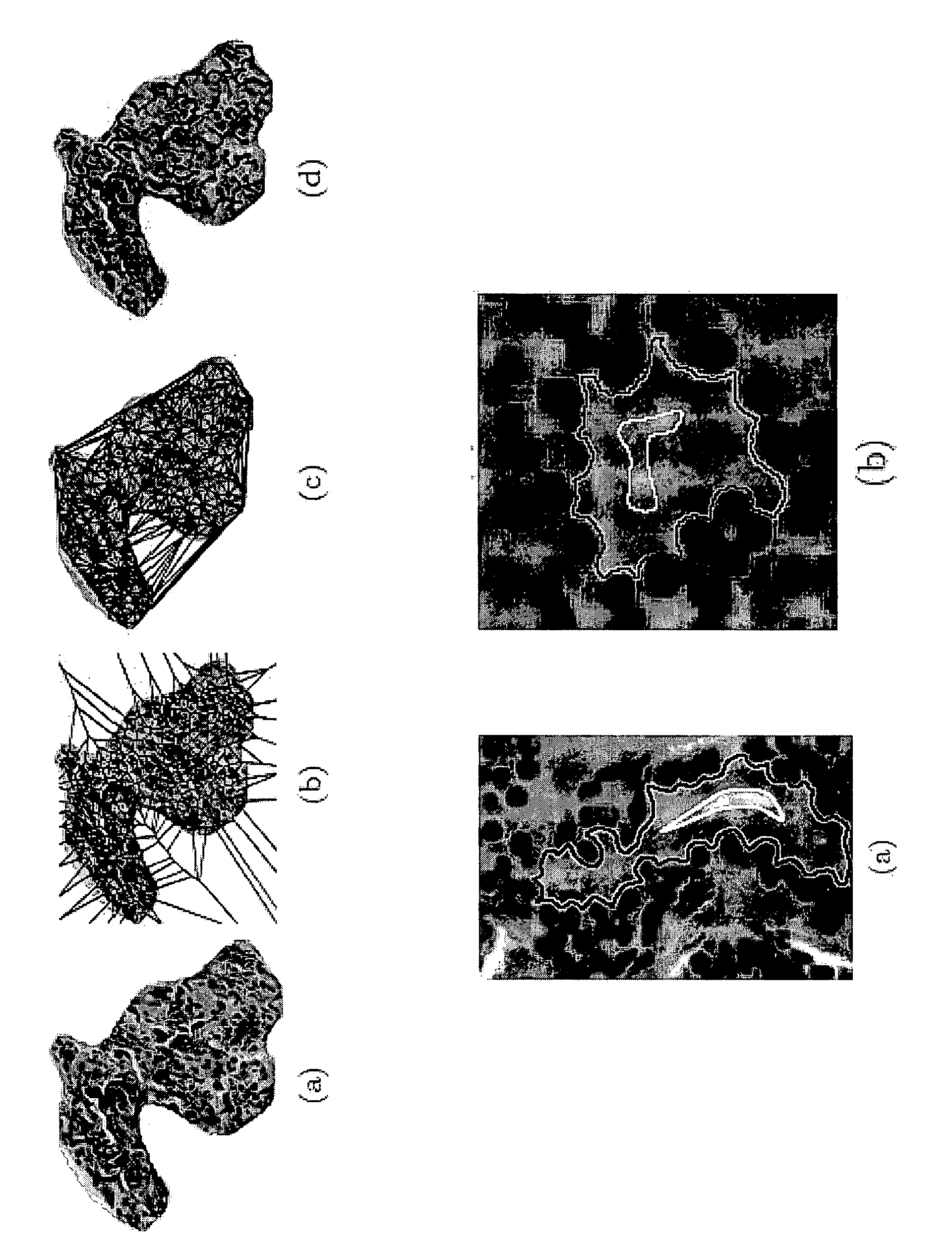
[0084]The current protocol for prostate cancer diagnosis involves manual analysis of prostate biopsy tissue by a pathologist to determine the presence or absence of cancer in a tissue sample, followed by Gleason grading to assign a number between 1 (early stage cancer) and 5 (highly infiltrative cancer) to grade the invasiveness of the cancer. Gleason grading is currently done by visual analysis of the arrangement, size, and shape of the gland structures and nuclei within the tissue sample. Recently however a number of problems have been identified with the Gleason system:

[0085]Variability: Significant levels of inter and intra-observer variability in manual grading of prostate cancer was observed with the rates of undergrading tissue patterns (assigning a tissue a grade lower than its actual grade) as high as 47%. FIG. 6 illustrates these clinical problems, as the difference between intermediate cancer grades (grade 3 (FIG. 1 (a)) and grade 4 (F...

example 3

Grading of Breast Cancer Using CBIR

Computerized Feature Extraction to Develop Quantitative Signatures for Breast Cancer Grades

[0110]Breast tissue samples are scanned into the computer at 40× optical magnification using a whole-slide digital scanner, where they are deconstructed into constituent scales. The grading of breast tissue is done across multiple magnifications. At lower magnifications only textural attributes and, overall features of tissue architecture including tubule formation are apparent, while at higher magnifications nuclear and other cellular cytological details become visible. Texture features include 13 Haralick texture features, along with several first-order statistical texture features. Also extracted is a series of steerable Gabor filter features, which provide a varying filter response to textural differences at a series of scales and orientations. FIGS. 10(b)-(e), and 10(g)-(j) represent the corresponding texture feature representations (Gabor, Sobel, Gradie...


PUM


Abstract

This invention relates to computer-aided diagnostics using content-based retrieval of histopathological image features. Specifically, the invention relates to the extraction of image features from a histopathological image based on predetermined criteria and their analysis for malignancy determination.

Description

FIELD OF INVENTION[0001]This invention is directed to computer-aided diagnostics using content-based retrieval of histopathological image features. Specifically, the invention is directed to the extraction of image features from a histopathological tissue image based on predetermined criteria and their use in the diagnosis and prognosis of malignancy in that tissue.BACKGROUND OF THE INVENTION[0002]Detection and surgical treatment of the early stages of tissue malignancy are usually curative. In contrast, diagnosis and treatment in late stages often have deadly results.[0003]Histopathological examination, which is a diagnostic method of a tissue specimen, is one of the most important medical tool in process of diagnosis. The histopathological diagnosis of several cancer types is based upon architectural (symmetry, circumscription, maturation, nests, arrangement, distribution), cytological (atypicality, mitosis, necrosis) and other cancer-specific criteria. Nowadays, pathologists know...

Claims


Application Information

Patent Timeline
Patent Type & Authority Applications(United States)
IPC IPC(8): G06K9/00
CPCG06K9/0014G06T2207/30096G06T7/0081G06T7/0012G06T7/11G06V20/695
Inventor MADABHUSHI, ANANTDOYLE, SCOTTFELDMAN, MICHAEL D.TOMASZEWSKI, JOHN E.
Owner THE TRUSTEES OF THE UNIV OF PENNSYLVANIA
Why Eureka
Try Eureka