Diagnostic and prognostic assistance device for physiopathological tissue changes

a technology of physiopathological tissue and diagnostic assistance, which is applied in the field of diagnostic and prognostic assistance devices for physiopathological tissue changes, can solve the problems of inability to make a device, the radiosensitivity difference of each type of cell involved and their intercellular communication, and the clinical effects of cutaneous radiological burns are known but difficult to predi

Inactive Publication Date: 2010-05-13
INSTITUT DE RADIOPROTECTION & DE SURETE NUCLEAIRE
View PDF22 Cites 11 Cited by
  • Summary
  • Abstract
  • Description
  • Claims
  • Application Information

AI Technical Summary

Benefits of technology

[0033]It should be noted that the means for varying the angle enable the acquisition of the speckle at several observation angles and thus the exploration of the tissue at several depths and, consequently, enable physiopathological changes of different layers of the tissue to be taken into account.
[0035]According to a preferred embodiment of the device, object of the invention, the means for varying the angle between the first and second directions are capable of varying said angle in the interval ranging substantially from 0° to 180° and making it possible to observe the speckle outside of the specular reflection, at several angles with respect to the direction of said specular reflection. This embodiment thus enables the preferential exploration of the different tissular layers, at various depths.
[0039]the electronic processing means are capable of processing the electrical signals in the form of non-compressed images, and enabling the first and second areas to be compared.

Problems solved by technology

The complexity of this tissular response results from radiosensitivity differences of each type of cell involved and their intercellular communications.
Cutaneous radiological burns are a syndrome whose clinical effects are known but are difficult to predict, whether in the short or in the long term.
At present, no device exists that makes it possible to assure a reliable diagnostic.
In certain cases, it is possible, by mathematical modelling, to establish a mapping of the irradiated area, but this requires a very precise knowledge of the nature and the localisation of the source, the volume of irradiated tissue and the exposure time, given that such information is not generally available in the case of an accident.
However, biopsy constitutes an invasive surgical act that surgeons are apprehensive about, since it is capable of aggravating the condition of the tissue already weakened by the irradiation.
When the dose is above 20-25 Gy, the irradiation causes serious cutaneous lesions and, even though the pathogeny of ionising radiation effects on cutaneous tissues is well documented, the medical response still remains extremely complex and delicate, particularly because the diagnostic remains difficult.
The risk of secondary complications linked to the exposure of sound tissues to ionising radiation is thus inevitable.
The acute toxicity of the radiotherapy vis-à-vis cutaneous tissues can bring about the stoppage of the treatment.
Several devices enable thermal burns to be diagnosed but they cannot be applied to the diagnostic of radiological burns since such tools do not provide any diagnostic element during the clinically silent phase of the radiological burn.
After the forty eighth hour following irradiation, these techniques do not enable sound skin to be differentiated from irradiated skin.
But, in a radiological burn context, such weighty and costly techniques do not make it possible to distinguish an irradiated tissue within a sound tissue during the clinically silent phase.
However, none of these techniques has ever made it possible to reveal an irradiated tissue compared to a sound tissue in the absence of visible clinical signs.
Consequently, such techniques cannot be used clinically for diagnostic and prognostic assistance for cutaneous irradiation.
The device is not currently applicable to living media on account of its physiological mobility (breathing, heart beat, etc.), which leads to problems of recycling of images.
In addition, the device used is heavy and thus difficult to transport and quite costly.

Method used


Image

  • Diagnostic and prognostic assistance device for physiopathological tissue changes
  • Diagnostic and prognostic assistance device for physiopathological tissue changes
  • Diagnostic and prognostic assistance device for physiopathological tissue changes

Examples


Embodiment Construction

[0022]Thus, no non-invasive system that can be used in vivo is currently capable of assisting the diagnosis of the serious pathology constituted by cutaneous irradiation, even though it shows no clinical sign.

[0023]The present invention aims to overcome this drawback.

[0024]The technique, object of the invention, and its valorisation on a pre-clinical model constitute a progress for early diagnosis and prognosis and the health of the patient.

[0025]As will be seen, the device, object of the invention, enabling the acquisition and the processing of speckle figures particularly by a fractal approach, constitutes an advantageous device for in vivo diagnostic assistance for radiological burns and the prognostic of their evolution. The diagnostic and prognostic value of this device has been validated.

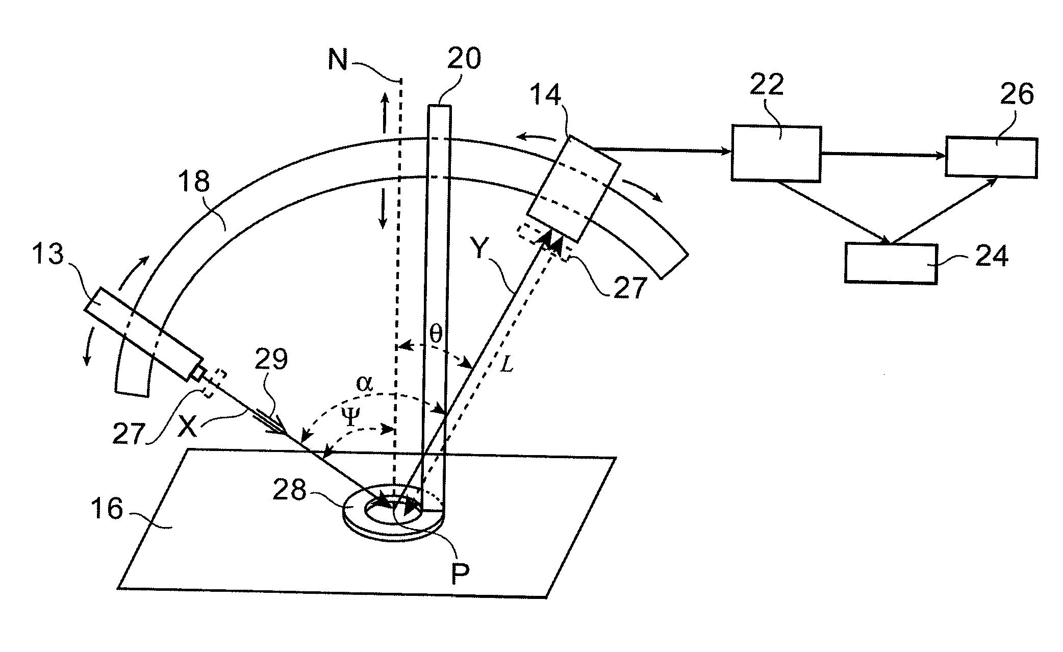
[0026]More precisely, the object of the present invention is a device for measuring, in vivo, properties of biological tissues, in particular for diagnostic and prognostic assistance for physi...


PUM


Abstract

Said device applies in particular to in vivo diagnostic and prognostic assistance for physiopathological changes to biological tissues. It comprises a coherent light source (13) for emitting coherent light along a first direction (X), for the purpose of illuminating a biological tissue (16) in first and second areas thereof, the first area being sound and the second area liable to include changes, the tissue thus illuminated generating a speckle phenomenon, means (14) for observing the speckle field in a second direction (Y) and for acquiring the speckle, and means (18) for varying the angle between the first and second directions, in order to observe the speckle field at different angles, so as to acquire information about the tissue at various depths in this tissue, support and absorption means (20, 28) to maintain a constant distance between the surface of the tissue and the observation and acquisition means and to absorb possible movements of the tissue, electronic means (22) for processing the speckle figures obtained by means of the observation and acquisition means, said processing being carried out in particular by a fractal method, in order to compare the first and second areas, and electronic means (24) for analysing the processing of the figures, by statistical methods, making it possible to validate the comparison made between the first and second areas.

Description

TECHNICAL FIELD[0001]The present invention relates to a device for measuring, in vivo, properties of biological tissues for diagnostic and prognostic assistance for physiopathological changes to said tissues.[0002]It especially applies to cutaneous tissular lesions and in particular to damage linked to irradiation.STATE OF THE PRIOR ART[0003]Radiological burns result from a cascade of complex biological and molecular mechanisms that can lead to their non-repair and to the destruction of cutaneous tissue (see document [1] which, like the other documents cited hereafter, is detailed at the end of the present description).[0004]The progressive instauration of a chronic inflammation, an angiogenesis defect, an abnormal remodelling of the extracellular matrix and a re-epithelisation defect is at the origin of radio-induced damage. The complexity of this tissular response results from radiosensitivity differences of each type of cell involved and their intercellular communications.[0005]C...

Claims


Application Information

Patent Timeline
Patent Type & Authority Applications(United States)
IPC IPC(8): A61B6/00
CPCA61B5/0059A61B2562/0242A61B5/445A61B5/444
Inventor CARVALHO, ODILEROY, LAURENCEBENDERITTER, MARC
Owner INSTITUT DE RADIOPROTECTION & DE SURETE NUCLEAIRE
Why Eureka
Try Eureka