Image processing apparatus and image processing method

a technology of image processing and image diagnosis, which is applied in the field of image processing apparatus and image processing method, can solve the problems of inability to display clinical reports or clinical charts about the constriction of a blood vessel, inability to label the blood vessel branches of non-interest, and inability to detect and display the constricted parts, etc., and achieve the effect of increasing the efficiency of image diagnosis of lesion parts

Active Publication Date: 2010-05-27
TOSHIBA MEDICAL SYST CORP
View PDF22 Cites 23 Cited by
  • Summary
  • Abstract
  • Description
  • Claims
  • Application Information

AI Technical Summary

Benefits of technology

[0007]An object of this invention is to provide an image processing apparatus and an image processing method, both capable of increasing the efficiency of image diagnosis of lesion parts of blood vessels.

Problems solved by technology

However, this technique inevitably labels the blood-vessel branches of non-interest, too.
The technique (3), i.e., detection and display of the constricted part, cannot accomplish the display of the clinical reports or clinical charts about the constriction of a blood vessel.
Consequently, it requires cumbersome operations to prepare clinical reports and clinical charts.

Method used


Image

  • Image processing apparatus and image processing method
  • Image processing apparatus and image processing method
  • Image processing apparatus and image processing method

Examples


Embodiment Construction

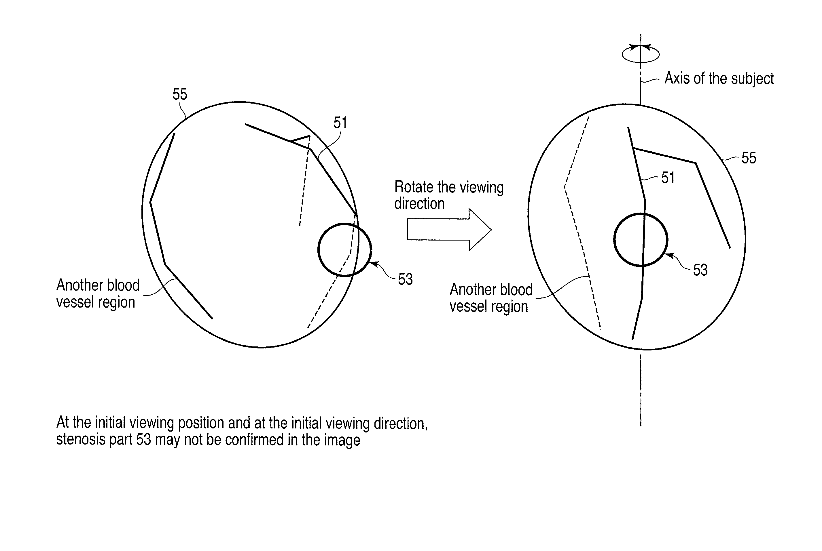
[0024]An image processing apparatus and an image processing method, both according to an embodiment of this invention, will be described with reference to the accompanying drawings. The image processing apparatus and image processing method, according to the embodiment, are applied to the image diagnosis of lesion parts of blood vessels. The blood vessels may exist in any parts of the human body, such as the heart and the brain. For the convenience of explanation, however, the following description relates to the blood vessels in the heart, because the images concerning the heart are believed to be diagnosed well by the technique according to this embodiment.

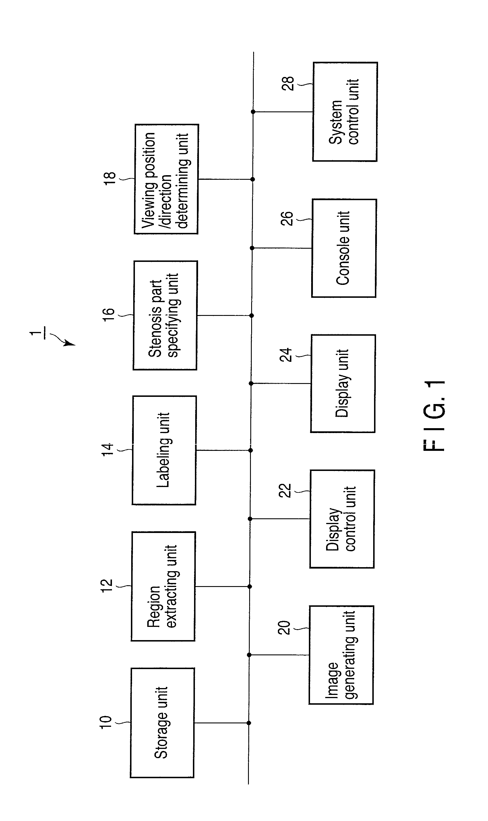
[0025]FIG. 1 is a diagram showing the configuration of an image processing apparatus 1 according to an embodiment of this invention. As shown in FIG. 1, the image processing apparatus 1 includes a storage unit 10, a region extracting unit 12, a labeling unit 14, a stenosis part specifying unit 16, a viewing position / direction de...


PUM


Abstract

A storage unit stores volume data including a blood vessel region. An extracting unit extracts the blood vessel region from the volume data. A specifying unit specifies a position of a region of interest in the blood vessel region and a deflection direction of a blood vessel region included in the region of interest. A determining unit determines a viewing position and a viewing direction based on the position of the region of interest and the deflection direction. A generating unit generates image data concerning the viewing position and the viewing direction based on the volume data. A display unit displays an image represented by the image data.

Description

CROSS-REFERENCE TO RELATED APPLICATIONS[0001]This application is based upon and claims the benefit of priority from prior Japanese Patent Application No. 2008-298534, filed Nov. 21, 2008, the entire contents of which are incorporated herein by reference.BACKGROUND OF THE INVENTION[0002]1. Field of the Invention[0003]The present invention relates to an image processing apparatus and an image processing method for use in image diagnosis of lesion parts in blood vessels.[0004]2. Description of the Related Art[0005]In recent years, various techniques have been developed for image processing apparatuses that are designed for image diagnosis of lesion parts in blood vessels, particularly lesion parts in coronary artery. These techniques include, for example, (1) extraction of images of the heart or the coronary artery (see, for example, Jpn. Pat. Appln. KOKAI Publication No. 2004-230086 and Jpn. Pat. Appln. KOKAI Publication No. 2006-246941); (2) labeling and classification of the branche...

Claims


Application Information

Patent Timeline
Patent Type & Authority Applications(United States)
IPC IPC(8): G06K9/00A61B6/03
CPCA61B5/0073A61B5/02007G06T2211/404G06T15/08G06T19/00G06T11/008
Inventor WAKU, TOSHIYAYAMAGATA, HITOSHI
Owner TOSHIBA MEDICAL SYST CORP
Why Eureka
Try Eureka