Membrane array and analytical device

a membrane array and membrane technology, applied in the field of analytical devices and methods, can solve the problems of inability to achieve rapid and high sensitivity analysis of analyte concentrations with small sample sizes, inability to rapid and high sensitivity analysis of analyte concentrations using whole blood, etc., to achieve rapid, high sensitivity and high sensitivity detection. , the effect of high sensitivity and high efficiency

Active Publication Date: 2010-06-03
ZBX CORP
View PDF36 Cites 4 Cited by
  • Summary
  • Abstract
  • Description
  • Claims
  • Application Information

AI Technical Summary

Benefits of technology

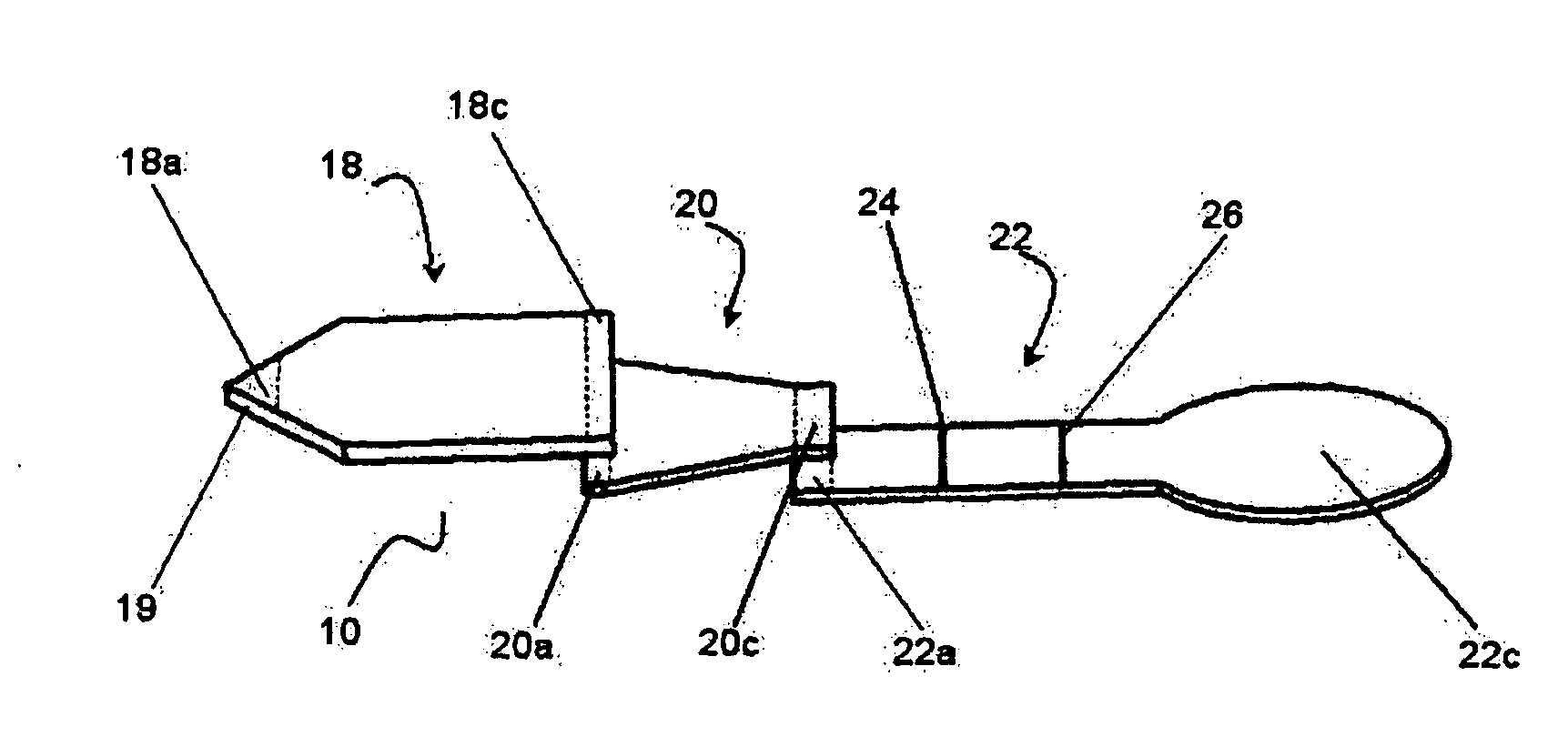
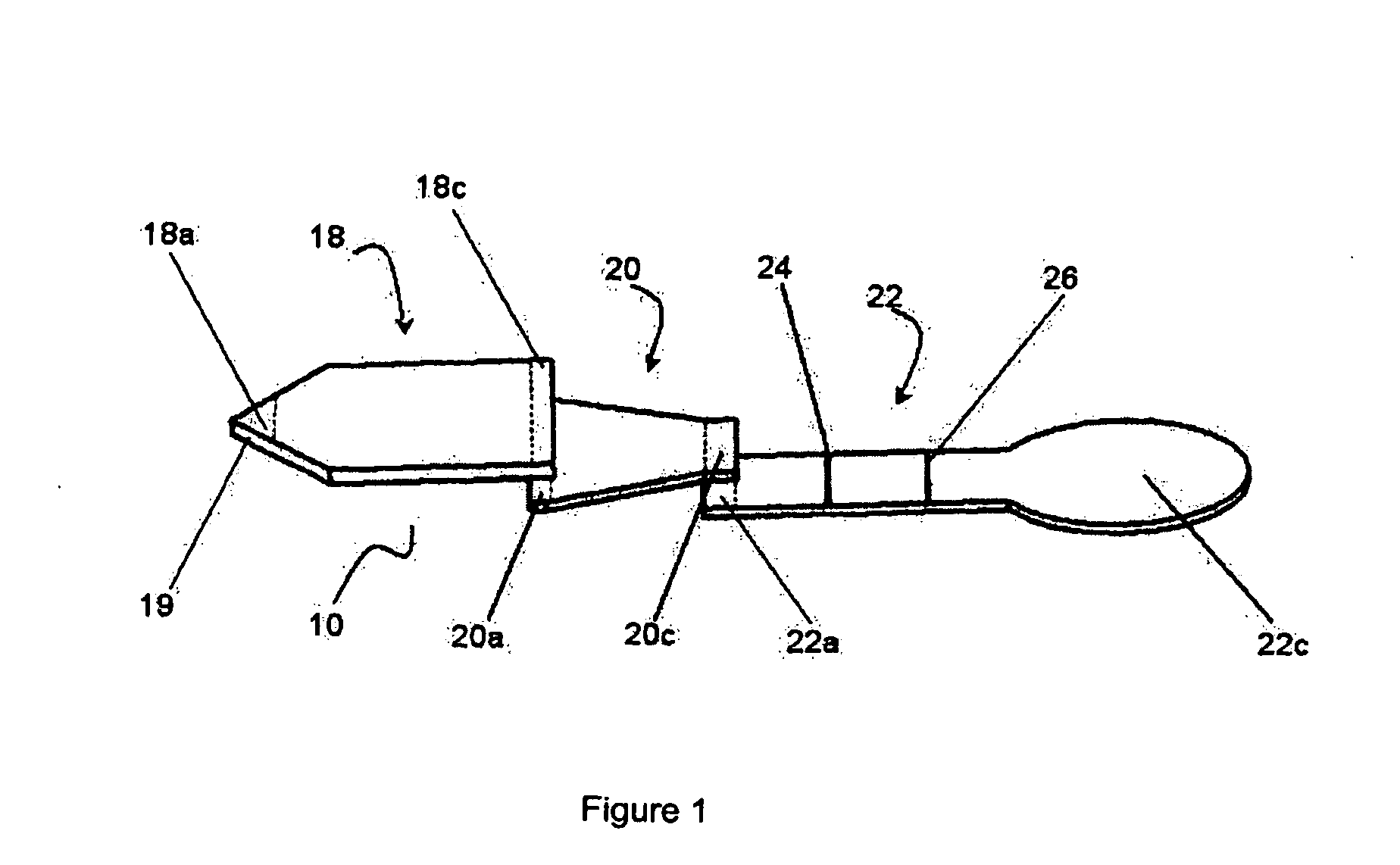
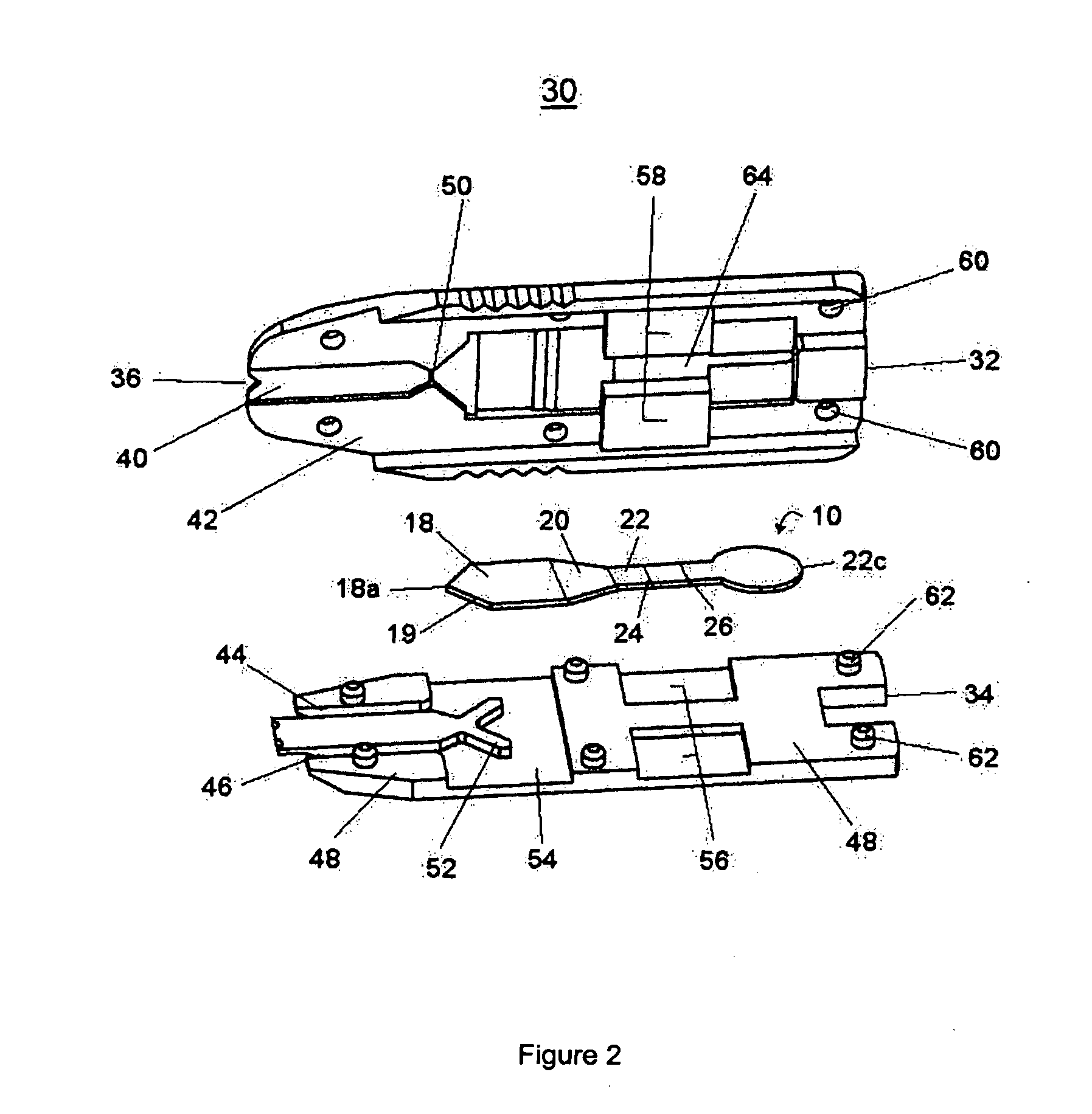
[0009]According to an aspect of the present invention is an improved membrane array that accommodates small sample volumes and provides rapid, highly efficient and highly sensitive detection of one or more analytes in the small sample volume.
[0085]a sample flow channel in fluid communication with said sample application means, said sample flow channel dimensioned to provide for substantially uniform horizontal flow of said sample into said membrane array.

Problems solved by technology

In operation of the device, multiple transfer steps are required before analysis and further this device is only designed for receiving samples of serum or plasma and as such is not suitable for analyses using whole blood.
In the use of this device, rapid and high sensitivity analysis of analyte concentrations cannot be achieved with small sample sizes.
However, rapid and high sensitivity detection of analytes using whole blood is not achievable in all circumstances and membrane array constructions with small sample volumes without background interference caused by hemolysis (the liberation of hemoglobin from the red blood cell).

Method used


Image

  • Membrane array and analytical device
  • Membrane array and analytical device
  • Membrane array and analytical device

Examples


example 1

[0139]A human cardiac troponin I test (TnI) device using one drop of whole blood sample is prepared according to current invention. For the analytical membrane, nitrocellulose (Whatman) with a pore size of about 5 μm was impregnated with both control and capture solutions using a conventional liquid dispenser. Control solution contains 1 mg / mL of goat anti-mouse IgG polyclonal antibodies (Arista Biologicals), and capture solution contains 2 mg / mL of an anti-troponin I monoclonal antibody (HyTest). Impregnated nitrocellulose was incubated at 37° C. for 30 minutes to immobilize the antibodies. The first separation membrane (Whatman) was sprayed with colloidal gold conjugate solution and then freeze dried to remove the water. The colloidal gold conjugate with a final OD of 2.2 at 540 nm was prepared from 40 nm gold particles (Arista Biologicals) and a monoclonal antibody specific to human cardiac troponin I (HyTest). An 8 μm nitrocellulose membrane (Whatman) was used as the second sepa...

example 2

[0140]A human procalcitonin (PCT) test device using one drop of whole blood sample is prepared according to current invention. For the analytical membrane, nitrocellulose (Millipore) with a pore size of 5 μm was impregnated with both control and capture solutions using a conventional liquid dispenser. Control solution contains 1 mg / mL of goat anti-mouse IgG polyclonal antibodies (Arista Biologicals), and capture solution contains 2 mg / mL of anti-calcitonin sheep polyclonal antibodies (Brahms). Impregnated nitrocellulose was incubated at 37° C. for 30 minutes to immobilize antibodies. Detection membrane or plasma separator (Whatman) was sprayed with colloidal gold conjugate solution and then freeze dried to remove water. Gold conjugate, prepared from 40 nm gold particles (Arista Biologicals) and a monoclonal antibody specific to PCT (Brahms), had a final OD 1.5 at 540 nm. A 8 μm nitrocellulose membrane (Whatman) was used as the separation membrane. The test strip is covered by a 25 μ...


PUM

PropertyMeasurementUnit
volumeaaaaaaaaaa
pore sizeaaaaaaaaaa
sizeaaaaaaaaaa

Abstract

A membrane array used to detect one or more analytes from a small sample of fluid with high sensitivity is provided. The membrane array can be employed in various analytical devices and is especially useful for identifying analytes from whole blood with minimal or negligible background interference.

Description

FIELD OF THE INVENTION[0001]This invention relates to analytical devices and methods useful for analytical assays of fluid samples. More specifically, the invention is directed to a novel membrane array and analytical devices incorporating same. The invention is useful for rapidly determining the presence of one or more analytes in small volumes of sample.BACKGROUND OF THE INVENTION[0002]Immunoassay devices and procedures currently exist for detecting the presence of an analyte in a sample of biological fluid. Typically, immunochemical reactions involving antigen / antibody reactions take place on dry porous carriers such as cellular membranes through which the sample to be analyzed flows by capillary action. The presence of an analyte in the sample can be detected either visually or by using reflectance or fluorescence based detection systems and instruments. Oftentimes, the label is an enzyme label or a particulate direct label, for instance a gold sol label.[0003]Typical immunochro...

Claims


Application Information

Patent Timeline
Patent Type & Authority Applications(United States)
IPC IPC(8): C40B30/00C40B40/00C40B60/12B01L99/00
CPCB01L3/5023B01L2200/027B01L2300/0681B01L2300/0825B01L2300/087Y10S435/971B01L2400/065G01N33/54366G01N33/558Y10S435/97B01L2400/0406G01N33/54388
Inventor QINWEI, SHI
Owner ZBX CORP
Why Eureka
Try Eureka