
Magnetic device for guiding catheter and method of use therefor

a magnetic device and catheter technology, applied in the field of magnetic devices for guiding catheters, can solve the problems of increasing the time, cost and inefficiency involved in this procedure, not having a design for feeding tubes to ensure the catheter's placement, etc., and achieve the effect of preventing the catheter from being inserted into the patient's lungs

Inactive Publication Date: 2010-06-10
SYNCRO MEDICAL INNOVATIONS +1
View PDF99 Cites 42 Cited by
  • Summary
  • Abstract
  • Description
  • Claims
  • Application Information

AI Technical Summary

Benefits of technology

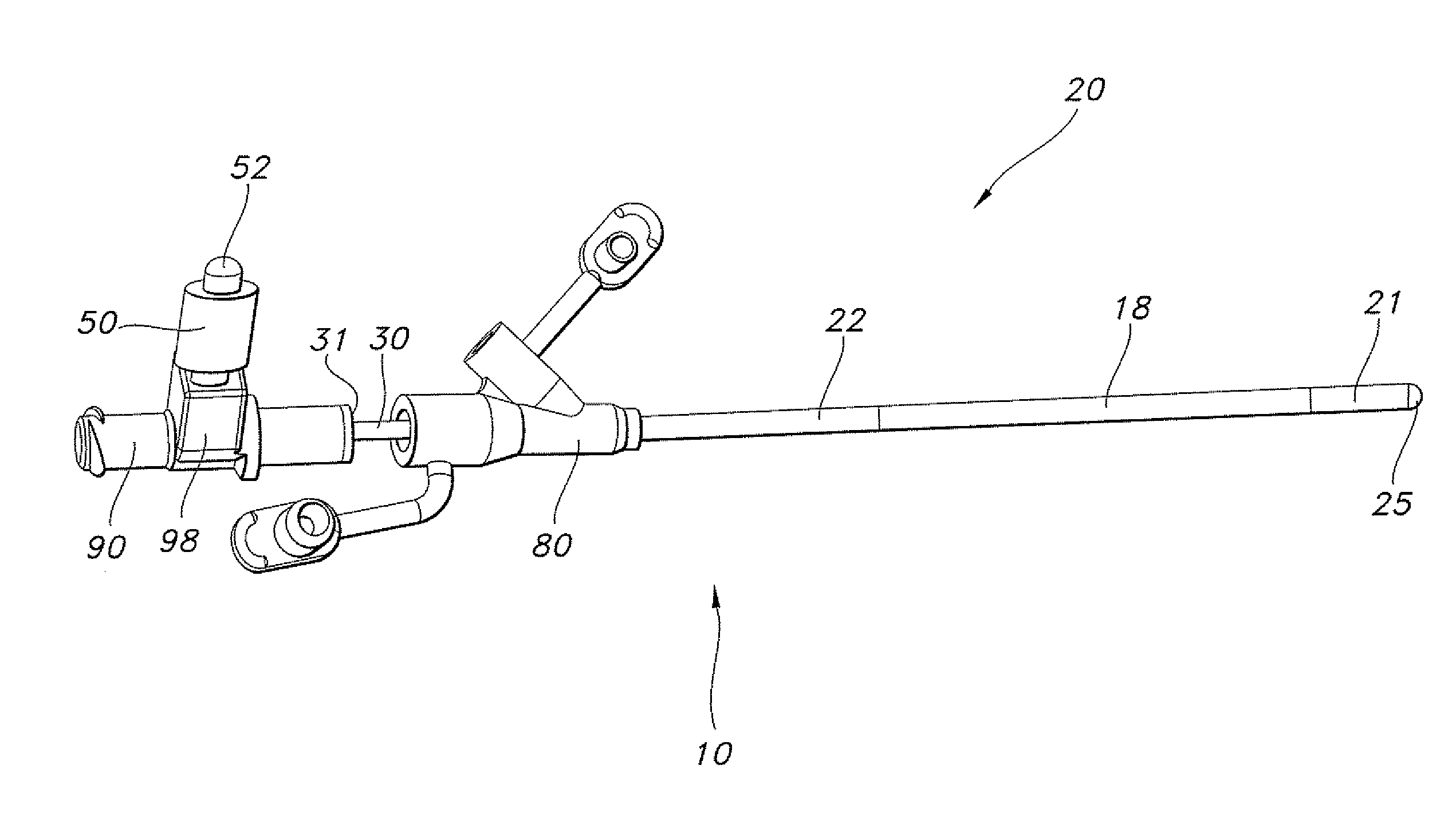
[0010]One or more external magnets that are suitable for placement on the back or the front of a patient's neck and methods for using the external magnet(s) to guide a feeding tube through the esophagus are described herein. The external magnet(s) are designed to be placed on the back or the front of the patient's neck and may be used with a magnetic feeding tube. The magnetic feeding tube may contain one or more magnet(s) or magnetically attractive material(s) (the “internal magnets”) that is attached to the feeding tube or one that is removable from the feeding tube. In one preferred embodiment, the feeding tube contains one or more magnet(s) or magnetically attractive material(s) (also referred to as the “internal magnets”) that are removable from the catheter. The external magnet is applied to the back or the front of the patient's neck and the feeding tube is inserted into the patient's nose and advanced within the patient's body. The magnetic field of the external magnet combines with the magnetic field of the internal magnet to produce a sufficient magnetic force to pull or push the feeding tube apparatus against the posterior wall of the esophagus to prevent placement of the stylet in the patient's trachea or windpipe to prevent insertion into the patient's lungs. Once the catheter is advanced beyond the patient's neck, the external magnet may be removed from the patient's neck. In some embodiments, the same or a second, different external magnet is used to advance the feeding tube apparatus to the desired location for administering food and / or medicine to the patient. In the embodiments utilizing a one or more magnet(s) or magnetically attractive material(s) that are removable from the catheter, the one or more magnet(s) or magnetically attractive material(s) (e.g. a magnet stack) may be removed when the feeding tube reaches the desired location. This allows for the catheter to remain in place while the patient undergoes diagnostic testing, such as magnetic resonance imaging.

Problems solved by technology

A reported problem with all feeding tubes is misplacement of the tube in the airway.
Both airway penetration and introduction of various chemicals into the lung and pleural spaces may occur prior to recognition of tube misplacement and can be fatal.
This requires stopping the procedure to perform the x-ray and thereby increases the time, cost and inefficiency involved in this procedure.
Although each of the above-described catheters provide improvements over other feeding tubes that are available, none of the feeding tubes is designed to ensure that the catheter is not accidentally placed in the trachea without the need for x-rays to confirm the location of the feeding tube.

Method used


Image

  • Magnetic device for guiding catheter and method of use therefor
  • Magnetic device for guiding catheter and method of use therefor
  • Magnetic device for guiding catheter and method of use therefor

Examples


Embodiment Construction

I. External Magnet for Guiding Feeding Tube

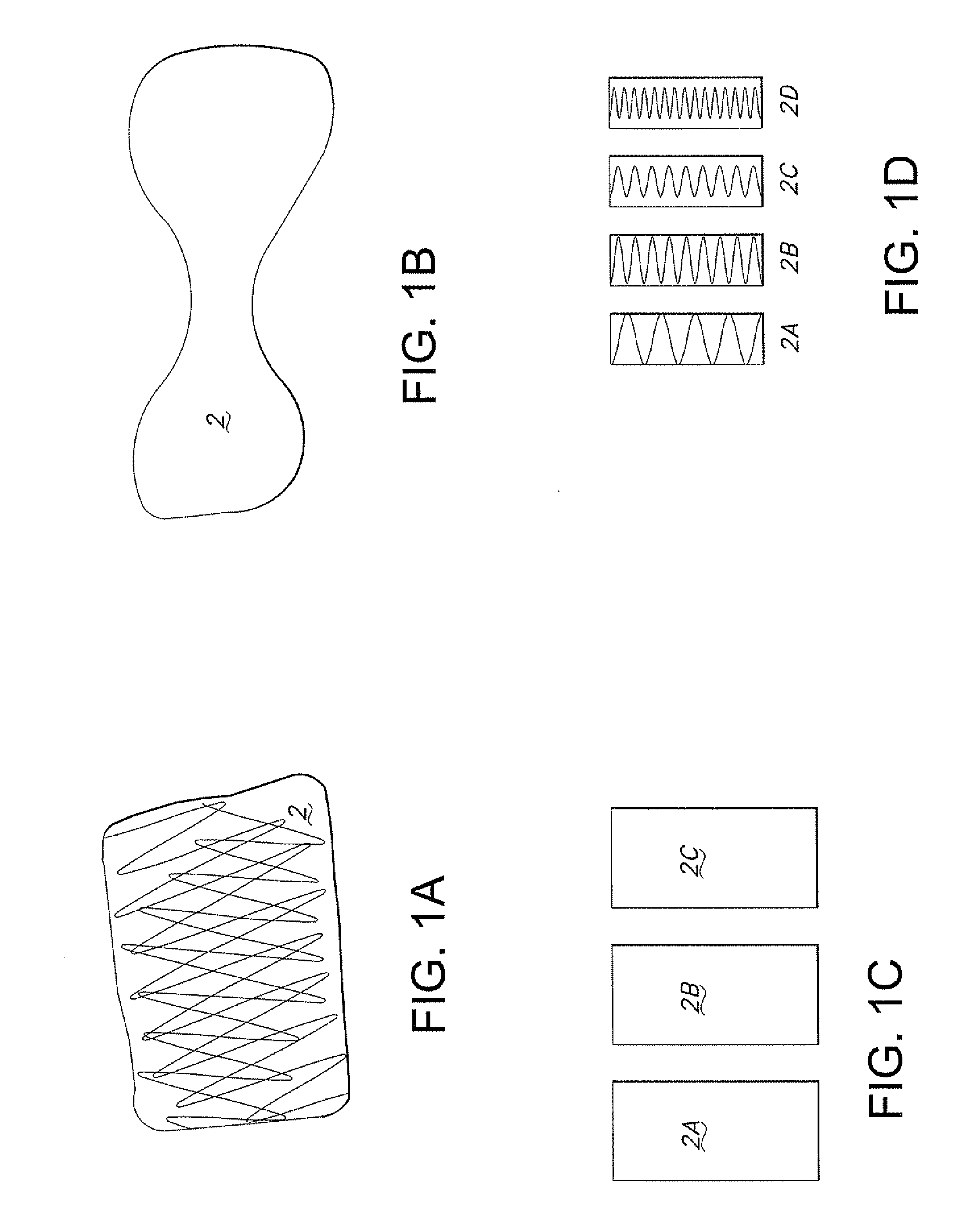
[0018]One or more external magnets (2) (see e.g. FIGS. 1A-D) that are suitable for placement on a patient's neck may be used to guide a feeding tube through the esophagus by directing the distal tip of the feeding tube to the posterior wall of the esophagus.

[0019]A. Neck Brace or Collar

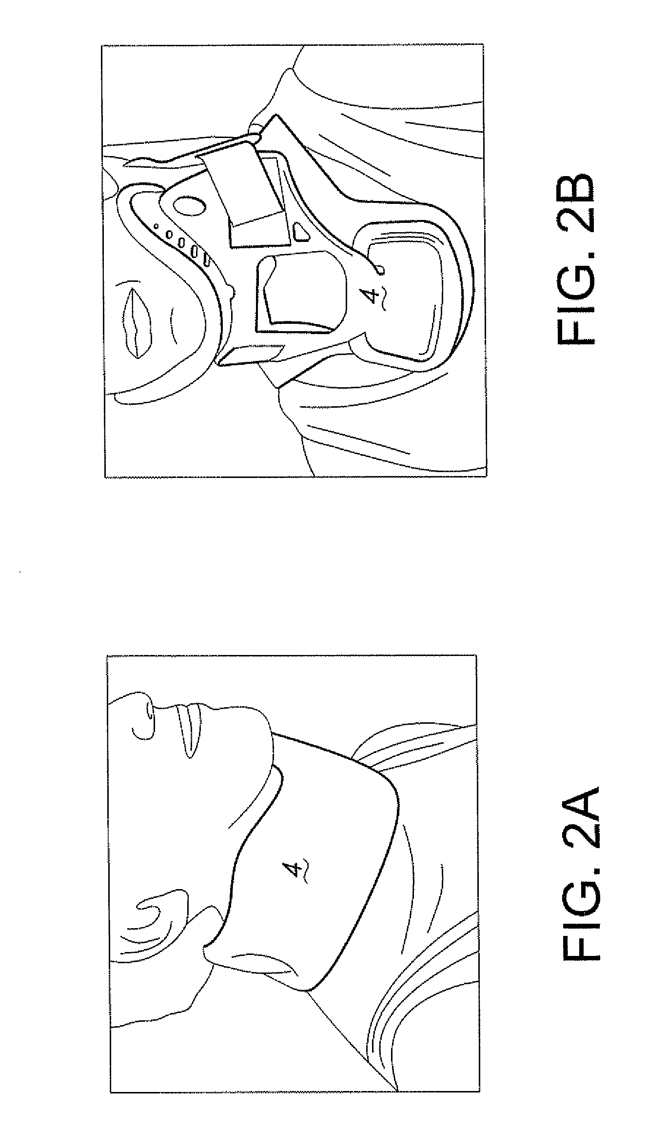
[0020]The one or more external magnets (2) may be located within a material that is designed to be placed on the neck, such as a neck brace or cervical collar (4). Any standard neck brace or cervical collar can be modified to contain one or more magnets. Optionally, the neck brace or cervical collar contains one or more pockets designed to contain one or more magnets. Optionally, the external magnet(s) are insertable into and removable from the neck brace or collar.

[0021]In one embodiment, the neck brace or collar is formed from a soft pliable material (see e.g. FIG. 2A). In another embodiment, the neck brace or collar is formed from a rigid plastic material ...


PUM


Abstract

One or more external magnets that are suitable for placement on the back or front of a patient's neck and methods for using the external magnet(s) to guide a feeding tube containing one or more internal magnets through the esophagus are described herein. The external magnet is applied to the back or the front of the patient's neck and the feeding tube is inserted into the patient's nose and advanced within the patient's body. The magnetic field of the external magnet combines with the magnetic field of the internal magnet to produce a sufficient magnetic force to pull or push the feeding tube apparatus against the posterior wall of the esophagus to prevent placement of the stylet in the patient's trachea or windpipe to prevent insertion into the patient's lungs.

Description

CROSS-REFERENCE TO RELATED APPLICATION[0001]This application claims priority to U.S. Provisional Application Ser. No. 61 / 093,628, entitled “Magnetic Device for Guiding Catheter”, filed Sep. 2, 2008. The disclosure of this application is incorporated herein by reference.FIELD OF THE INVENTION[0002]The present invention relates to methods and devices for placement of feeding tubes.BACKGROUND OF THE INVENTION[0003]A reported problem with all feeding tubes is misplacement of the tube in the airway. Inadvertent insertion of nasogastric tubes into the trachea and distal airways is reported to range from 0.3% to 15% of insertions. (see Thomas et al., Journal of the American College of Nutrition, 17(2):195-197 (1998)). Both airway penetration and introduction of various chemicals into the lung and pleural spaces may occur prior to recognition of tube misplacement and can be fatal. Currently, nursing staff checks the location of the feeding tube using x-ray during the insertion process. This...

Claims


Application Information

Patent Timeline
Patent Type & Authority Applications(United States)
IPC IPC(8): A61B1/00
CPCA61M25/0127
Inventor PINSKY, MICHAEL RAYMONDGAUTIER, JUAN BERNARDO OCHOAWINKLER, JOSEFWAKEFORD, GARYGABRIEL, SABRYFRANKHOUSER, PAULSANDOSKI, AARON
Owner SYNCRO MEDICAL INNOVATIONS
Learn More