
Method for contrast-agent-free angiographic imaging in magnetic resonance tomography

a magnetic resonance tomography and contrast-free technology, applied in the field of magnetic resonance tomography, or mrt, can solve the problems of inability to use the method, the sensitivity of the patient movement, and the inability to contrast the agen

Active Publication Date: 2010-06-24
SIEMENS HEALTHCARE GMBH
View PDF12 Cites 24 Cited by
  • Summary
  • Abstract
  • Description
  • Claims
  • Application Information

AI Technical Summary

Benefits of technology

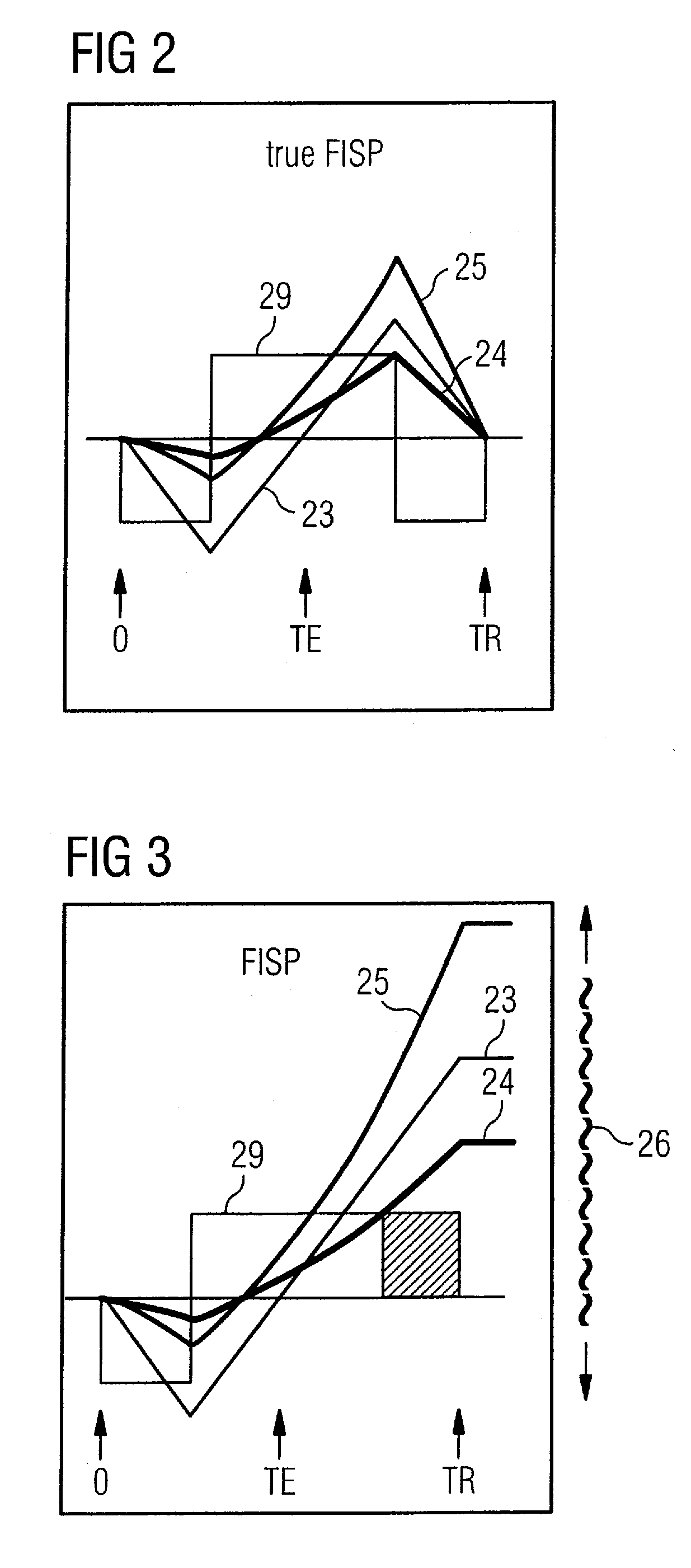
[0023]It is also advantageous for the SSFP sequences that are to be implemented, to use the optimal flip angle α that maximizes the respective SSFP signal, and for all geometric MRT parameters—of both the flow-sensitive and the flow-insensitive SSFP sequences—to be identical.

Problems solved by technology

A disadvantage of the TOF method is its sensitivity to patient movement, in particular in the cervical region.
A disadvantage of this method is that it can be applied only to one particular speed interval at a time, in order to avoid ambiguities.
However, as free ions all these substances are highly toxic, and thus cannot be used in this form.
However, recently there have been increased reports of (kidney) ailments (Nephrogenic Systemic Fibrosis, or NSF) caused by contrast agents containing gadolinium.
Nonetheless, FBI and SPACE represent very complicated measurement methods, because a triggering, in particular triggering by breath and EKG, as is required both in FBI and in SPACE, is generally very expensive:

Method used


Image

  • Method for contrast-agent-free angiographic imaging in magnetic resonance tomography
  • Method for contrast-agent-free angiographic imaging in magnetic resonance tomography
  • Method for contrast-agent-free angiographic imaging in magnetic resonance tomography

Examples


Embodiment Construction

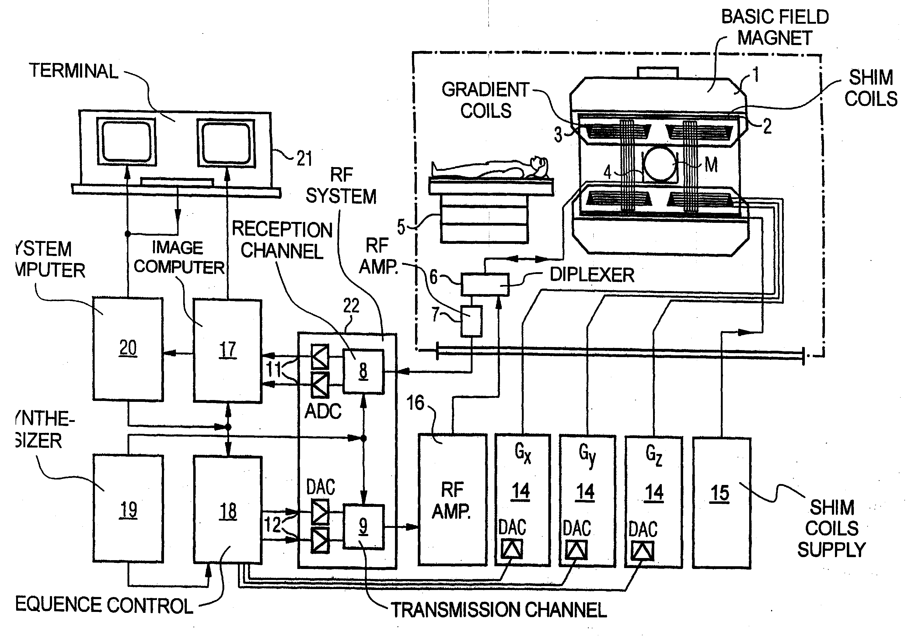
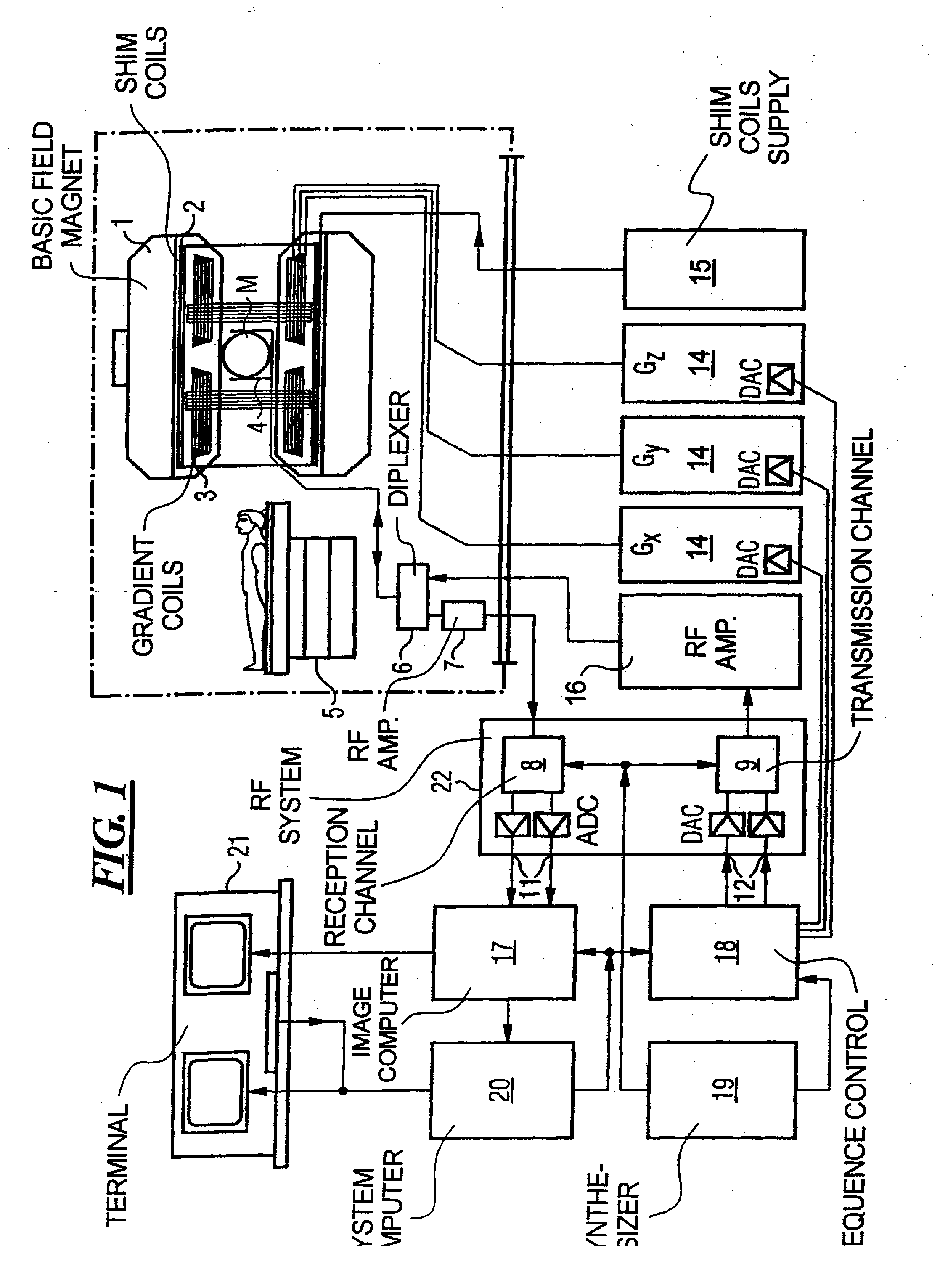
[0035]FIG. 1 schematically shows a magnetic resonance tomography device for producing a magnetic resonance image of a subject according to the present invention. The basic design of the tomography device corresponds to the design of a conventional tomography device, with differences noted below. A basic field magnet 1 produces a strong magnetic field that is constant over time for the polarization or orientation of the nuclear spins in the examination region of a subject, such as a part of a human body that is to be examined. The high degree of homogeneity of the basic magnetic field required for a magnetic resonance measurement is defined in a spherical measurement volume M into which the parts of the human body that are to be examined are brought. In order to support the homogeneity requirements, and in particular in order to eliminate chronologically invariable influences, shim plates made of a ferromagnetic material are attached at a suitable location. Temporary variable influen...


PUM


Abstract

A method for contrast-agent-free non-triggered angiographic imaging in magnetic resonance tomography that includes the steps of (S1) 2D or 3D measurement of a bodily region having a flow of blood, using a flow-insensitive SSFP sequence, (S2) measurement of the same bodily region using a flow-sensitive SSFP sequence, (S3) registration of the measurement results obtained in steps S1 and S2 to one another, (S4) unweighted or self-weighted subtraction of the registered measurement result obtained in step S2 from the registered measurement result obtained in step S1, (S5) execution of a 2D or 3D image correction of the image obtained in step S4 by removing image distortions caused by gradient field inhomogeneities and / or magnetic basic field inhomogeneities, and (S6) representation of the angiogram obtained in step S5 in the form of an MIP or segmented 2D or 3D vessel tree representation.

Description

BACKGROUND OF THE INVENTION[0001]1. Field of the Invention[0002]The present invention relates in general to magnetic resonance tomography, or MRT as is used in the field of medicine in order to examine patients. In particular, the present invention relates to a method for the magnetic resonance tomographic representation of vessels (angiography) without the use of contrast agent, and without physiological synchronization with the execution of the MR sequence.[0003]2. Description of the Prior Art[0004]The term “angiography” refers in general to the representation of blood vessels (arteries and / or veins) using medical imaging methods (x-ray, CT, MRT). In magnetic resonance angiography (MRA) there are two classical methods that make use of the property of the flow of blood in order to represent the vessels (time of flight methods, or TOF, and phase-contrast angiography, or PC angio). There is also MRA supported by contrast agent, which uses contrast agent that increases spin relaxation...

Claims


Application Information

Patent Timeline
Patent Type & Authority Applications(United States)
IPC IPC(8): A61B5/055
CPCG01R33/5613G01R33/5635G01R33/5614
Inventor DEIMLING, MICHAEL
Owner SIEMENS HEALTHCARE GMBH
Learn More