Thorascopic Heart Valve Repair Method and Apparatus

a heart valve and thoracic valve technology, applied in the direction of surgical forceps, instruments, catheters, etc., can solve the problems of prolonged hospital stay, high trauma degree, and significant risk of complications

Active Publication Date: 2010-07-08
MAYO FOUND FOR MEDICAL EDUCATION & RES
View PDF112 Cites 47 Cited by
  • Summary
  • Abstract
  • Description
  • Claims
  • Application Information

AI Technical Summary

Benefits of technology

[0053]In addition to the fiberoptic visualization system that insures that a valve leaflet is properly captured, other real-time visualization systems are employed to help guide the instrument 10 to the valve leaflet 16. Preferably a transesophageal or intravascular color-Doppler echocardiography system is used for this purpose. As explained above, this imaging system is also used to determine the length of the neo-implanted artificial chordae in real-time by observing reduction or disappearance of regurgitation by transesophageal or intravascular color-Doppler echocardiography.

Problems solved by technology

However, these invasive, open-chest procedures produce a high degree of trauma, a significant risk of complications, an extended hospital stay, and a painful recovery period for the patient.
Moreover, while heart valve surgery produces beneficial results for many patients, numerous others who might benefit from such surgery are unable or unwilling to undergo the trauma and risks of current techniques.
Rupture or elongation of the chordae tendinae result in partial or generalized leaflet prolapse, which causes mitral (or tricuspid) valve regurgitation.

Method used


Image

  • Thorascopic Heart Valve Repair Method and Apparatus
  • Thorascopic Heart Valve Repair Method and Apparatus
  • Thorascopic Heart Valve Repair Method and Apparatus

Examples


second embodiment

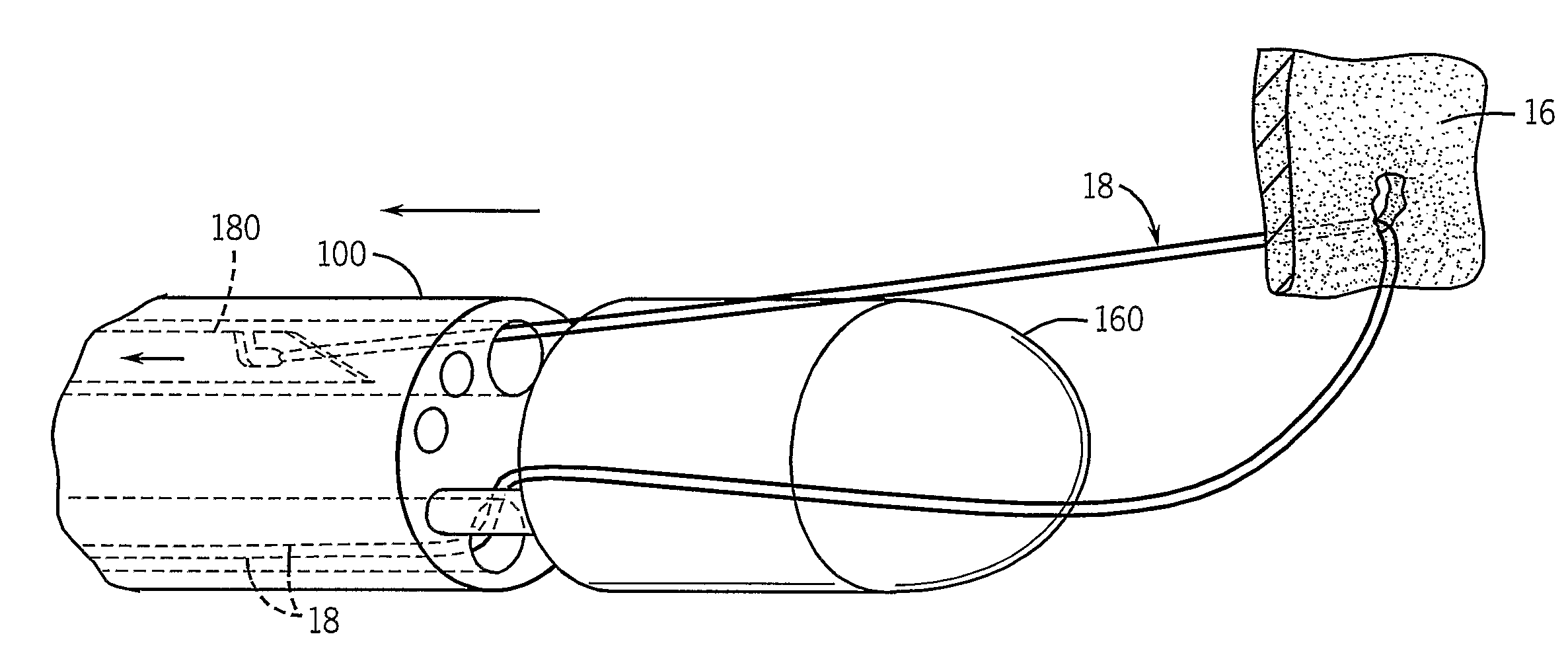
[0046]As shown in FIGS. 9A-9D, the suture deployment system at the distal end of the instrument 10 is positioned around a valve leaflet 16 to be repaired as shown in FIG. 9A. The suture 18 in this embodiment is a closed loop with one end of the loop disposed in the tip 160 and its other end disposed in the lumen 164 and wrapped around the needle 180. The needle 180 is extended through the grasped valve leaflet 16 into the instrument tip 160 where it hooks one end of the looped suture 18 in a notch 166 formed on one side of the needle as shown in FIG. 9B. The needle 180 is then retracted to pull the looped suture 18 through the puncture opening in the leaflet 16. The leaflet is then released as shown in FIG. 9C by sliding the tip 160 to its open position. The instrument 10 is then withdrawn as shown in FIG. 9D to slide the unhooked end of the looped suture 18 along the length of the needle toward the leaflet 16 where it forms a Larks head around the leaflet edge.

[0047]The instrument ...

third embodiment

[0048]As shown in FIGS. 10A-10D, the suture deployment system at the distal end of the instrument 10 is positioned around a valve leaflet 16 to be repaired as shown in FIG. 10A. The midpoint 17 of the suture 18 is looped around the lumen 164 and its two loose ends 20 are coiled up in the tip 160. After the tip 160 is closed to capture the valve leaflet 16, the needle 180 is extended through the grasped valve leaflet 16 into the instrument tip 160. The free ends 20 of the suture 18 are positioned in the tip 160 to form a loop 19 and a notch 166 formed on one side of the needle extends through this loop 19 and “hooks” the free ends of the suture 18 as shown in FIG. 10B. The needle 180 is then retracted back into the lumen 164 to pull the hooked ends of the suture 18 through the puncture opening in the leaflet 16. The leaflet is then released as shown in FIG. 10C by sliding the tip 160 to its open position. The instrument 10 is then withdrawn from the heart as shown in FIG. 10D to pull...


PUM


Abstract

An instrument for performing thorascopic repair of heart valves includes a shaft for extending through the chest cavity and into a heart chamber providing access to a valve needing repair. A movable tip on the shaft is operable to capture a valve leaflet and a needle is operable to penetrate a capture valve leaflet and draw the suture therethrough. The suture is thus fastened to the valve leaflet and the instrument is withdrawn from the heart chamber transporting the suture outside the heart chamber. The suture is anchored to the heart wall with proper tension as determined by observing valve operation with an ultrasonic imaging system.

Description

CROSS-REFERENCE TO RELATED APPLICATIONS[0001]This application is a divisional application of U.S. patent application Ser. No. 11 / 813,695, filed Jul. 11, 2007, which is a 371 of PCT / US06 / 01699 filed Jan. 19, 2006, which claims the benefit of U.S. Provisional Patent Application No. 60 / 645,677 filed Jan. 21, 2005.BACKGROUND OF THE INVENTION[0002]Various types of surgical procedures are currently performed to investigate, diagnose, and treat diseases of the heart and the great vessels of the thorax. Such procedures include repair and replacement of mitral, aortic, and other heart valves, repair of atrial and ventricular septal defects, pulmonary thrombectomy, treatment of aneurysms, electrophysiological mapping and ablation of the myocardium, and other procedures in which interventional devices are introduced into the interior of the heart or a great vessel.[0003]Using current techniques, many of these procedures require a gross thoracotomy, usually in the form of a median sternotomy, t...

Claims


Application Information

Patent Timeline
Patent Type & Authority Applications(United States)
IPC IPC(8): A61B17/062
CPCA61B17/0469A61B17/29A61B17/0625A61B17/295A61B2017/00057A61B2017/00783A61B2017/0608A61B2019/5206A61B1/00167A61B1/07G02B23/2469A61B5/0084A61B5/02028A61B5/4836A61B17/0483A61B2090/306A61B2090/0807A61B5/0036A61B17/0482A61B17/06A61B17/0491A61B2017/00243
Inventor SPEZIALI, GIOVANNI
Owner MAYO FOUND FOR MEDICAL EDUCATION & RES
Why Eureka
Try Eureka