Minimally-invasive measurement of esophageal inflammation

a technology of esophageal inflammation and measurement method, applied in the field of medicine, can solve the problems of difficult diagnosis of inflammatory conditions of the gastrointestinal tract, eosinophilic gastroenteritis (ege), inflammatory bowel disease (ibd), etc., and achieves the effect of not being useful for cell-mediated reactions, serologic and radiographic assessments are not diagnostic, and not being useful for diagnosis

Active Publication Date: 2010-09-23
UNIV OF COLORADO THE REGENTS OF +1
View PDF7 Cites 14 Cited by
  • Summary
  • Abstract
  • Description
  • Claims
  • Application Information

AI Technical Summary

Benefits of technology

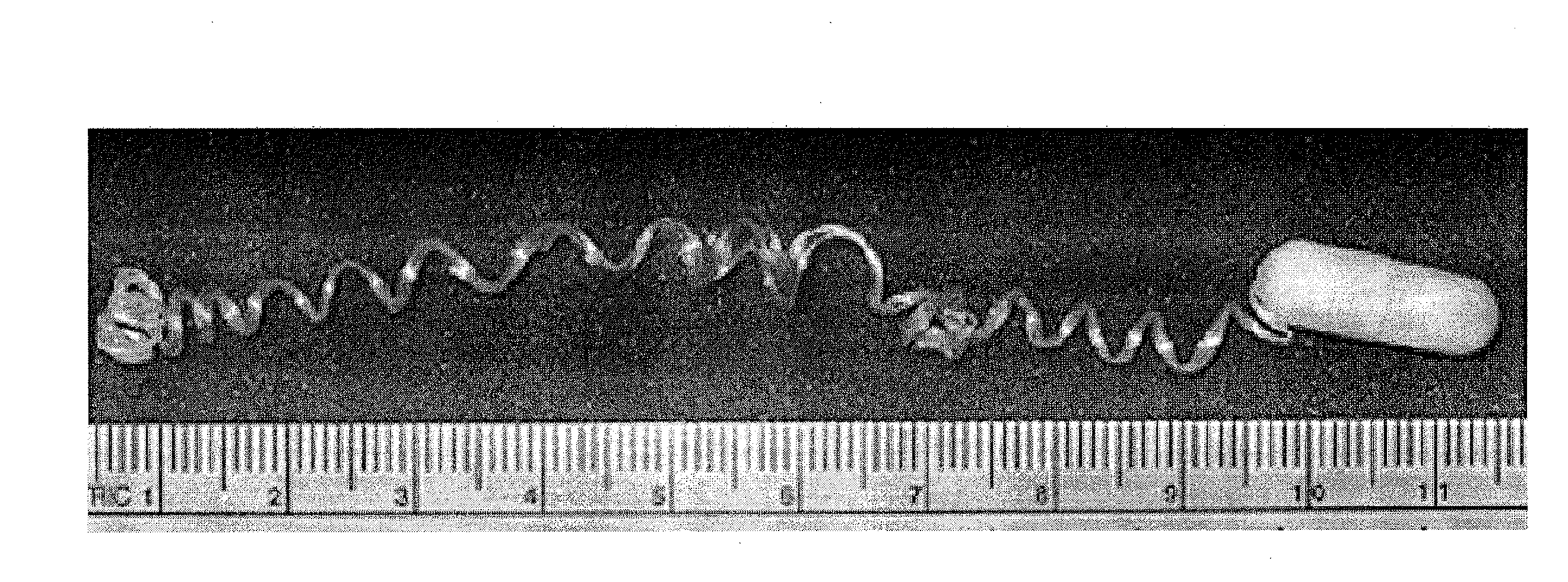
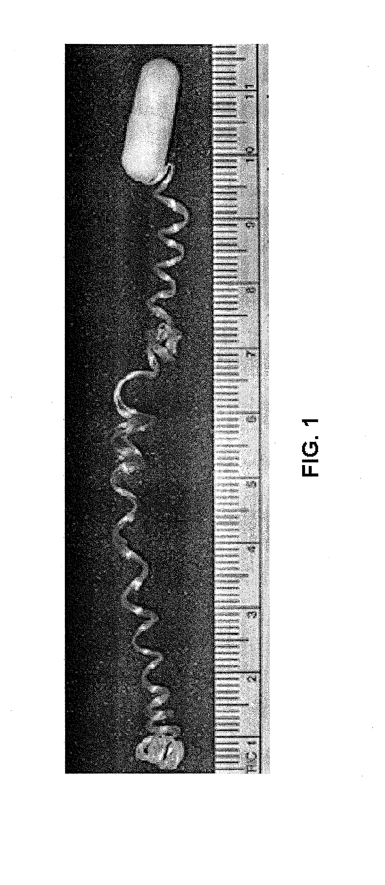
[0016]The device may be any apparatus which allows the capture of the inflammatory mediators and other cells of the esophagus. In one embodiment, the device comprises a pharmaceutical capsule having an opening, a drag material within the capsule, and a line embedded in the drag material that runs through the opening of the capsule. The pharmaceutical capsule may be dissolvable or it may pass through the subject's system. In another embodiment, the capsule may be comprised of two parts, a base and a cap, and the opening of the capsule may be a perforated opening. The drag material may be constructed of various materials. In some embodiments, the drag material is malleable. The line may be of various lengths and may be made of various materials to be more or less abrasive and more or less absorptive. In a particular embodiment, the line comprises two components, a string and a sampling cloth. The string component may be referred to as the proximal segment, whereas the sampling cloth may be referred to as the distal segment. In some embodiments, after deployment of the string, the proximal segment is located in the esophagus of the subject. The string may be made of, for example, an absorbent, mesh, or textured fiber. In one embodiment, the string and sampling cloth are constructed of the same material. In some embodiments, a portion of the string may be pulled out of the capsule prior to swallowing the capsule and the end of the string may be attached to the cheek. After the predetermined period of time, the string may be removed from the esophagus.
[0017]In another embodiment, the device further comprises a capture agent for one or more diagnostic indicators. These capture agents may be located on the line. In a particular embodiment, the capture agents may be focused on the proximal segment of the line. The capture agent may be any agent that binds an analyte through an interaction that is sufficient to permit the agent to bind and concentrate the analyte from a homogeneous mixture of different analytes. The binding interaction may be mediated by an affinity region of the capture agent. Representative capture agents include antibodies, and more specifically monoclonal antibodies. Further non-limiting examples include eosinophil granule protein antibodies.
[0018]The predetermined period of time may be various lengths of time. For example, the predetermined time may be between 15 minutes and 12 hours. In some embodiments, the predetermined period of time may be 1, 2, 3, 4, 5, 6, 7, 8, 9, 10, 11, 12, 18, 24 or more hours, or any increment thereof. In one embodiment, the predete

Problems solved by technology

Making the diagnosis of many inflammatory conditions of the gastrointestinal tract such as severe gastroesophageal reflux (GERD), eosinophilic gastroenteritis (EGE), food allergic enteropathy (FAE), and inflammatory bowel disease (IBD) is often difficult.
Serologic and radiographic assessments are not diagnostic for any of these conditions.
In addition, these tests are beneficial only in identifying IgE mediated reactions and are not useful for cell-medi

Method used


Image

  • Minimally-invasive measurement of esophageal inflammation
  • Minimally-invasive measurement of esophageal inflammation
  • Minimally-invasive measurement of esophageal inflammation

Examples


example 1

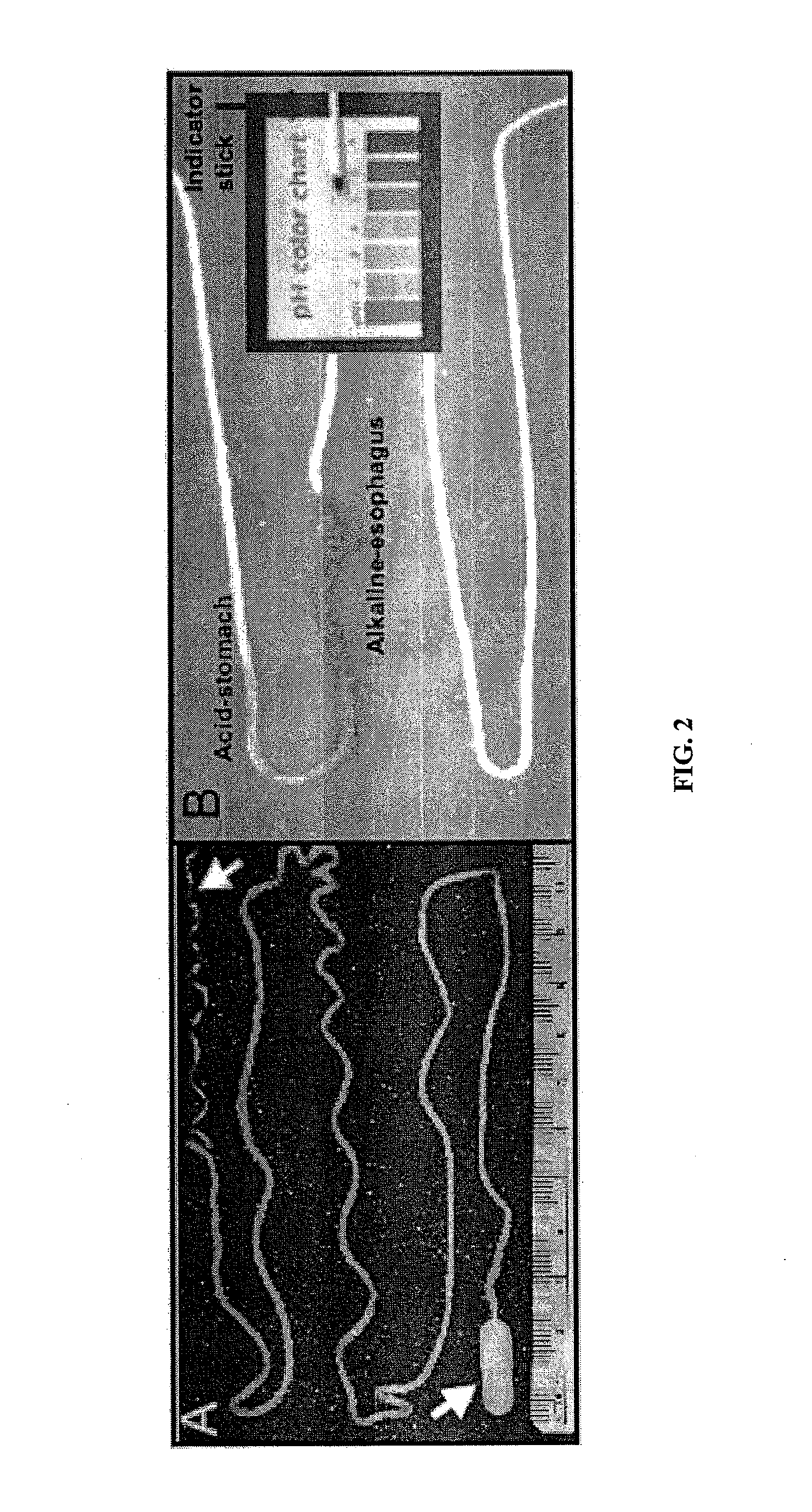
[0101]The string test was administered to a patient with reflux esophagitis and left in place for 1 and 12 hours. The first 10-20 cm of the string was pulled out from the capsule and the end of the string was secured between the fingers. The capsule and remaining string were swallowed with water and the string in the fingers was taped to the cheek. After the predetermined period, the taped string was removed from the cheek and the string retrieved from the intestinal tract. Following removal, the contents of the string were assessed for the presence of inflammatory proteins and RNA. The entire string was placed either in protein buffer, RNA stalizing solution or formalin. After 1 and 12 hours in the esophagus, elevations of interleukin-8 and TNF α-protein were identified by Mesoscale and ELISA technology. In these studies, protein was extracted from the string, and the soluble product was placed in the well of a 96-well plate for analysis. Following placement of the soluble product ...

example 2

[0102]Identification of microbial flora from EST. Identification and enumeration of esophageal and oral bacterial population by selective culture.

[0103]Microbial culture. Strings were swallowed or remained in the mouth for 15 min. A 5 cm piece of esophageal or oral string was aseptically transferred to 5 ml of sterile Hank's Buffered Salt Solution (HBSS) and vortexed for 30 sec before a 5 min incubation at RT. For nasal secretion collection swap was humidified in sterile saline; a nasal swab was collected using a BBL™ culture swab. String and nasal solution was vortexed again for 30 sec and the string removed. The solution was centrifuged for 15 min at 4750 rpm. Supernatant was aspirated and the pellet was resuspended in 1 ml of sterile HBSS. Dilutions up to 10−5 were performed and 50 ml of non diluted samples or 10−5 dilution was plated in duplicate onto selective media. Plates were incubated at 37° C. overnight in aerobic or anaerobic (BD Bioscience Gas Packs) conditions. Plate co...

example 3

[0105]Measurements of eosinophil derived granule proteins from the Esophageal String Test (EST) can detect histological evidence of esophageal inflammation associated with EE. Preliminary results show that supernatants derived from activated eosinophils adhere to the EST and that eosinophil derived granule proteins within these samples can be measured by Western blot and ELISA analyses. These findings support the technical feasibility for the EST to be used to measure eosinophil derived granule proteins contained in the esophageal lumen of patients with EE.

[0106]Detection of eosinophil granule major basic protein-1 (MBP1) on EST strings incubated with acidic lysates of resting eosinophils. In order to demonstrate the ability of EST string to detect eosinophil granule cationic proteins, purified blood eosinophils from normal subjects (>99% eosinophils) were used to prepare soluble acidic lysates by sonication of different numbers of eosinophils (1×10⁵ or 1×10⁶ cells) in either 0.1N H...


PUM

PropertyMeasurementUnit
Timeaaaaaaaaaa
Timeaaaaaaaaaa
Timeaaaaaaaaaa

Abstract

The methods and apparatus of the present invention allow the evaluation of inflammation of the esophagus. Measurements may be utilized, for example, to diagnose a disease of the esophagus, to monitor inflammation of the esophagus, or to access the treatment of a disease of the esophagus. In one embodiment, the invention comprises a method for measuring esophageal inflammation comprising deploying a device into the esophagus of a subject, removing the device after a predetermined period of time, analyzing the device for a diagnostic indicator of esophageal inflammation and evaluating the diagnostic indicator to diagnose esophageal inflammation.

Description

[0001]This application claims the benefit of U.S. Provisional Application No. 60 / 985,386, filed Nov. 5, 2007, the contents of which are hereby incorporated by reference.[0002]This invention was made with government support under grant numbers R21-AI079925 awarded by the National Institutes of Health (NIH). The government has certain rights in the invention.BACKGROUND OF THE INVENTION[0003]A. Field of the Invention[0004]The present invention relates generally to the field of medicine. More particularly, it concerns methods of diagnosing and monitoring diseases of the esophagus.[0005]B. Description of Related Art[0006]Making the diagnosis of many inflammatory conditions of the gastrointestinal tract such as severe gastroesophageal reflux (GERD), eosinophilic gastroenteritis (EGE), food allergic enteropathy (FAE), and inflammatory bowel disease (IBD) is often difficult. Serologic and radiographic assessments are not diagnostic for any of these conditions. In the instance of food allerg...

Claims


Application Information

Patent Timeline
IPC IPC(8): A61B5/00
CPCA61K31/437G01N33/5308C12Q1/6883A61K31/4375C12Y406/01002A61P1/04Y02A90/10A61B1/00156A61B1/2733C12Q1/04C12Q1/527G01N33/6863G01N33/6866G01N33/6869G01N33/6893G01N2333/521G01N2333/988G01N2800/14G01N2800/7028G01N2800/7095
Inventor FURUTA, GLENN T.ACKERMAN, STEVEN J.
Owner UNIV OF COLORADO THE REGENTS OF
Why Eureka
Try Eureka