Low-cost, compact, & automated diabetic retinopathy diagnostics & management device

a diabetic retinopathy, management device technology, applied in the direction of diagnostics, instruments, medical science, etc., can solve the problems of inconvenient use, high cost, bulky, etc., and achieve the effect of reducing the number of patients, and improving the accuracy of diagnosis

Inactive Publication Date: 2010-09-30
KULKARNI MANISH D +1
View PDF2 Cites 24 Cited by
  • Summary
  • Abstract
  • Description
  • Claims
  • Application Information

AI Technical Summary

Problems solved by technology

Most of the industrial and clinical OCDR and OCT machines are expensive, cumbersome to use, bulky, not very efficient and are fragile.
Abnormalities in blood-flow circulation due to diabetes is the root cause behind retinal microstructure damage.
However, no clinical tools exist that can perform functional and velocity mapping of blood vessels in the retina for tracking early development of diabetic eye diseases.
This increases system complexity and bulkiness.
Ruled gratings are cumbersome & expensive.

Method used


Image

  • Low-cost, compact, & automated diabetic retinopathy diagnostics & management device
  • Low-cost, compact, & automated diabetic retinopathy diagnostics & management device
  • Low-cost, compact, & automated diabetic retinopathy diagnostics & management device

Examples


Embodiment Construction

5.1 Optical Coherence Tomography (OCT)

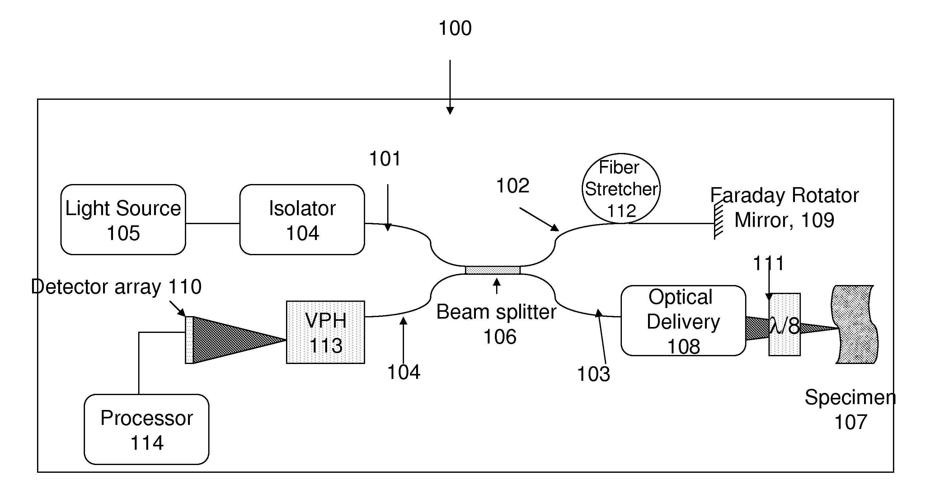
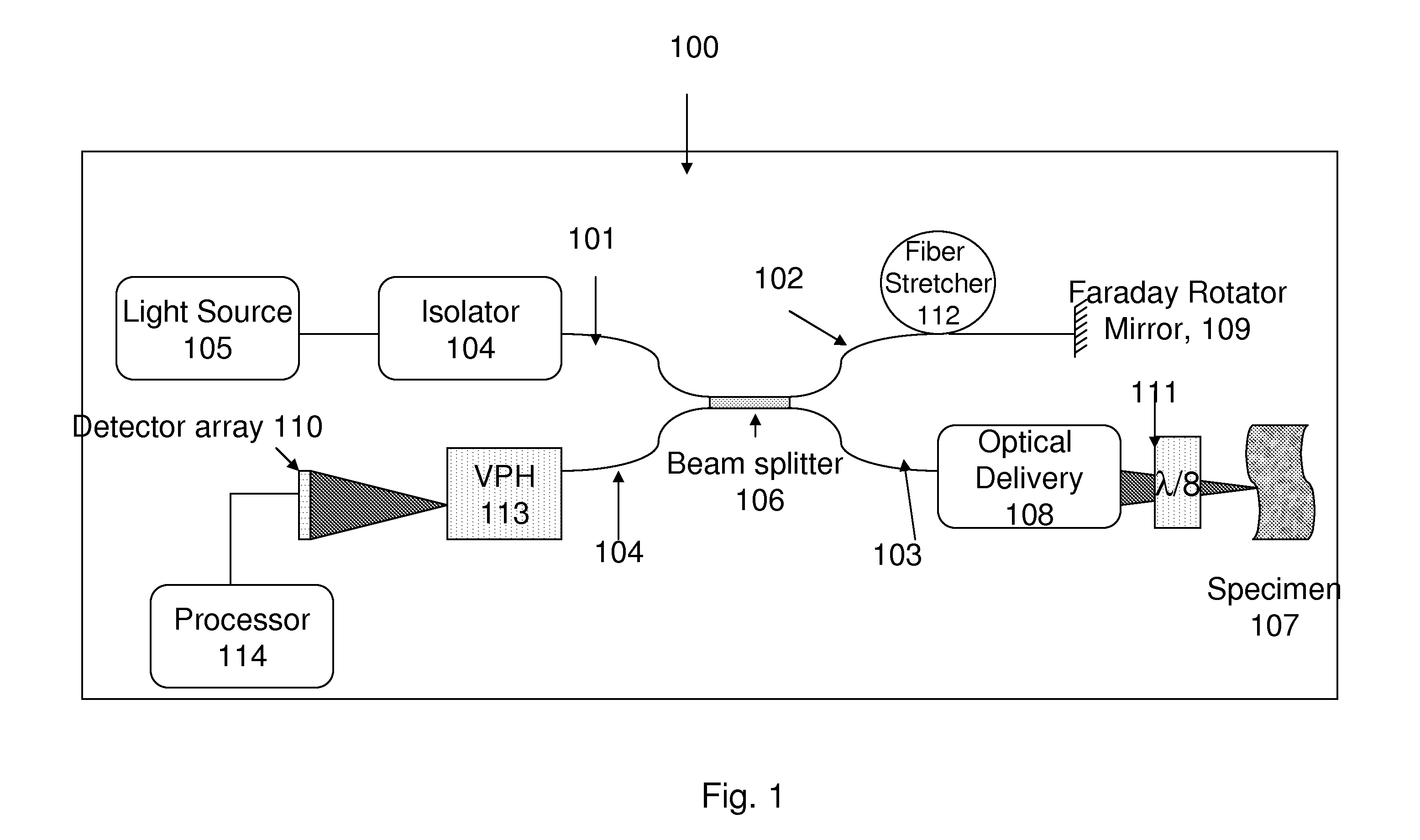
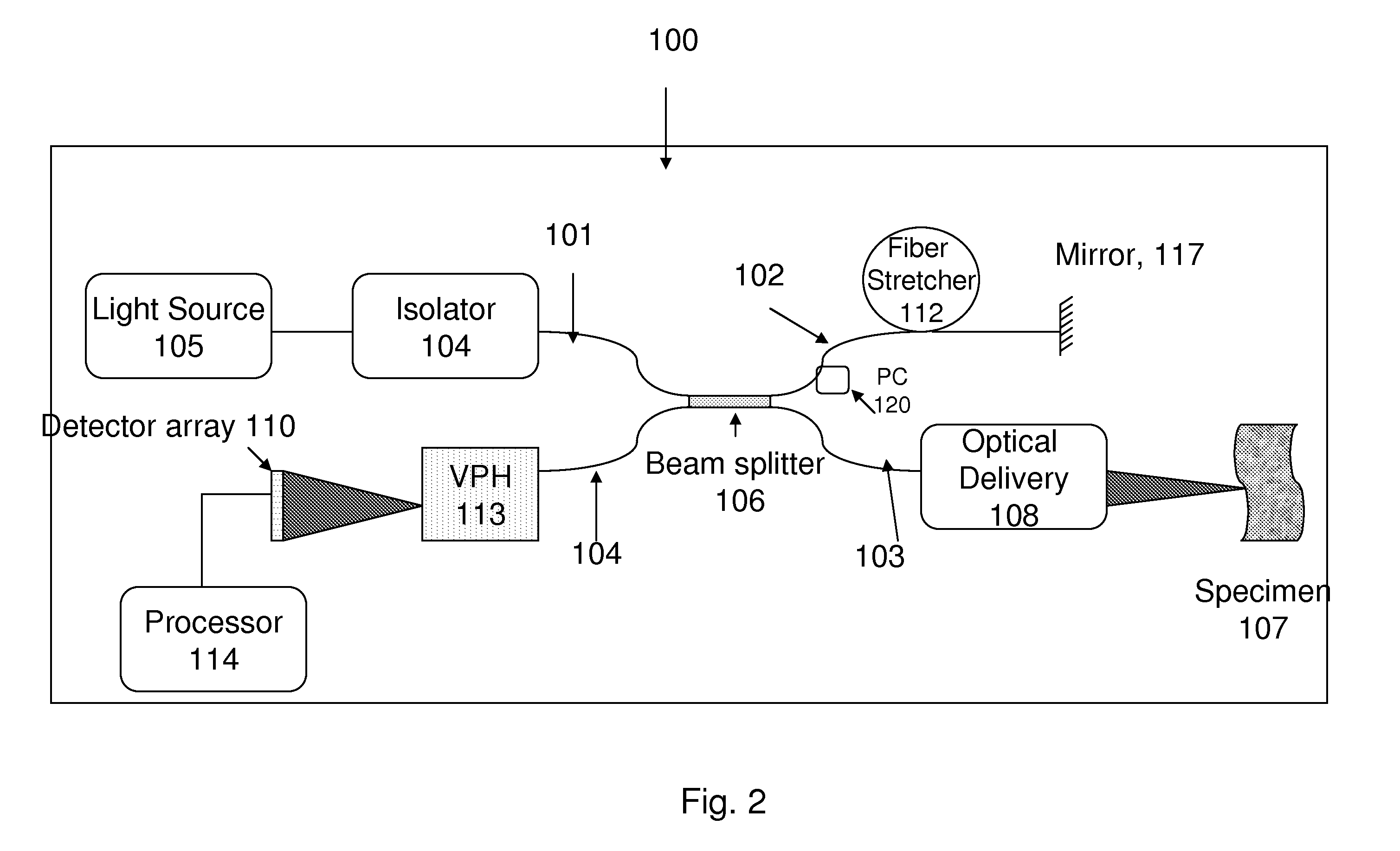
[0016]Optical coherence tomography (OCT) is similar to ultrasound imaging in that cross-sectional images of micro-features are acquired from adjacent depth resolved reflectivity profiles of the tissue (FIG. 1). OCT employs a fiber optically integrated Michelson interferometer illuminated with a short coherence length light source such as a superluminiscent diode (SLD). The interferometric data are processed in a computer and displayed as a gray scale image. In an OCT image, the detectable intensities of the light reflected from human tissues range from 10−5 to 10−11 th part of the incident power.

[0017]Recent OCT systems use spectroscopic detection. Basically the interferometric light exiting the detector arm is dispersed via a grating. The spectra are acquired using a line-scan camera. The resulting spectra are typically (by way of example, not by limitation) transferred to a processor for inverse Fourier transforming and relevant signal process...


PUM


Abstract

A Diabetic Retinopathy Diagnostic system based on OCT which will map 3-D blood circulation including velocity information with micron-scale resolution in the retina is disclosed here. The system leverages the advancements in telecommunication and device technologies and employs novel Doppler algorithms. For example, the reference arm in the interferometric system can be a fiber-optically integrated Faraday rotating mirror. By way of example, but not limitation, typically, the light in the detection arm of the Michelson interferometer can be measured using a Volume phase holographic dispersion grating. By way of example, but not limitation, the dispersed light can be focused on a line-scan camera or multi-line 2-D camera.

Description

1 CROSS-REFERENCE TO RELATED APPLICATIONS[0001]This patent application claims the priority filing date of the provisional US patent application (Application No. 61 / 163,872) titled “Low-cost, compact, & automated diabetic retinopathy diagnostics & management device,” filed on Mar. 27, 2009 by the inventor Manish D. Kulkarni. This benefit is claimed under 35. U.S.C. $119 and the entire disclosure of the Provisional U.S. patent Application No. 61 / 163,872 is incorporated here by reference.2 BACKGROUND[0002]2.1 Field[0003]The following description relates to optical test & measurement, interferometry, optical ranging and imaging, optical coherence domain reflectometry (OCDR), optical frequency domain reflectometry (OFDR), optical coherence tomography (OCT), Doppler processing and Doppler OCT in general.[0004]2.2 Background[0005]Optical Coherence Domain Reflectometry (OCDR) has been playing a major role in industrial and scientific metrology and medical diagnostics. Optical Coherence Tomo...

Claims


Application Information

Patent Timeline
Patent Type & Authority Applications(United States)
IPC IPC(8): G01B9/02
CPCA61B3/102G01B2290/70G01B9/02091G01B9/02045
Inventor KULKARNI, MANISH D.DEVARAJ, BALASIGAMANI
Owner KULKARNI MANISH D
Why Eureka
Try Eureka