Microscopy control system and method

a microscopy and control system technology, applied in the field of laser scanning microscopy automation control, can solve the problems of poor temporal resolution of the events under investigation, limited manpower, and light employed in microscopy can harm the cell, and achieve the effect of increasing the lifetime of the hardwar

Inactive Publication Date: 2010-09-30
NAT UNIV OF IRELAND MAYNOOTH +1
View PDF2 Cites 26 Cited by
  • Summary
  • Abstract
  • Description
  • Claims
  • Application Information

AI Technical Summary

Benefits of technology

[0019]Some implementations of the invention allow parts of the hardware not required in an expe

Problems solved by technology

However, the laser light employed in microscopy can harm the cell before a desired event occurs, a process known as phototoxicity.
As experiments may run for hours or days, manpower restrictions apply when controlling and evaluating the experiments.
This poses a ch

Method used


Image

  • Microscopy control system and method
  • Microscopy control system and method
  • Microscopy control system and method

Examples


Embodiment Construction

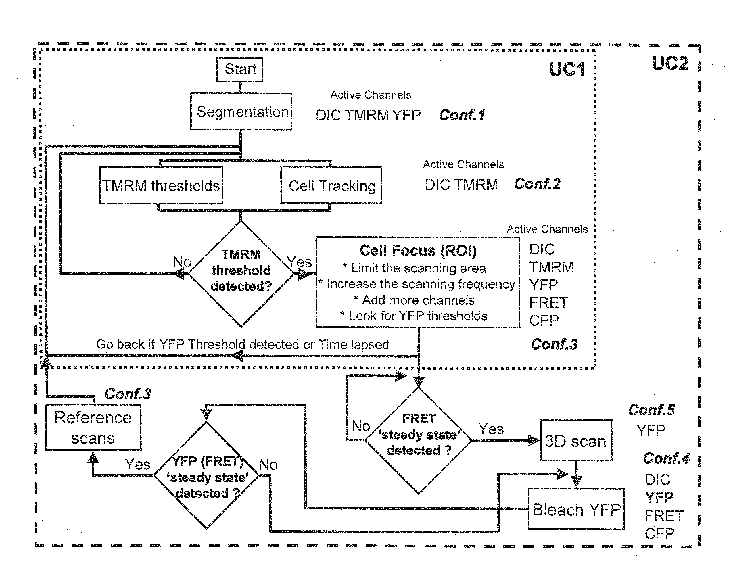
[0031]In a first aspect of the present embodiment, image analysis techniques are used for cell segmentation and tracking to extract time series of fluorescent signals from cells under laser scanning microscopy analysis. As these signal changes indicate biologically relevant information, their changes are compared to user-defined criteria. These are subsequently used as triggers to adapt microscope modalities including sampling rates, laser excitation, magnification, during single cell measurements.

[0032]In a further aspect of the present embodiment, a graphical framework is provided to enable the application of the above criteria based mechanism to a large class of single cell experiments. This allows the time course of an experiment to be determined through criteria and subsequent control actions, based on a-priori biological models of the experiment.

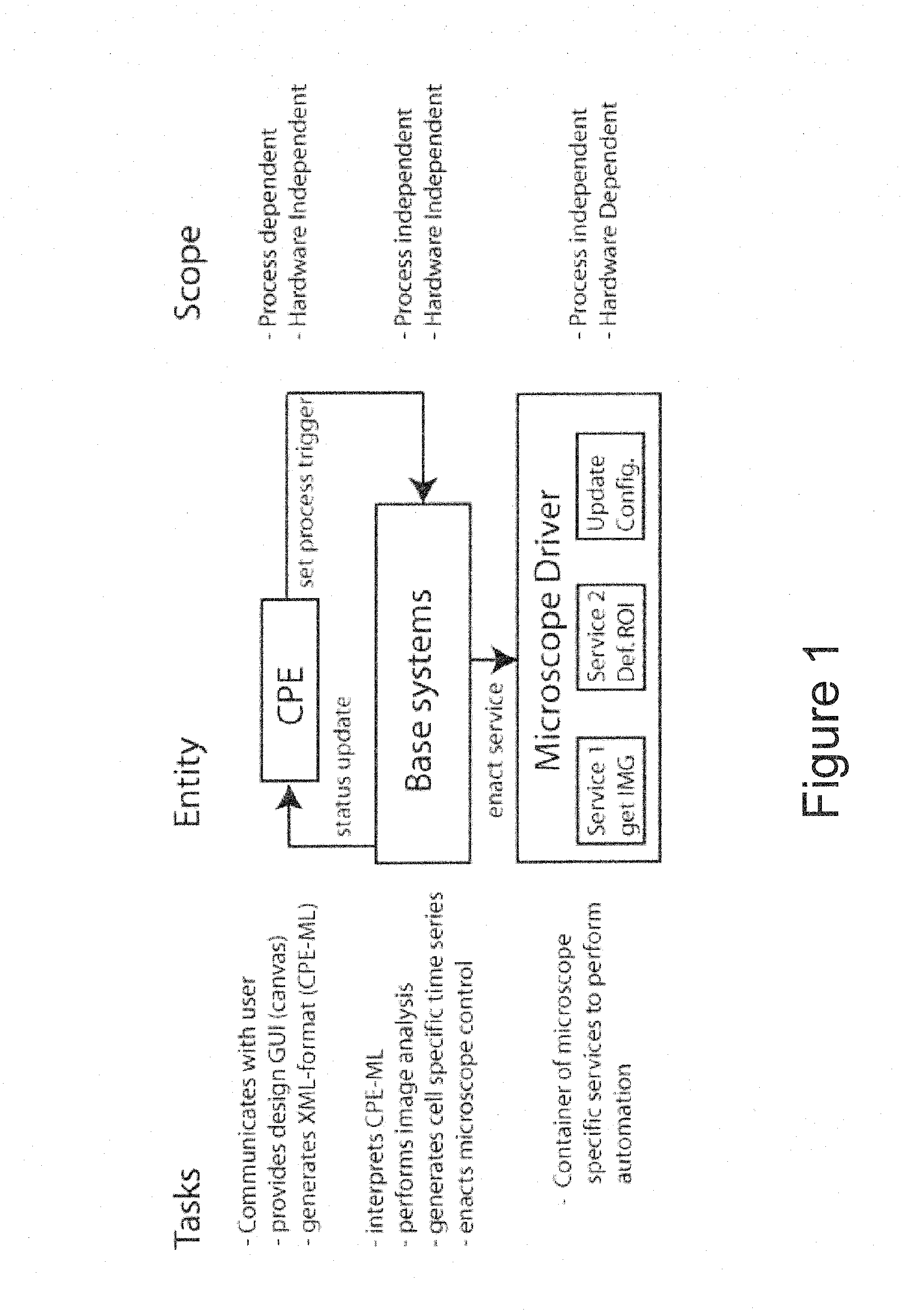
[0033]Referring now to FIG. 1, a system for automated control of laser microscopy according to a preferred embodiment of the present ...


PUM


Abstract

A method for controlling laser scanning microscopy of a probe comprising at least one cell is disclosed. The method comprises the steps of acquiring at least one initial image of the probe and identifying at least one cell within an initial probe image. Using a pre-defined grammar, a first set of scanning mode parameters for monitoring the cell(s); a first set of trigger parameters including at least one physiological parameter defining an event in the cell(s); and a second set of scanning mode parameters for monitoring at least one cell of the probe after an occurrence of the event is defined. A successive set of probe images acquired according to the first set of scanning mode parameters is provided and processed to determine if the event has occurred. Responsive to the event occurring, microscope modality is changed to the second set of scanning mode parameters.

Description

[0001]This application claims priority to Irish application S2009 / 0230 filed Mar. 25, 2009, the disclosure of which is incorporated herein by reference in its entirety.FIELD OF THE INVENTION[0002]This invention relates to a method and system for automated control of laser scanning microscopy.BACKGROUND[0003]Microscopy of living cells is heavily used in modern research to understand cellular processes and drug action in cell tissue. Artificial fluorescent dyes and also fluorescent proteins can be excited in volumes down to the resolution limit by microscopy lasers to support the visualization of events that can be identified by changes in fluorescent intensity and can in turn be studied by a biologist.[0004]Different events may require different set-ups for an experiment including selecting: dyes, laser excitation and detection channels, sampling speed and spatial magnification, all being influenced by the biologist's view of the underlying process.[0005]However, the laser light empl...

Claims


Application Information

Patent Timeline
IPC IPC(8): G01Q10/02
CPCG01N15/1475G02B21/365G02B21/008
Inventor HUBER, HEINRICHDUSSMANN, HEIKOPERRINE, PAULWENUS, JAKUBKALAMATIANOS, DIMITRIOSWUERSTLE, MAXIMILIAN
Owner NAT UNIV OF IRELAND MAYNOOTH
Why Eureka
Try Eureka