
Image analysis of brain image data

a brain image and image analysis technology, applied in the field of system of analyzing image data, can solve the problems of difficult to detect hemorrhagic stroke in early stage brain images, difficult to distinguish accurately between many, and difficult to identify areas which have undergone only subtle changes, so as to reduce the time delay involved, efficient extraction of information, and efficient visualization

Inactive Publication Date: 2010-10-14
KONINKLIJKE PHILIPS ELECTRONICS NV
View PDF12 Cites 7 Cited by
  • Summary
  • Abstract
  • Description
  • Claims
  • Application Information

AI Technical Summary

Benefits of technology

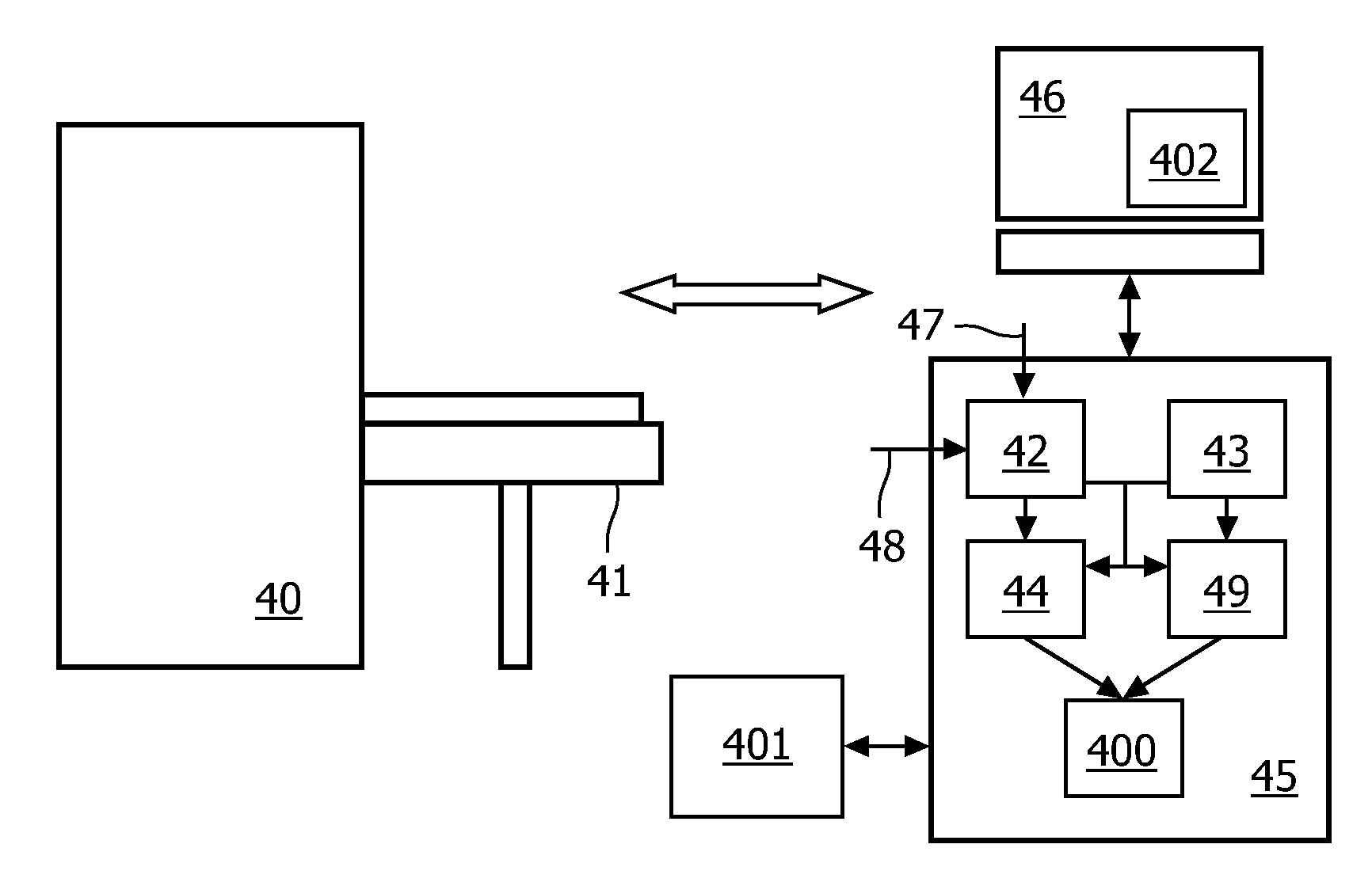
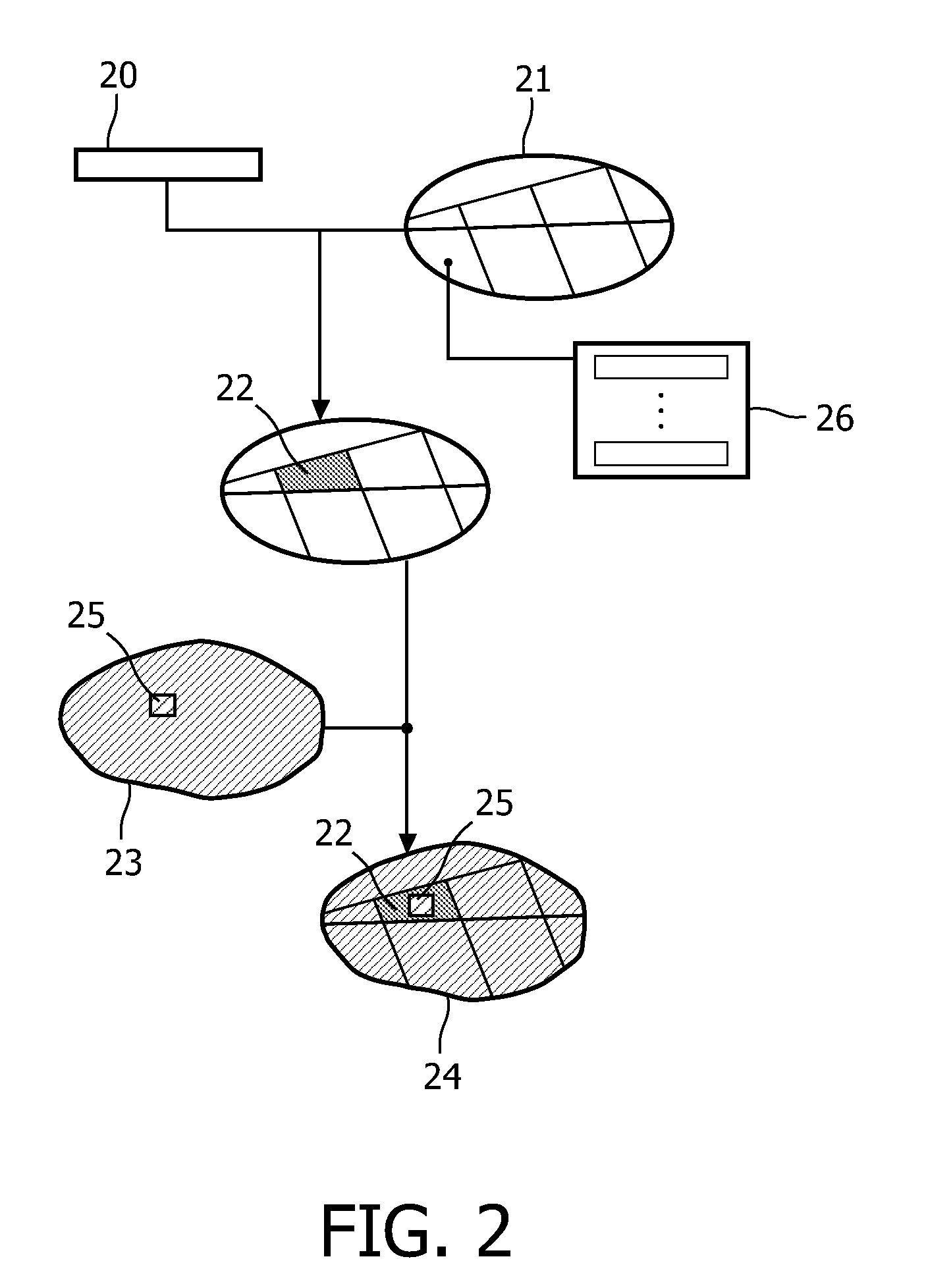
[0012]In general it may be difficult to detect and interpret subtle changes in image data, and it is an advantage of the present invention that from a combination of the non-image-based clinical or functional findings and the image-based information, the medical practitioner is directed to the relevant area of the image data to assist the medical practitioner in making the diagnosis. In general a region of interest is the region under investigation or examination.
[0013]In an advantageous embodiment, the identified one or more regions of interest in the target image data are visualized. In general, despite of the availability of detailed 3D images of the object it is still challenging for the medical practitioner to efficiently extract information from the data. By identifying the one or more regions of interest in the target image data the visualization process is rendered efficient for the medical practitioner.
[0014]In advantageous embodiments, an image analysis system is used for analyzing brain image data. The object is a brain and the deficit is a neurological deficit. This is a very useful application of the image analysis system of the invention.
[0015]In an advantageous embodiment, image-based computations are automatically performed on at least the part of the image data pertaining to the one or more regions of interest. Computations only in relevant areas of the image data may thus be ensured.
[0016]In advantageous embodiments, a number of labels may be assigned to the voxels or group of voxels of the object, e.g. the brain model, thereby providing a more comprehensive information tool to the medical practitioner. In an embodiment, the one or more labels further comprise a functional label, and / or a label indicative of the probability of a structural defect such as a lesion.
[0017]In an advantageous embodiment, the system may further comprise or be connected to a decision support system. A decision support system may advise the medical practitioner, based on existing knowledge, as well as provide a prediction of the course of the disease, thereby reducing the time delay involved in obtaining a diagnosis as well as increasing the certainty of a given diagnosis. The decision support system may also advise the user or the system on any parameters to be used in the visualization of image processing of the region of interest.

Problems solved by technology

In image analysis of suspected malignant tissue it may be difficult to distinguish accurately between many diseases that can produce a similar or even identical effect in the image data.
Likewise it may be difficult to identify areas which have undergone only subtle changes.
As an example, it may be difficult, especially for the inexperienced practitioner, to detect an early stage hemorrhagic stroke in brain images.
Therefore, the early detection and differential diagnosis of a brain lesion can typically not be based on image data alone.
However, in clinical practice such a setting can not be guaranteed.

Method used


Image

  • Image analysis of brain image data
  • Image analysis of brain image data
  • Image analysis of brain image data

Examples


Embodiment Construction

[0033]Embodiments of the invention will be illustrated with references to exemplary brain image data. The image analysis system in these embodiments is adapted to perform the brain image data analysis based on a neurological deficit resulting from a brain defect such as a lesion. Those skilled in the art will understand, however, that the invention may be applied to analyzing image data describing other regions of the human or animal anatomy, e.g. the heart, liver, lungs, femur or cardiac arteries. The brain example should not be construed as limiting the scope of the invention.

[0034]The diagnostics of lesions of the brain is a multi-disciplinary task where information from different sources is gathered and combined. For example, information from clinical investigations, neurological tests, imaging and laboratory tests is combined and evaluated by the medical practitioner to arrive at a diagnosis. An important tool in arriving at the diagnosis is the use of image data. However, it m...


PUM


Abstract

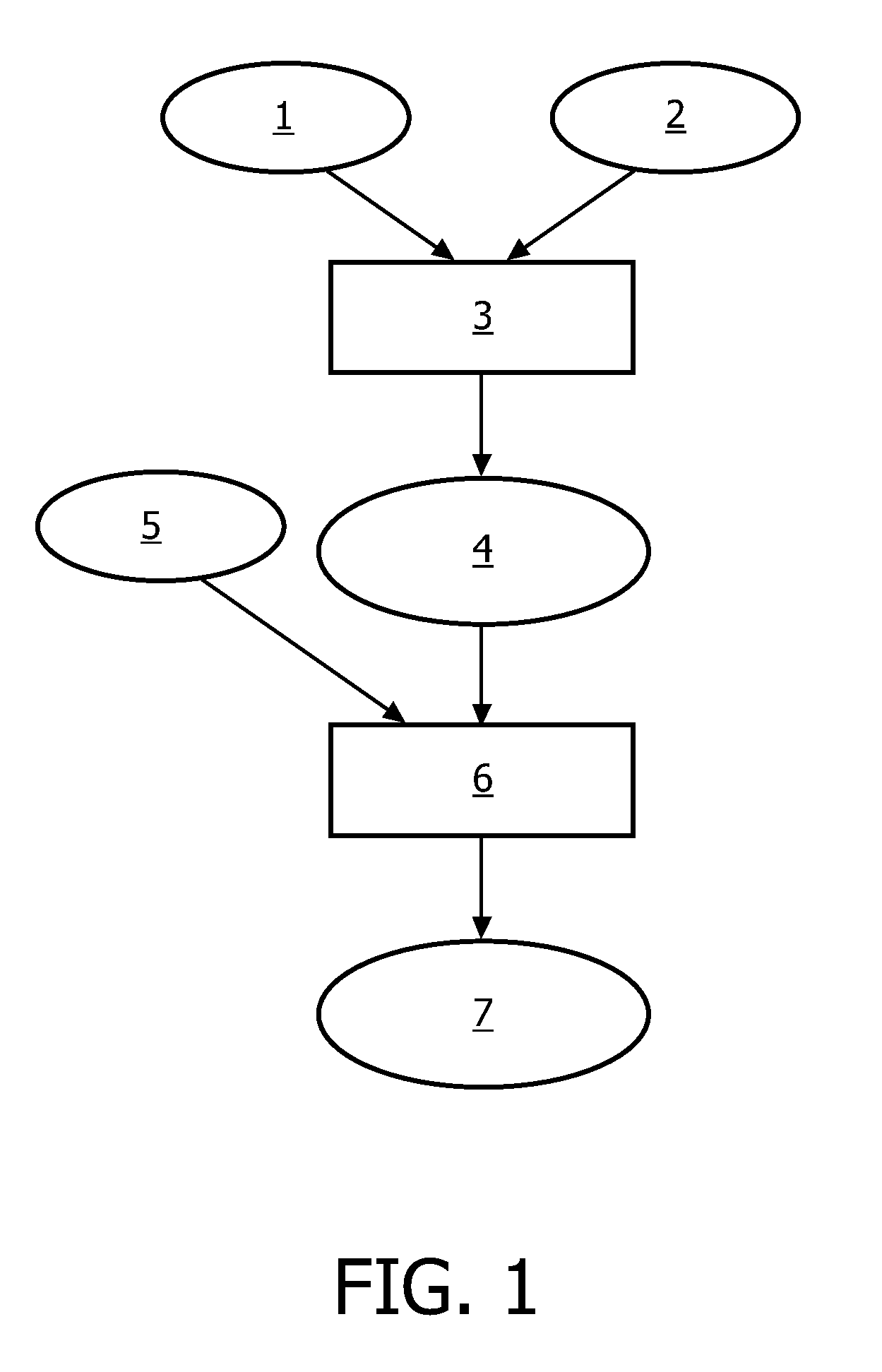
The present invention relates to analysis of image data, e.g. brain image data, where regions of interest are identified in patient specific image data based on non-image data. The brain image data is analyzed by correlating non-image data (20) in the form of data indicative of a neurological deficit and an object model (21) to identify one or more regions of interest (22) in the brain model, mapping the brain model to patient specific brain image data to obtain target image data (24), and identifying the one or more regions of interest in the target image data.

Description

FIELD OF THE INVENTION[0001]The present invention relates to a system of analyzing image data, and in particular to a system for identifying regions of interest in patient specific image data based on non-image data.BACKGROUND OF THE INVENTION[0002]In image analysis of suspected malignant tissue it may be difficult to distinguish accurately between many diseases that can produce a similar or even identical effect in the image data. Likewise it may be difficult to identify areas which have undergone only subtle changes. As an example, it may be difficult, especially for the inexperienced practitioner, to detect an early stage hemorrhagic stroke in brain images. The brain is a very sensitive organ regarding the loss of neurons and recovering from damage. In connection with brain lesions it is therefore crucial to detect and diagnose any lesions as early as possible, and ideally even before any anatomical changes occur. Therefore, the early detection and differential diagnosis of a bra...

Claims


Application Information

Patent Timeline
IPC IPC(8): G06T7/00G06K9/56G16Z99/00
CPCG06F19/3437G06T7/0012G06T19/00G06T2210/41G06T2207/10116G06T2207/10132G06T2207/30016G06T2207/10072G16H50/50G16Z99/00
Inventor MEETZ, KIRSTENBUELOW, THOMAS
Owner KONINKLIJKE PHILIPS ELECTRONICS NV
Learn More