Medical diagnosis support device, virtual microscope system, and specimen support member

Active Publication Date: 2010-12-30
EVIDENT CORP
View PDF5 Cites 8 Cited by
  • Summary
  • Abstract
  • Description
  • Claims
  • Application Information

AI Technical Summary

Benefits of technology

[0045]According to the medical diagnosis support device of the present invention, an image processing method and a photographing method which are the most suitable for a specimen are automatically selected, whereby a specimen can be analyzed without causing a user so much trouble.
[0046]According to the virtual microscope syste

Problems solved by technology

However, some analysis methods such as “detection of cancer invasion” and “detection of signals” are specific to each particular med

Method used


Image

  • Medical diagnosis support device, virtual microscope system, and specimen support member
  • Medical diagnosis support device, virtual microscope system, and specimen support member
  • Medical diagnosis support device, virtual microscope system, and specimen support member

Examples


first embodiment

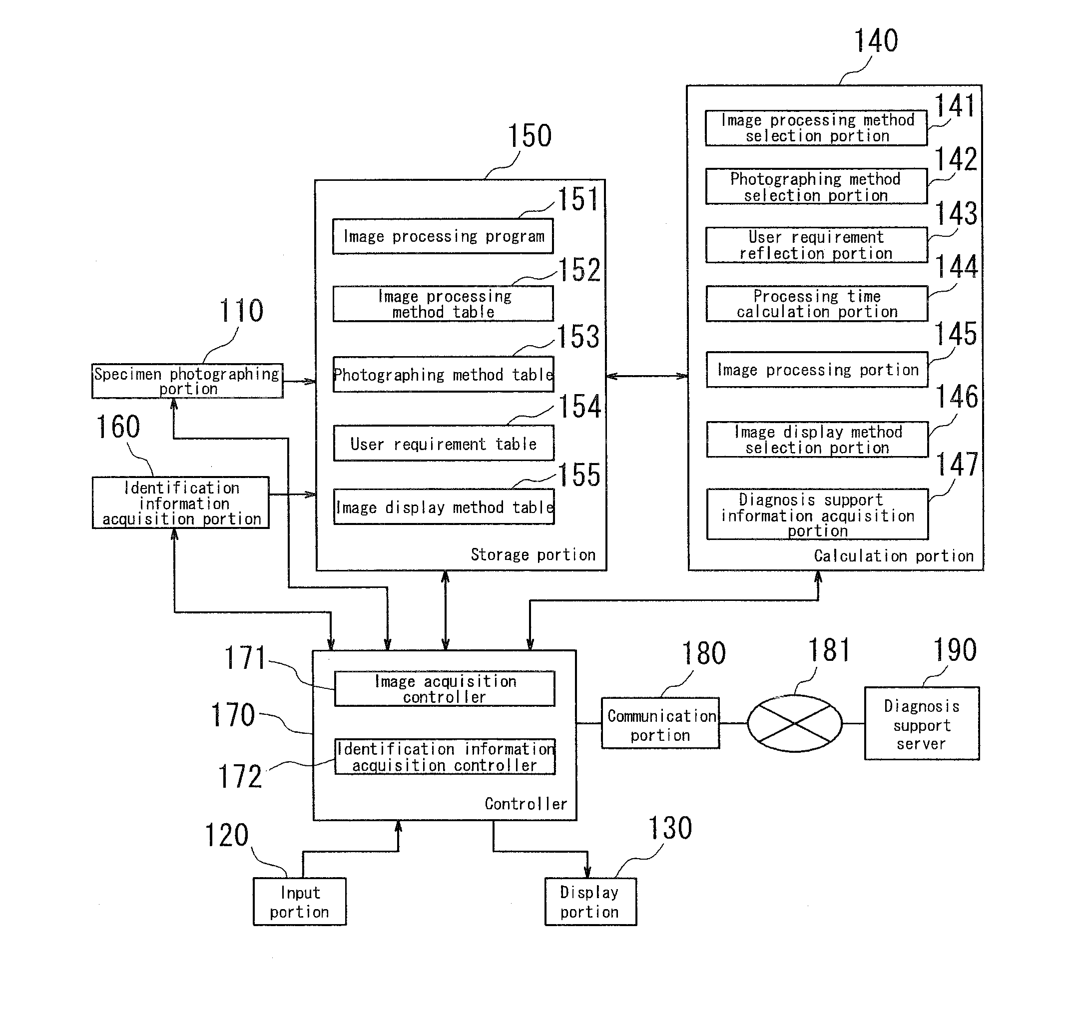
[0074]FIG. 1 is a block diagram showing a functional constitution of main parts of a medical diagnostics support device according to a first embodiment of the present invention. The medical diagnosis support device is structured to include a computer such as a personal computer and provided with a specimen photographing portion 110 including a microscope, an input portion 120, a display portion 130, a calculation portion 140, a storage portion 150, an identification information acquisition portion 160, and a controller 170 for controlling the respective portions. The controller 170 is capable of communicating, by way of a communication portion 180, with a local medical system which shares medical data with the present medical diagnosis support device. The controller 170 is also capable of communicating with a diagnosis support server 190 or other external medical system by way of network 181.

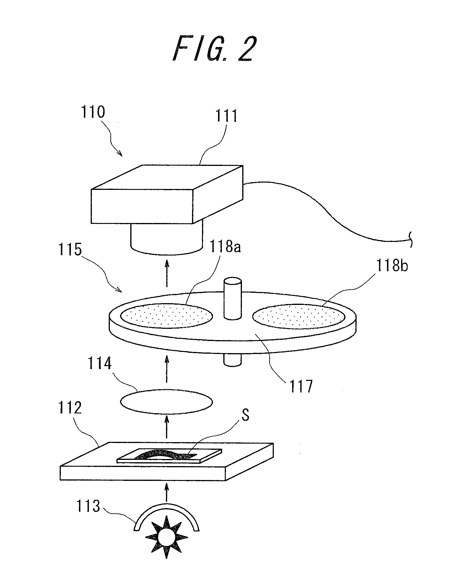
[0075]The image acquisition portion 110 acquires a multiband image of a target stained speci...

second embodiment

[0111]FIG. 14 and FIG. 15 are views each showing a structure of the main parts of a virtual microscope system according to a second embodiment of the present invention. This virtual microscope system has a function of the medical diagnosis support device described in the first embodiment and is provided with a host system 400, a microscope device 200 connected to the host system 400, an input portion 510 having, for example, a keyboard and a mouse, a display portion 520, and an identification information acquisition portion 530 such as a barcode reader. In the present embodiment, the microscope device 200 corresponds to the specimen photographing portion 110 in FIG. 1, the input portion 510 corresponds to the input portion 120 in FIG. 1, the display portion 520 corresponds to the display portion 130 in FIG. 1, and the identification information acquisition portion 530 corresponds to the identification information acquisition portion 160 in FIG. 1. FIG. 14 primarily shows a schematic...

third embodiment

[0129]FIG. 16 is a block diagram showing a functional constitution of main parts of a medical diagnostics support device according to a third embodiment of the present invention. The medical diagnosis support device of the present embodiment is substantially the same as the medical diagnosis support device shown in FIG. 1, except that an identification information calculation portion 148 is added to the calculation portion 140 in the former. Since other structures of the medical diagnosis support device in FIG. 16 is substantially the same as those shown in FIG. 1, the same structural components having the same effects are designated by the same reference numbers and detailed explanations thereof will be omitted.

[0130]The identification information calculation portion 148 calculates identification information of a specimen from a specimen image for acquiring identification information, which specimen image is acquired by photographing the specimen according to a predetermined photog...


PUM


Abstract

The present invention provides a medical diagnosis support device which enables a user to acquire the most appropriate information to support medical diagnosis without causing the user so much trouble. Specifically, the medical diagnosis support device comprises: an image processing method storage portion 152 for memorizing plural types of image processing methods; a photographing method storage portion 153 for memorizing plural types of photographing methods; an identification information acquisition portion 160 for acquiring identification information of a specimen S;
    • an image processing method selection portion 141 for selecting, based on identification information thus acquired, a corresponding image processing method from the image processing method storage portion 152; a photographing method selection portion 142 for selecting, based on the acquired identification information or the image processing method thus selected, a corresponding photographing method from the photographing method storage portion 153; a specimen photographing portion 110 for photographing the specimen S according to the selected photographing method to acquire a specimen image; and an image processing portion 145 for subjecting the specimen image acquired by the specimen photographing portion 110, to image processing, according to the image processing method selected by the image processing method selection portion 141.

Description

CROSS-REFERENCE TO RELATED APPLICATION[0001]The present application claims priority from Japanese Application No. 2009-151502, filed on Jun. 25, 2009, the content of which is incorporated herein by reference.BACKGROUND OF THE INVENTION[0002]1. Field of the Invention[0003]The present invention relates to a medical diagnosis support device, a virtual microscope system, and a specimen support member for use therewith, for acquiring information to support medical diagnosis from analysis of a specimen.[0004]2. Description of Related Art[0005]There has been known as one of pathological examination methods tissue diagnosis which makes diagnosis of a disease or examines how much a lesion site has grown by collecting tissue of the lesion site and observing it by a microscope. Such tissue diagnosis as this is also referred to as biopsy. It is widely practiced by biopsy to observe an enlarged view of a thin slice of several micron thickness, of a block specimen obtained from a removed organ an...

Claims


Application Information

Patent Timeline
IPC IPC(8): G06K9/62G06V10/141G06V10/20
CPCG06K9/2027G06K9/2063G06T2207/30024G06T7/408G06K9/36G06T7/90G06V10/225G06V10/141G06V10/20
Inventor OTSUKA, TAKESHI
Owner EVIDENT CORP
Why Eureka
Try Eureka