Endovascular Optical Coherence Tomography Device

a technology of optical coherence tomography and endovascular vein, which is applied in the field of endovascular oct devices, can solve the problems of low resolution imaging and achieve the effect of increasing the rotational and translational scanning stability of the oct prob

Inactive Publication Date: 2011-01-13
RGT UNIV OF CALIFORNIA
View PDF6 Cites 22 Cited by
  • Summary
  • Abstract
  • Description
  • Claims
  • Application Information

AI Technical Summary

Benefits of technology

[0011]The endovascular access probe has a distal end which is used for imaging tissue, and where the optic element includes a GRIN lens and prism disposed on the distal end of the optical fiber to direct light in a beam perpendicular to the longitudinal axis of the fiber, and a glass ferrule encasing the GRIN lens and prism to protect the optic element and distal tip of the optical fiber from mechanical damage.
[0012]The endovascular OCT probe further includes a hollow steel tube disposed within the axial lumen of the hollow coil wire within which tube the optical fiber is disposed, the hollow steel tube being provided in at least a proximal of the probe whereby torsional stiffness of the OCT probe is increased to increase rotational and translational scanning stability of the OCT probe.
[0016]The endovascular OCT probe further comprises a hollow steel tube disposed within the axial lumen of the hollow coil wire within which tube the optical fiber is disposed, the hollow steel tube being provided only in a proximal portion of the probe whereby torsional stiffness of the OCT probe is increased to increase rotational and translational scanning stability of the OCT probe, and a second more distal hollow coil wire of smaller diameter than the proximal hollow coil, the second distal smaller coil wire including the optical fiber extending from the proximal coil wire and terminating in the optic element, the proximal hollow coil wire being characterized by greater stiffness than the distal hollow coil wire.

Problems solved by technology

The main disadvantage of IVUS is low resolution imaging.

Method used


Image

  • Endovascular Optical Coherence Tomography Device
  • Endovascular Optical Coherence Tomography Device
  • Endovascular Optical Coherence Tomography Device

Examples


Embodiment Construction

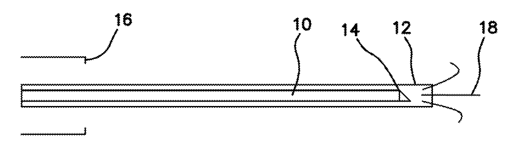
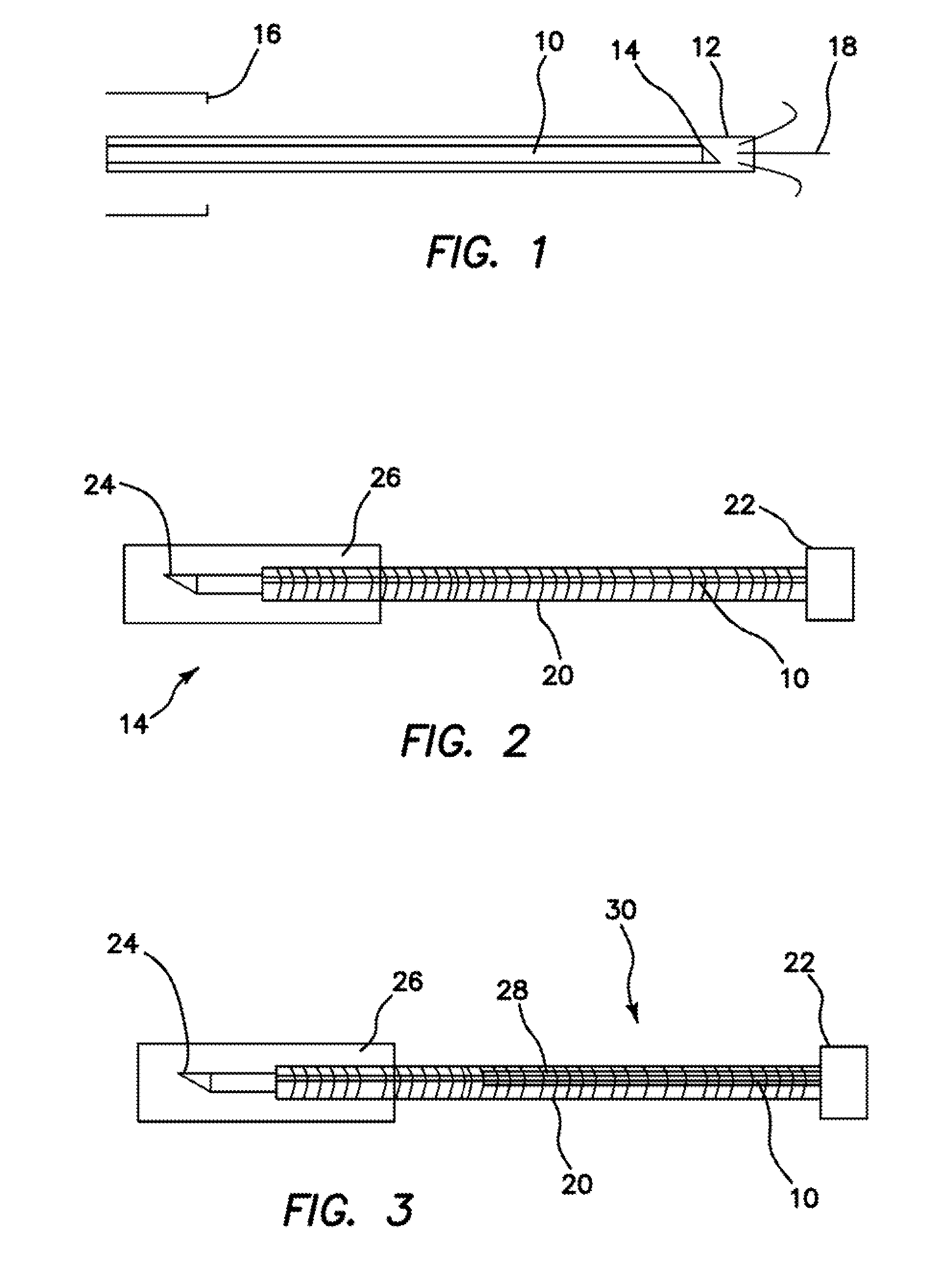
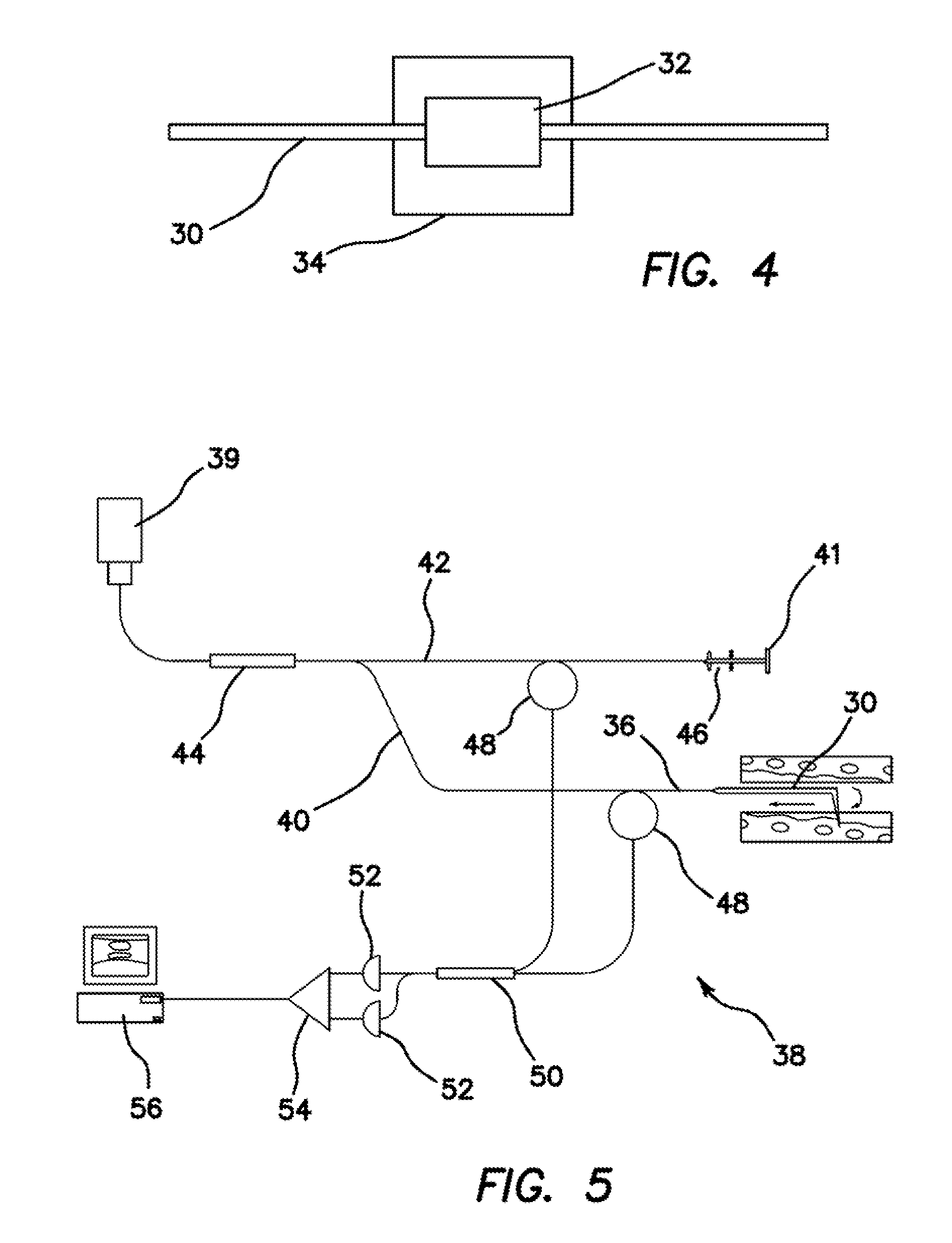
[0040]The illustrated embodiment of the invention is an endovascular OCT device incorporated onto the framework of an intracranial endovascular access device for intravascular imaging. This construct is obtained by removing the solid core of an intracranial access wire while retaining the outer coiled shell. A single mode optical fiber is inserted into the hollow coil wire. In the proximal end that remains outside the body a FC / APC or FC / PC single mode fiber adaptor is assembled. In the distal end which is used for imaging tissue, a gradient-index (GRIN) lens and prism are glued together to focus and guide the light into a beam perpendicular to the fiber's longitudinal axis. A glass ferrule with strong medical glue inside protects the tip from mechanical damage. This construct confers the OCT device with adequate mechanical, biological, radiological, and optical properties necessary for in vivo endovascular imaging in patients. FC / PC and FC / APC connectors are most commonly found in ...


PUM


Abstract

An endovascular OCT probe is included in an endovascular access device for intravascular imaging. The probe includes a hollow coil wire defining an axial lumen of the endovascular access device. A single mode optical fiber for transmitting light is disposed in the axial lumen of the hollow coil wire so that translation and rotation of the hollow coil wire carrying the optical fiber within the endovascular access device is stabilized for scanning endovascular tissue with at least 5 microns resolution. An optic element directs light from and into the optical fiber at a distal tip of the optical fiber and is coupled to or fixed to the distal end of the optical fiber. The optic element and the distal end of the optical fiber is disposed within a glass ferule to protect it from damage.

Description

RELATED APPLICATIONS[0001]The present application is related to U.S. Provisional Patent Application, Ser. No. 61 / 022,776 filed on Jan. 22, 2008, and U.S. Provisional Patent Application, Ser. No. 61 / 022,501 filed on Jan. 21, 2008, which are incorporated herein by reference and to which priority is claimed pursuant to 35 USC 119.BACKGROUND OF THE INVENTION[0002]1. Field of the invention[0003]The invention relates to the field of endovascular OCT device incorporated in the context of an intracranial endovascular access device for intravascular imaging.[0004]2. Description of the Prior Art[0005]Optical coherence tomography (OCT) is a technology that permits high resolution in vivo imaging. Imaging is carried out at near histology resolution thus yielding clinically relevant information without the need for surgical biopsy. OCT has been used in vivo to image coronary artery disease. The in vivo clinical use of OCT has never been reported to study cerebral vasculature. To study cerebral, ...

Claims


Application Information

Patent Timeline
Patent Type & Authority Applications(United States)
IPC IPC(8): A61B6/00
CPCA61B5/0062A61B5/0066A61B5/0084A61B5/6852A61B5/02007A61B5/02014A61B5/0086
Inventor MATTHEWS, MARLON S.SU, JIANPINGNWAGWU, CHIEDOZIE I.CHEN, ZHONGPINGLINSKEY, MARK E.
Owner RGT UNIV OF CALIFORNIA
Why Eureka
Try Eureka