If a preparation wall or an area does not conform to this axis of
insertion, there is no possibility of positioning the therapeutic agent to give a precise fit on the tooth and fix it with
dental cement.
Excessive conicity or
divergence of opposing preparation surfaces causes unnecessary loss of material and thus an
increased risk of devitalizing the tooth.
Additionally, the tooth is weakened to masticatory forces.
Especially metallic restorations have thereby insufficient adhesiveness relative to the tooth and decementation shortly follows.
Contrariwise,—in the case of opposing surfaces that are too steep or almost parallel—integration is hampered or made impossible by reason of the high
frictional resistance between the tooth and the restoration.
If preparation surfaces are present which represent an
undercut relative to the axis of
insertion, the corresponding surfaces of the restoration can no longer be accurately fitted or show a thin
cement gap.
But rather, for example in the case of a
solid crown, it will be impossible to design an accurately fitting crown border.
Thus, depending on size, undercuts count as clear faulty sites or errors in a preparation for
accommodation of an extraorally fabricated restoration.
In
dentistry there is a ubiquitous conflict of aims which has very serious medical consequences and relates to the removal of neither too little nor too much dental hard substance when forming a preparation.
Above all, the unnecessary removal of too much hard substance endangers both the
vitality of the tooth and the
mechanical stability of the residual tooth substance.
Mortification of the pulp tissue (dental pulp) constitutes a serious complication which usually calls for endodontic treatment (
root canal treatment) and the subsequent production of the restoration from scratch.
Should the endodontic treatment fail, the tooth becomes useless and must be removed.
The same fate applies to necrotic teeth which are not subjected to endodontic treatment on account of the very high cost of such treatment.
Otherwise correct
insertion of the restoration will be hampered or not possible.
A drawback of this checking
system is that for assessment the measured preparation is superimposed on a template showing a desired preparation.
This method is unsuitable for the assessment of preparations of real teeth, because real teeth differ in shape and size and the optimal preparation varies individually for each
clinical case, whilst a plurality of preparation forms may be clinically indicated within a tolerance range.
Moreover, when the desired preparation and the measured information are superimposed,
superimposition errors may lead to incorrect assessment of the preparation.
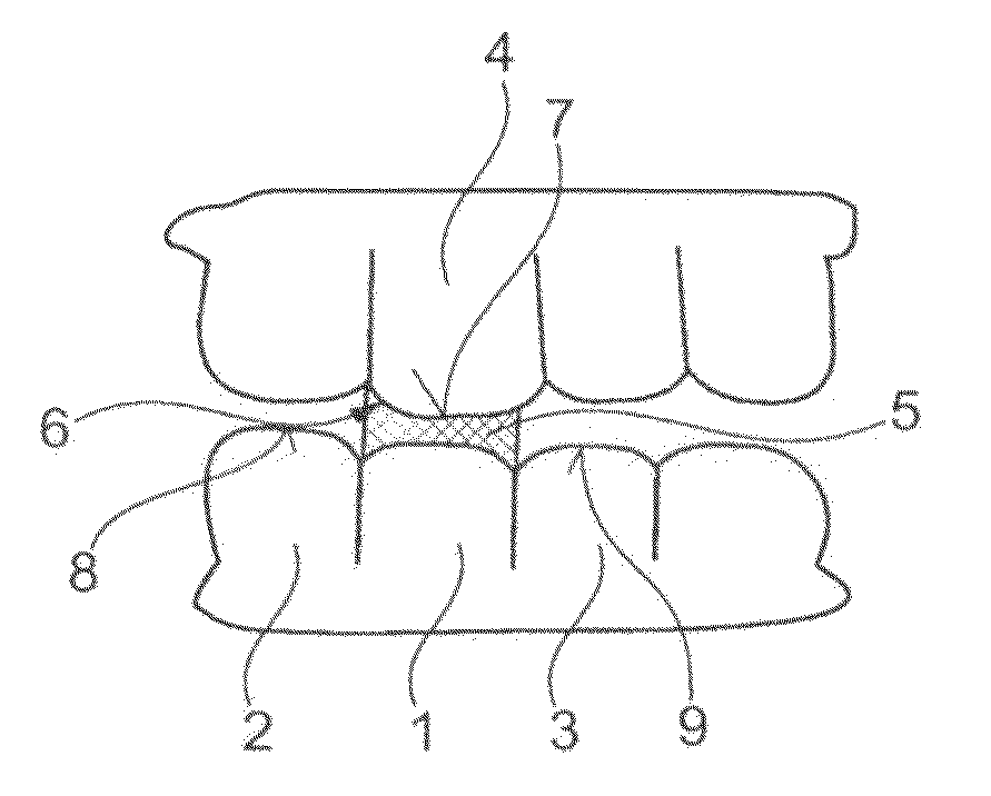
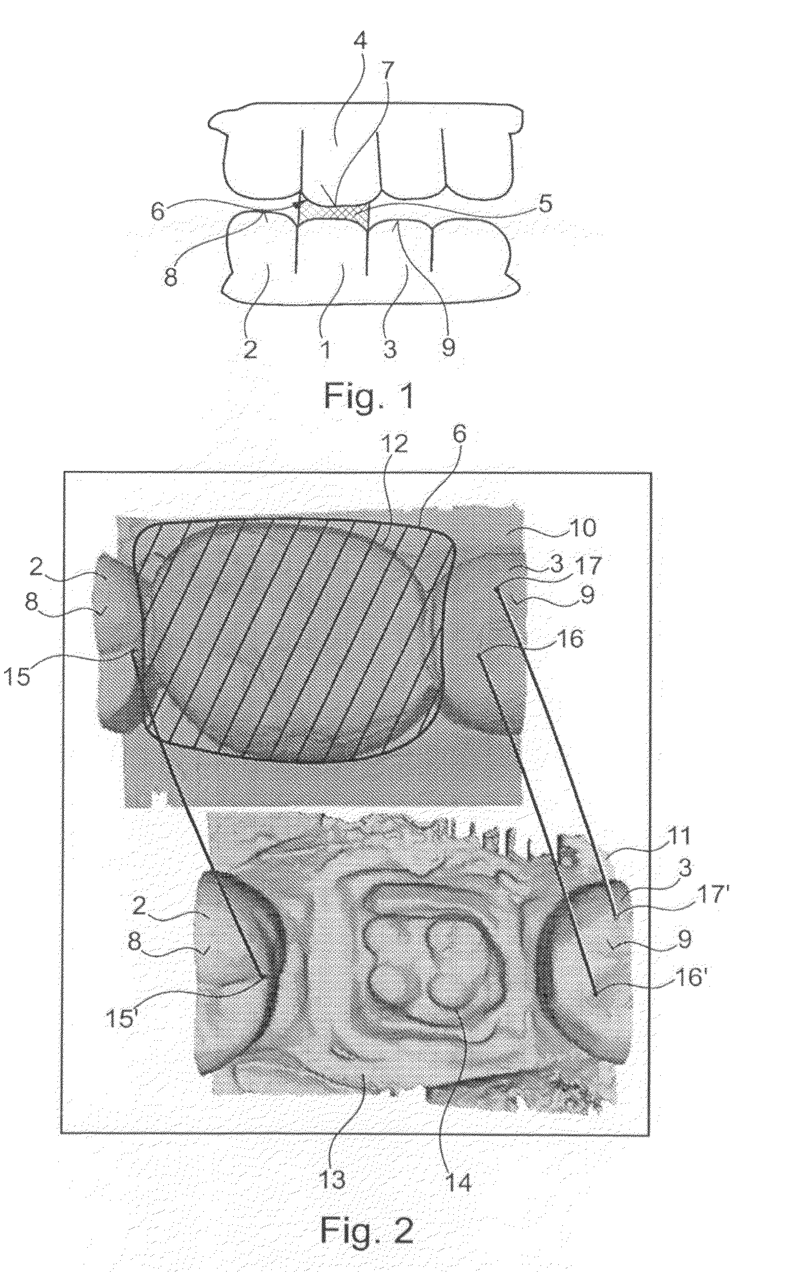
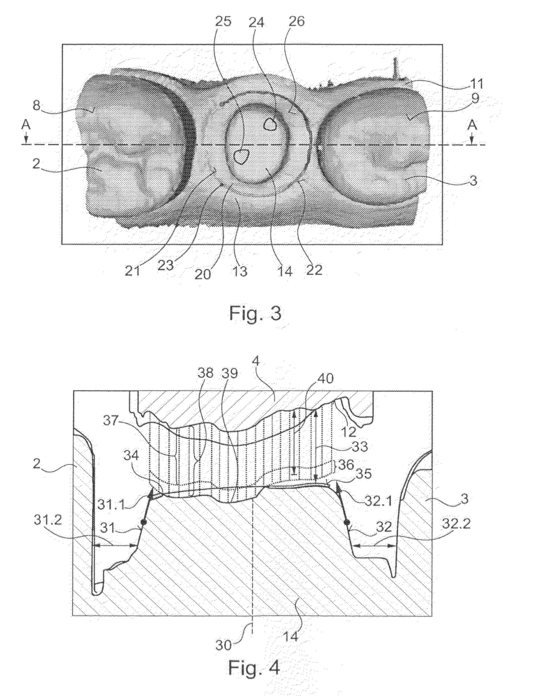
Another drawback is that the preparation is not checked for its position relative to the antagonistic teeth and to the adjacent teeth, but exclusively for shape.
The considerable additional time expenditure, the consequent enormous cost, and the increased load on the patient caused by the repeated impressions results in the method for making impressions for checking a preparation being used only extremely rarely in dental practices or dental clinics.
However, it is precisely this unfavorable method involving the creation of an impression of the preparation that forms the basis for all known novel CAD-based methods for evaluation of preparations.
Both represent an unreasonable load on the patient and on the dentist carrying out routine treatment.
This set of problems involving enormous time expenditure for CAD-based checking of a preparation following impression-making is additionally intensified by interactive evaluation of the
data set.
This interactive measurement of the preparation takes much time due to this procedure.
There is additionally the risk of unsatisfactory regions of the preparation remaining undiscovered on account of the user-dependent selection of the evaluated areas of the preparation.
The use of this method on a patient is, however, greatly restricted on account of movements of the head and / or the lower jaw, and the process is thus extremely hazardous.
However, the registration of head / lower jaw movements, for example by simply sticking reference points to the
skin, is not possible to the required
degree of precision.
Fixed anchorage of the reference points on the teeth is very time-consuming and problematical, since it additionally hinders the rather tight access to the tooth to be prepared.
However, there are no neighboring structures to provide a complete assessment of the preparation, which structures can in turn only be used for assessment with the aid of the elaborate method of making an impression and fabricating a model.


