Intraocular lens system

Active Publication Date: 2011-02-17
EMMETROPIA
View PDF80 Cites 52 Cited by
  • Summary
  • Abstract
  • Description
  • Claims
  • Application Information

AI Technical Summary

Benefits of technology

[0054]Moreover, to allow for a smaller incision, the corrective power needed to correct the vision of a patient may be divided between additional lenses of the optical assembly and the base lens. Additionally, if only a single front lens is engaged with the base lens, the power may be divided between the front lens and the base lens. By reducing the power of the base lens, the base lens is smaller, thereby requiring a smaller incision for implantation of the intraocular lens system.
[0055]Further, the present invention includes a feature wherein the base lens has a diameter different from the diameter of the front lens or optical assembly. As such, the capsule of the eye is more likely to “cellophane wrap” the base lens and less likely to “cellophane wrap” the front lens or optical assembly. For example, the front lens or optical assembly may have a larger diameter than the base lens. Furthermore, the front lens or optical assembly may have rounded edges and the base lens may have sharp edges to encourage cellophane wrapping of the base lens and discourage cellophane wrapping of the front lens or optical assembly.
[0058]In another aspect, a base lens has a simpler and easier to manufacture structure while maintaining flanges for engaging a front lens assembly.

Problems solved by technology

Current procedures and methods used by refractive surgeons may not satisfy the total refractive needs of the patient.
Particularly, the most commonly performed refractive surgical procedures, such as, for example, cataract extraction with intraocular lens implantation, in addition to the most recently popularized corneal refractive surgical procedures, such as eximer laser photoblation, exhibit limitations.
One reason for the limitations is the lack of post-operative refractive accuracy.
The lack of post-operative refractive accuracy renders the commonly known refractive surgical procedures uncompetitive with currently available non-surgical alternatives for patients, for example, glasses and contact lenses.
Difficulties arise in treating presbyopia because typically once a person manifests symptoms of presbyopia, the symptoms worsen as the person ages.
Conventional techniques for replacing an intraocular lens each time the patient's vision deteriorates do not always present a practical or cost-effective approach.
Patients suffering from eye trauma or other eye afflictions may have the iris or other portions of the eye distorted, destroyed, or discolored.
Such visualization, although not outside the scope of a surgeon skilled in the art, increases the degree of difficulty of the procedure, thus increasing the chance that a surgical error or other problem will arise in the surgical procedure, leading to unwanted complications.
Furthermore, analyses of current corneal refractive technologies indicate the presence of a significant amount of preexisting or naturally occurring post-operative, as well as preoperative, image distortion (optical aberration) or degradation, particularly under low light conditions, such as when driving at night.
Due to the practical limits of performing intraocular surgery, as well as the biological and physical behavior of the human eye during and after various types of intraocular surgery, predictability at the + / −0.25 D accuracy level with a single surgical procedure is difficult to achieve as a practical matter.
Furthermore, factors such as biometry errors, variable wound healing, and capsular contraction around the intraocular lenses contribute to decreasing the likelihood of achieving the desired refractive accuracy.
The engulfment typically makes the removal of the lenses and their haptics more difficult.
Exchange of iris fixated anterior chamber lenses does not typically guarantee precise position or orientation.
Posterior chamber lenses similarly cannot be removed because of posterior capsule fibrosis.
However, the surgical implantation procedure itself may create additional optical aberrations which cannot be anticipated preoperatively and thus the primary lens implant cannot account for these optical aberrations.
However, the design of current intraocular lenses typically makes such a procedure difficult and impractical.
Furthermore, after a period of time with normal healing, it becomes physically dangerous and / or nearly impossible for the patient to have the implanted lens removed once the eye tissue takes hold on the capsular fixation holes of the lens.
Therefore, such an argument is not realistic, practical, or safe.
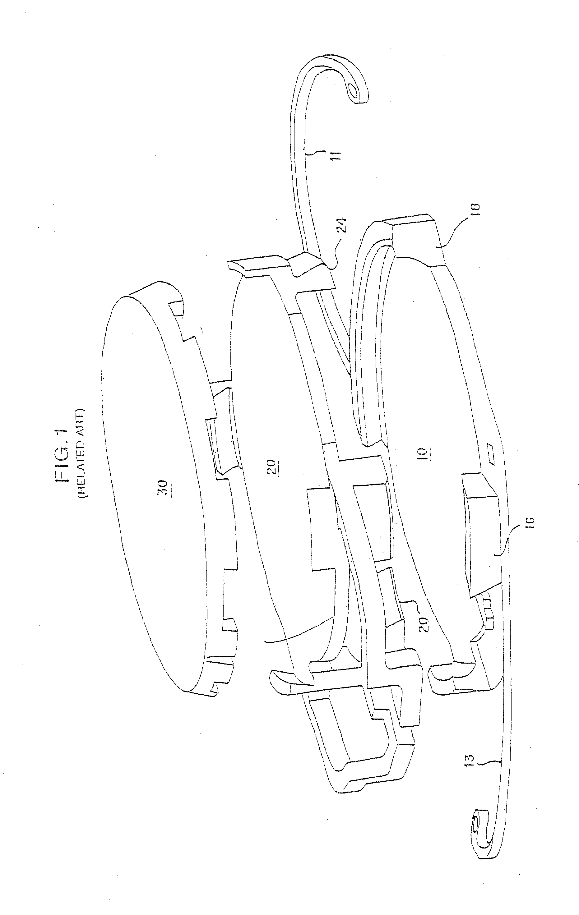
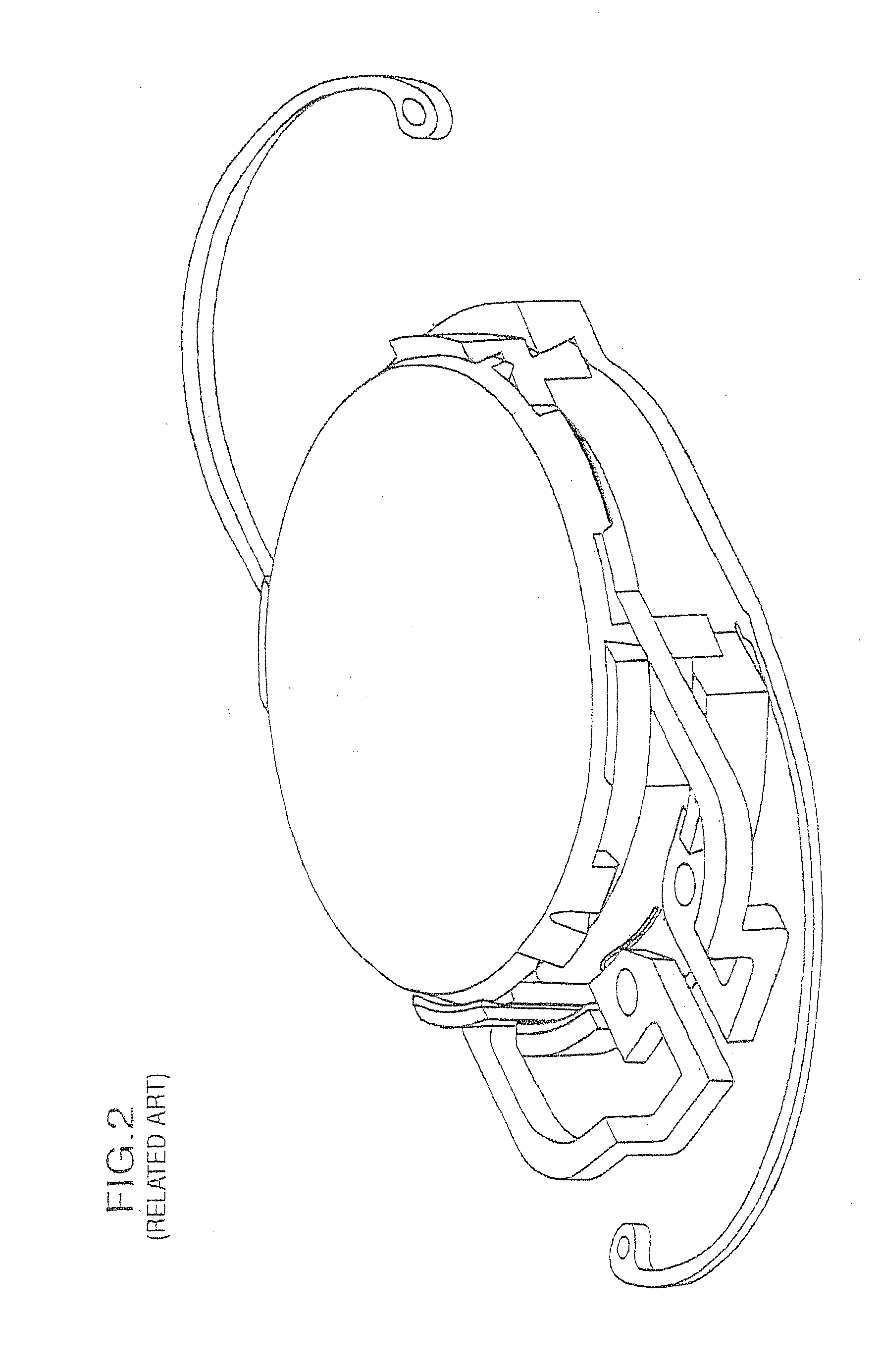
A single component intraocular lens, which in general is not designed to be removed and with only two optical surfaces, cannot accurately allow for compensation of sphere, cylinder, cylindrical axis, and all forms of optical aberrations that may be discovered after the initial implantation.
The easy exchangeability of optical elements is critical because the actual surgery of implanting the lens in the first place, as well as variances in the manner in which the eye heals after implantation, potentially create distortions which may not stabilize for several months after the operation.
Therefore, the ability to measure and to compensate for the distortion(s) optimally takes place several months after surgery and cannot typically be predicted prior thereto.
Since the same surgical wound is used for both the primary and secondary operations, additional distortion due to wound healing would not be anticipated as a result of the second operation.
From an optical standpoint, any time incisions are made to the cornea, the cornea loses some of its natural globularity due to imperfections caused by the incisions and the resultant trauma.
The imperfections in the cornea lead to induced astigmatism, or optical aberrations caused by irregularities in the shape of the cornea.
The “cellophane wrapping” makes it extremely difficult and dangerous for the surgeon to 
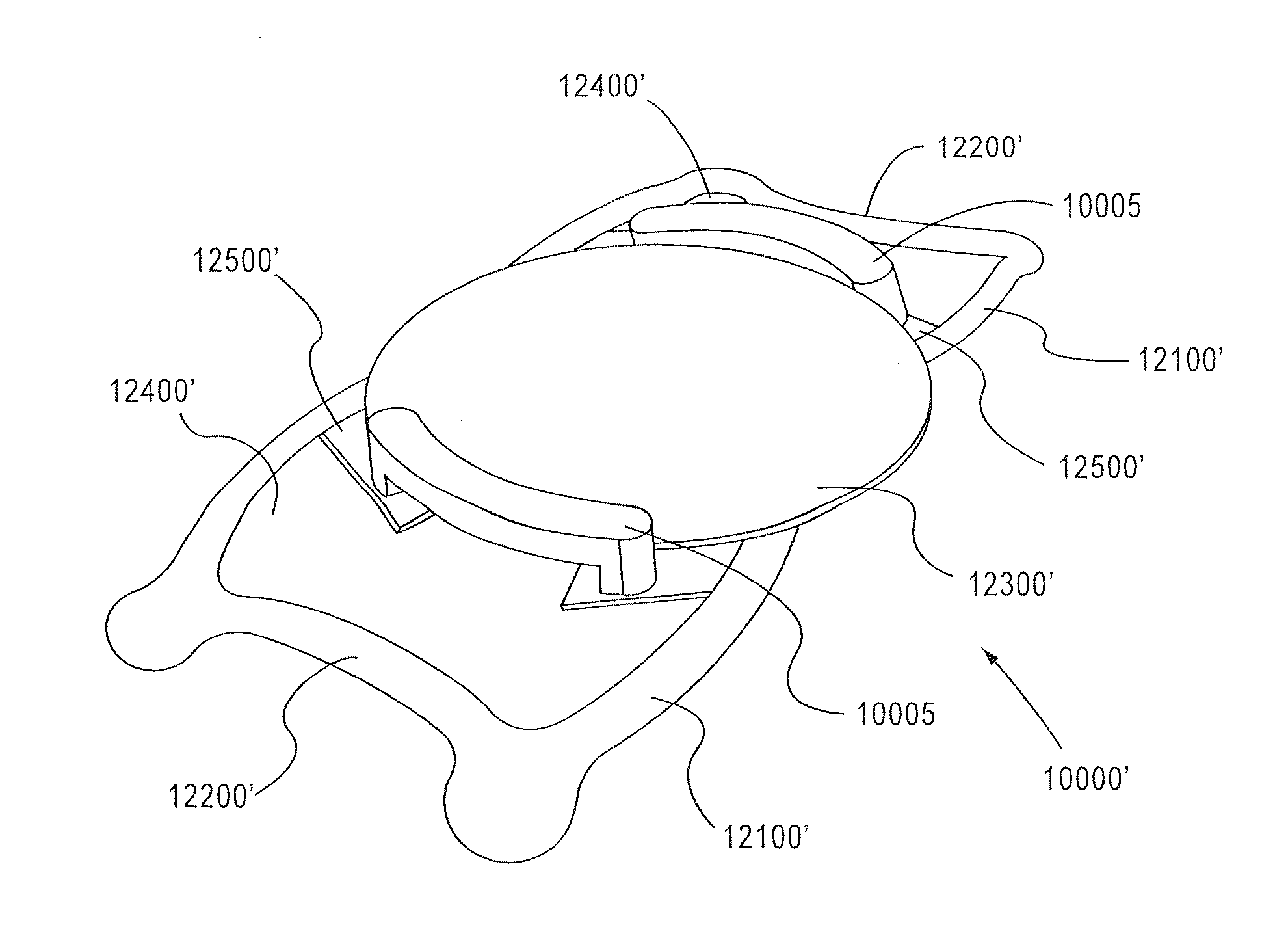
gain access to any surface of the base lens 100 (1000, FIG. 24) after the primary surgery heals, which is necessary for enhancement operations.

Method used


Image

  • Intraocular lens system
  • Intraocular lens system
  • Intraocular lens system

Examples


Embodiment Construction

[0103]As discussed above, and shown in FIG. 5A, in the applicant's prior intraocular lens assembly, axis orientation marks may be provided to aid in positioning and orienting a top lens of a MC-IOL. In an aspect of the present invention, instead of providing axis orientation marks, the MC-IOL is provided with an access bore 850. As shown in FIG. 29a, the access bore 850 may be provided at opposing points along the collar 225′ to indicate an axial orientation. The axial orientation indication of the access hole 850 serves essentially the same function as the orientation marking described above. However, as shown in FIG. 29b, unlike the orientation marking, the access bore 850 extends from the collar 225″ through an outer radial portion of a top lens 300″ and a mid lens 200″, and through a lower portion of the top lens 300″ and mid lens″. Accordingly, the access bore 850 serves an additional function of providing an access path to an abutting region defined between the mid lens 200′″ ...


PUM


Abstract

A multi-component intraocular lens implanted in an optical system of a human eye, includes one or more removable components. One component acts as a base lens and another component acts as the front lens. A front lens formed from two integral optical portions may be milled with tabs to establish an axial orientation of the front lens. The front lens may have a different diameter than the base lens. The base lens may have sharp or angled edges and the front lens may have rounded edges. Non-optical portions of the intraocular lens system may be manufactured from a material that is capable of releasing a pharmacological agent. A flange may be fused with the base lens to allow the front lens to engage with the base lens.

Description

CROSS-REFERENCE TO RELATED APPLICATION[0001]This is a continuation-in-part application of application Ser. No. 12 / 499,613, filed Jul. 8, 2009, which is a continuation-in-part application of application Ser. No. 12 / 000,364, filed Dec. 12, 2007, which is a continuation-in-part application of application Ser. No. 11 / 698,875, filed Jan. 29, 2007.BACKGROUND OF THE INVENTION[0002]1. Field of the Invention[0003]This invention relates to a method for correcting the optical system of an eye using an intraocular lens system. Particularly, this invention relates to a method of correcting focusing abnormalities and optical aberrations measured by wave front or similar technology to quantify optical aberrations in the optical system of the eye, using a laser, or other apparatus and / or methods of fabricating or modifying a lens, for the optical system of an eye having a foldable, interchangeable intraocular lens system provided therein.[0004]2. Description of Related Art[0005]The field of refract...

Claims


Application Information

Patent Timeline
IPC IPC(8): A61F2/16
CPCA61F2250/0039A61F2/1648A61F2002/1683A61F2002/1689A61F2220/0025A61F2250/006A61F2250/0067A61F2002/1686A61F2002/1682
Inventor WERBLIN, THEODORE P.
Owner EMMETROPIA
Why Eureka
Try Eureka