
Methods and devices for sternal closure

a technology for sternal closure and sternal artery, applied in the field of surgical procedures, can solve the problems of reducing lung capacity, pain and discomfort for patients, and little resistance to other forms of relative motion, and achieve the effect of inhibiting the adhesion of adhesives and removing contaminants

Active Publication Date: 2011-04-07
DOCTORS RES GROUP
View PDF4 Cites 13 Cited by
  • Summary
  • Abstract
  • Description
  • Claims
  • Application Information

AI Technical Summary

Benefits of technology

[0010]According to the present invention, a method for closing a sternum separated into at least a first sternum portion and a second sternum portion includes preparing at least one cut surface of at least one of the first sternum portion or second sternum portion to have an adhesive applied thereon. The adhesive is applied to at least a portion of the at least one cut surface and the at least one cut surface is approximated to a second cut surface of the other of the first sternum portion or second sternum portion. As the adhesive cures, it bonds with the first cut surface and the second cut surface. Preparing the at least one cut surface may advantageously remove contaminants that could inhibit adhesion of the adhesive.

Problems solved by technology

While these conventional devices restrain the sternum portions from pulling apart, they do little to resist other forms of relative motion, for example, sliding of one cut surface relative to the other or flexing motion about the incision.
These non-prevented relative movements may result in pain and discomfort for the patient, as well as more severe complications such as infections, all of which may lead to prolonged medication, decreased lung capacity and a longer return to normal functionality (e.g. a return to work).
Additionally, the relative movement may cause soft fibrous scar tissue to form across the incision, rather than the desired bone growth.
Relative motion between the two sternal portions may also cause the conventional devices to loosen.
More severely, the relative motion may cause the conventional devices to cut into and pull through the sternum bone, which also results in a loosening of the devices and separation of the sternal portions.
This separation may delay healing or may result in additional surgical procedures to tighten or replace the conventional closure devices.
The loosening or pulling through of the conventional devices may be caused by loading that the patient cannot practically control, such as cyclic loading due to normal respiration as well as less frequent high cycle loads generated during coughing or sneezing.
While the metal plate inhibits relative movement between the two sternal portions better than conventional wires and cables, the metal plate has its own limitations and disadvantages.
For instance, the thickness of metal plate closure devices results in discomfort to the patient.
Additionally, the relative movement between the sternal portions may result in a failed screw purchase.
The loose screw or screws allow the metal plate to loosen from the sternum causing pain and discomfort for the patient.
Furthermore, metal plate closure devices are costly and difficult to remove in the event there is a need for future access to the thoracic cavity.

Method used


Image

  • Methods and devices for sternal closure
  • Methods and devices for sternal closure
  • Methods and devices for sternal closure

Examples


Embodiment Construction

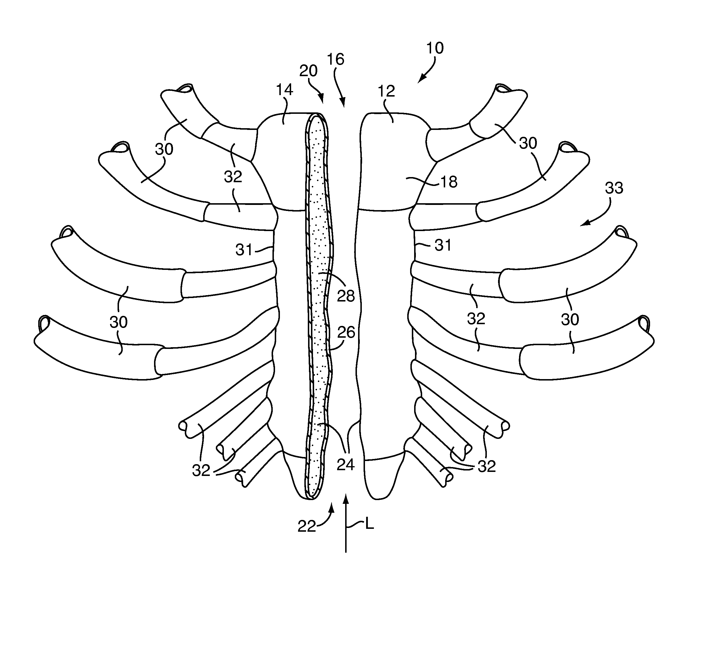
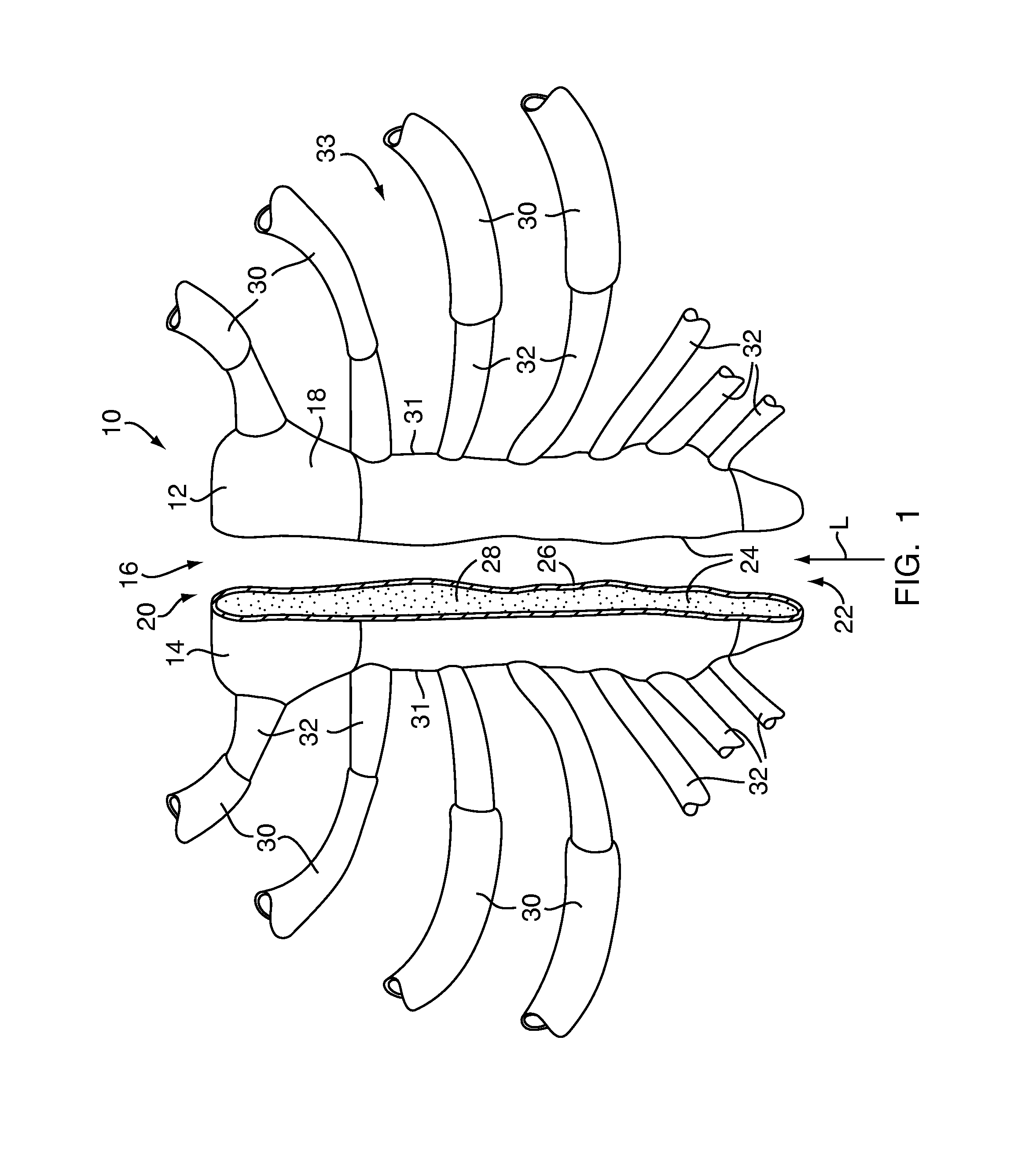
[0059]Referring to FIG. 1, the present invention provides a method for closure of a sternum 10 that has been separated into a first sternum portion 12 and a second sternum portion 14 by an incision 16, for example, from a surgical procedure such as a median sternotomy. The incision 16 extends in a longitudinal direction L approximately along a midline of the anterior surface 18 of the sternum 10 from an upper end 20 to a lower end 22. The incision 16 forms a cut surface 24 on each of the first and second sternum portions 12 and 14, exposing the sternum's composite bone structure having a shell of cortical bone 26 surrounding a core of cancellous bone 28. A plurality of rib bones 30 are connected to peripheral edges 31 of the sternum 10 by cartilage 32 to form a thoracic cage 33 for protecting internal organs from physical trauma.

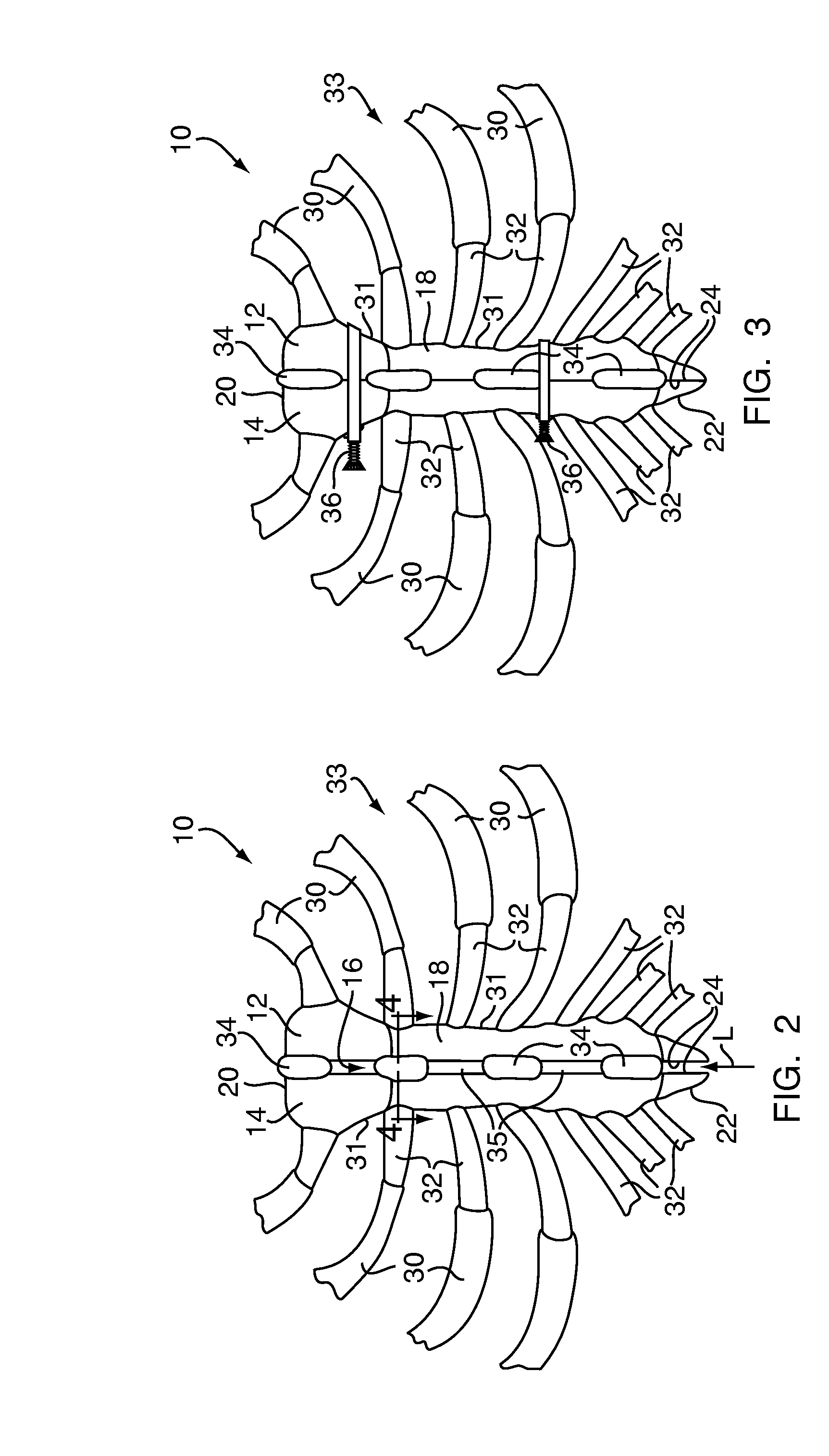
[0060]Referring to FIG. 2, at the end of the surgical procedure, such as a sternotomy, an adhesive 34 is applied to the at least one of the first and second...


PUM


Abstract

A method for closing a sternum separated into at least a first sternum portion and a second sternum portion includes preparing at least one cut surface of at least one of the first or second sternum portions to have an adhesive applied thereon. The adhesive is applied to at least a portion of the at least one cut surface. The at least one cut surface is approximated to a second cut surface of the other of the first sternum portion or second sternum portion. As the adhesive cures, it bonds with the first cut surface and the second cut surface. Preparing the at least one cut surface may remove contaminants that could inhibit adhesion of the adhesive. The adhesive may be applied through adhesive injection holes formed along an incision separating the first and second sternum portions. Devices for preparing the cut surfaces and applying the adhesive are also provided.

Description

CROSS REFERENCE TO RELATED APPLICATIONS[0001]This application is a continuation-in-part of U.S. patent application Ser. No. 12 / 575,060, filed Oct. 7, 2009, which is hereby incorporated by reference in its entirety.FIELD OF THE INVENTION[0002]The present invention relates to surgical procedures and, more particularly, to sternal surgical procedures.BACKGROUND OF THE INVENTION[0003]During surgical procedures, an incision is made to provide a surgeon with access to a patient's internal organs. At the end of the surgical procedure, the incision is closed to the external environment, thereby allowing the patient to heal and recover from the surgery. For surgical procedures to organs within a patient's thoracic cage, such as the heart and lungs, the patient's sternum may need to be separated to provide the surgeon with access to the organs.[0004]The sternum is an elongated, generally flat, bone located longitudinally in the center of the thorax. The sternum has a composite bone structure ...

Claims


Application Information

Patent Timeline
Patent Type & Authority Applications(United States)
IPC IPC(8): A61B17/08
CPCA61B17/00491A61B17/68A61B17/8811A61B17/823A61B17/8805A61B17/8076
Inventor DESLAURIERS, RICHARD J.KOLB, ERIC
Owner DOCTORS RES GROUP
Learn More