
System and Method for Normalized Diffuse Emission Epi-illumination Imaging and Normalized Diffuse Emission Transillumination Imaging

a diffuse emission and imaging system technology, applied in the field of medical imaging, can solve problems such as generating an un-normalized emitted light image of the tissue, and achieve the effect of improving images

Active Publication Date: 2011-04-14
THE GENERAL HOSPITAL CORP
View PDF24 Cites 19 Cited by
  • Summary
  • Abstract
  • Description
  • Claims
  • Application Information

AI Technical Summary

Benefits of technology

The patent describes a system and method for improving images generated by planar fluorescence epi-illumination imaging and planar fluorescence transillumination imaging. This normalization process can be used during surgery to identify tumors or other lesions and borders, as well as for fluorescence endoscopy and dental procedures. The normalized images provide significantly better quality and can be combined to create a more complete image of the tissue. Overall, this technology can help improve the accuracy and reliability of image-based medical diagnostics.

Problems solved by technology

The method further includes generating an un-normalized emitted light image of the tissue.

Method used


Image

  • System and Method for Normalized Diffuse Emission Epi-illumination Imaging and Normalized Diffuse Emission Transillumination Imaging
  • System and Method for Normalized Diffuse Emission Epi-illumination Imaging and Normalized Diffuse Emission Transillumination Imaging
  • System and Method for Normalized Diffuse Emission Epi-illumination Imaging and Normalized Diffuse Emission Transillumination Imaging

Examples


Embodiment Construction

[0048]Before describing the imaging system and method, some introductory concepts and terminology are explained. As used herein, the term “phantom” is used to describe a test object being imaged. A phantom is typically an article having diffuse light propagation characteristics similar to living tissue, for example, a piece of appropriately engineered resin block. For another example, a phantom can be a vial, which contains cells having fluorescent proteins therein, i.e. a fluorescent marker or a fluorochrome.

[0049]As used herein, the term “excitation light” is used to describe light generated by an “excitation light source” that is incident upon a biological tissue. The excitation light can interact with the tissue, and can be received by a light detector (e.g., a camera), at the same wavelength (excitation wavelength) as that at which it was transmitted by the excitation light source. The excitation light can be monochromatic, or it can cover a broader spectrum, for example, white...


PUM


Abstract

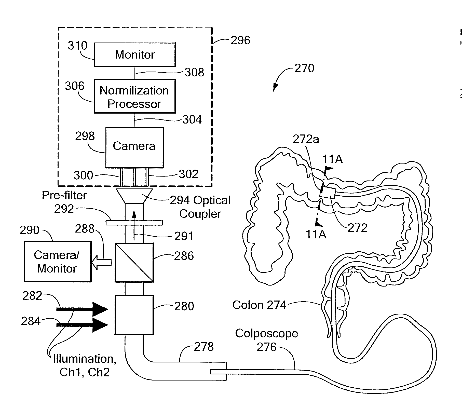
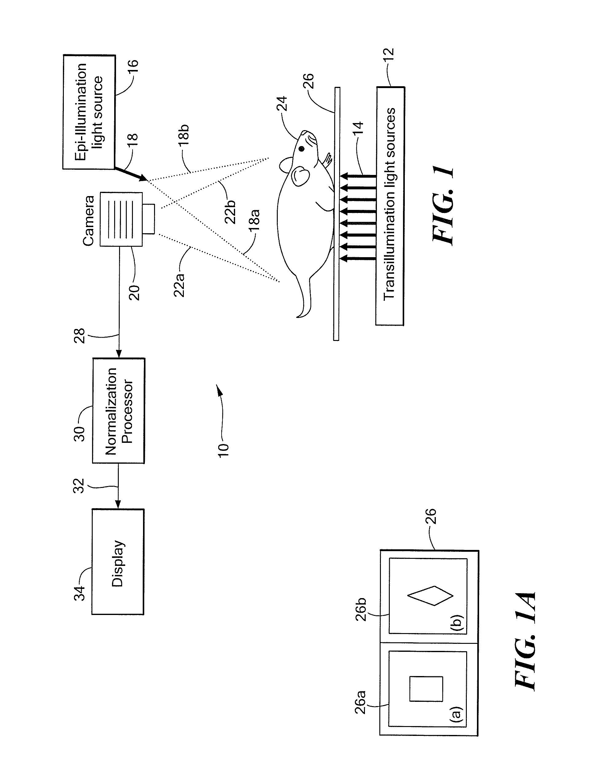
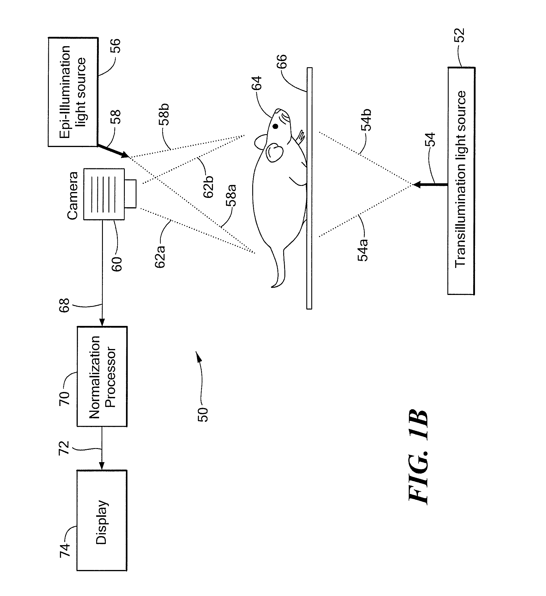
A system and method provide normalized fluorescence epi-illumination images and normalized fluorescence transillumination images. The normalization can be used to improve two-dimensional (planar) fluorescence epi-illumination images and two-dimensional (planar) fluorescence transillumination images. The system and method can also provide normalized bioluminescence epi-illumination images and normalized bioluminescence transillumination images. In some arrangements, the system and method can provide imaging of small animals, intro-operative imaging, endoscopic imaging, and / or imaging of hollow organs.

Description

CROSS REFERENCE TO RELATED APPLICATIONS[0001]This application is a Divisional Application of and claims the benefit of U.S. patent application Ser. No. 11 / 720,967 filed on Jun. 6, 2007, which is a U.S. National Stage application under 35 U.S.C. §371 of, and claims the benefit of, International Patent Application No. PCT / US2005 / 044651 filed on Dec. 8, 2005, all of which applications claim priority to U.S. Provisional Patent Application No. 60 / 634,369 filed on Dec. 8, 2004, all of which applications are hereby incorporated herein by reference in their entirety.FIELD OF THE INVENTION[0002]This invention relates generally to medical imaging and, more particularly, to a system and method for generating fluorescence epi-illumination images, fluorescence transillumination images, and bioluminescence images associated with small animal imaging, intra-operative imaging, endoscopic imaging and imaging of hollow organs.BACKGROUND OF THE INVENTION[0003]Fluorescence images can be generated in-vi...

Claims


Application Information

Patent Timeline
Patent Type & Authority Applications(United States)
IPC IPC(8): A61B6/00
CPCA61B1/00009A61B1/043A61B5/0064A61B5/418A61B5/0084A61B5/0088A61B5/415A61B5/0071A61B1/000094A61B1/000095
Inventor NTZIACHRISTOS, VASILIS
Owner THE GENERAL HOSPITAL CORP
Learn More