Automated Neuroaxis (Brain and Spine) Imaging with Iterative Scan Prescriptions, Analysis, Reconstructions, Labeling, Surface Localization and Guided Intervention

a neuroaxis and autonomic imaging technology, applied in the field of medical diagnostic imaging devices, can solve the problems of human error, mistake may arise in incorrect labeling of vertebrae and discs, mistake may arise in incorrectly visualizing the corresponding vertebrae under the skin, etc., and achieve the effect of avoiding surgical site mislocation

Active Publication Date: 2011-05-26
ABSIST +1
View PDF18 Cites 13 Cited by
  • Summary
  • Abstract
  • Description
  • Claims
  • Application Information

AI Technical Summary

Benefits of technology

[0013] The present invention addresses these and other problems in the prior art by providing an apparatus and method of localizing a spinal column of a patient with robust automated labeling of vertebrae across a population and across different imaging modalities facilitating autoprescription and follow-on therapeutic procedures. Thereby, human error in incorrectly identifying a vertebrae in an image, and thus mislocating a surgical site, is avoided.
[0015] In another aspect of the invention, this processing is in conjunction with a localized coil placed on the torso of the patient that provides an external reference correlated to the identification and labeling, enabling accurate insertion or aiming of therapeutic treatments.
[0016] By virtue of the foregoing, an entire spine can be effectively surveyed with sub-millimeter in-plane resolution MRI in less than one minute. All cervical-thoracic-lumbar vertebrae and discs can be readily identified and definitively numbered by visual inspection. All cervical-thoracic-lumbar vertebrae and discs can be readily identified and definitively numbered by semi-automated computer algorithm. Rapid technique should facilitate accurate vertebral numbering, improve patient care, and reduce the risk of surgical misadventure. Coupled with an integrated head and spine array coil, rapid computer automated iterative prescription and analysis of the entire neuro-axis may be possible.

Problems solved by technology

Unfortunately, human error may occur due to the variability in the patient population or due to an oversight.
The mistake may arise in incorrectly labeling vertebrae and discs in a diagnostic image.
The mistake may arise in incorrectly visually identifying the corresponding vertebrae under the skin before performing a surgical or therapeutic (e.g., radiation) treatment.
The mistake may arise in improperly identifying a normal, benign, or malignant condition because an opportunity is missed to correctly correlate information from a plurality of imaging systems (e.g., a type of tissue may be determined if an MRI and a CT image could be properly correlated and analyzed).
Vertebrae and discs may not be adequately captured in the image due to congenital defect, disease, injury or surgery.
Current methods, (e.g. bath oil / vitamin E capsules for MRI), have several limitations including single image modality utility requiring completely different and sometimes incompatible devices for each modality, complicating the procedure and adding potential error in subsequent multimodality integration / fusion.
They require a separate step to mark the skin / surface where the localizer is placed and when as commonly affixed to the skin by overlying tape, may artifactually indent / compress the soft tissue beneath the marker or allow the localizer to move, further adding to potential error.
Sterile technique is often difficult to achieve.
Furthermore, it may be impossible to discriminate point localizers from each other or directly attain surface coordinates and measurements with cross sectional imaging techniques.
In regards to the latter, indirect instrument values are subject to significant error due to potential InterScan patient motion, nonorthogonal surface contours, and technique related aberrations which may not be appreciated as current multipurpose spatial reference phantoms are not designed for simultaneous patient imaging.
Nothing “automatic” exists today and there are no real standards for how to characterize points of reference on the skull, let alone the spine.
Limited coverage, resolution and contrast of conventional MRI localizers coupled with a high prevalence of spinal variance make definitive numbering difficult and may contribute to the risk of spinal intervention at the wrong level.
Numbering difficulties are often heightened in patients referred for spine MRI.
Moreover, these patients are often unable to lie still within the magnet for more than a short period of time due to a high prevalence of back pain and spasms.
Resultant intrascan motion confounds image interpretation and interscan motion renders scan coordinates and positional references unreliable.
Such surgical misadventures may lead to needless pain and suffering, as well as contribute to accelerating medical malpractice costs.
Although several research techniques have been described to automate spine image analysis, to the authors' best knowledge, none has successfully addressed the need for accurate and unambiguous numbering.
Computer characterization of a vertebrae or disc is of limited clinical value if that structure can not be accurately identified and named.

Method used


Image

  • Automated Neuroaxis (Brain and Spine) Imaging with Iterative Scan Prescriptions, Analysis, Reconstructions, Labeling, Surface Localization and Guided Intervention
  • Automated Neuroaxis (Brain and Spine) Imaging with Iterative Scan Prescriptions, Analysis, Reconstructions, Labeling, Surface Localization and Guided Intervention
  • Automated Neuroaxis (Brain and Spine) Imaging with Iterative Scan Prescriptions, Analysis, Reconstructions, Labeling, Surface Localization and Guided Intervention

Examples


Embodiment Construction

Spine Localization, Automated Labeling, and Data Fusion Diagnostic System.

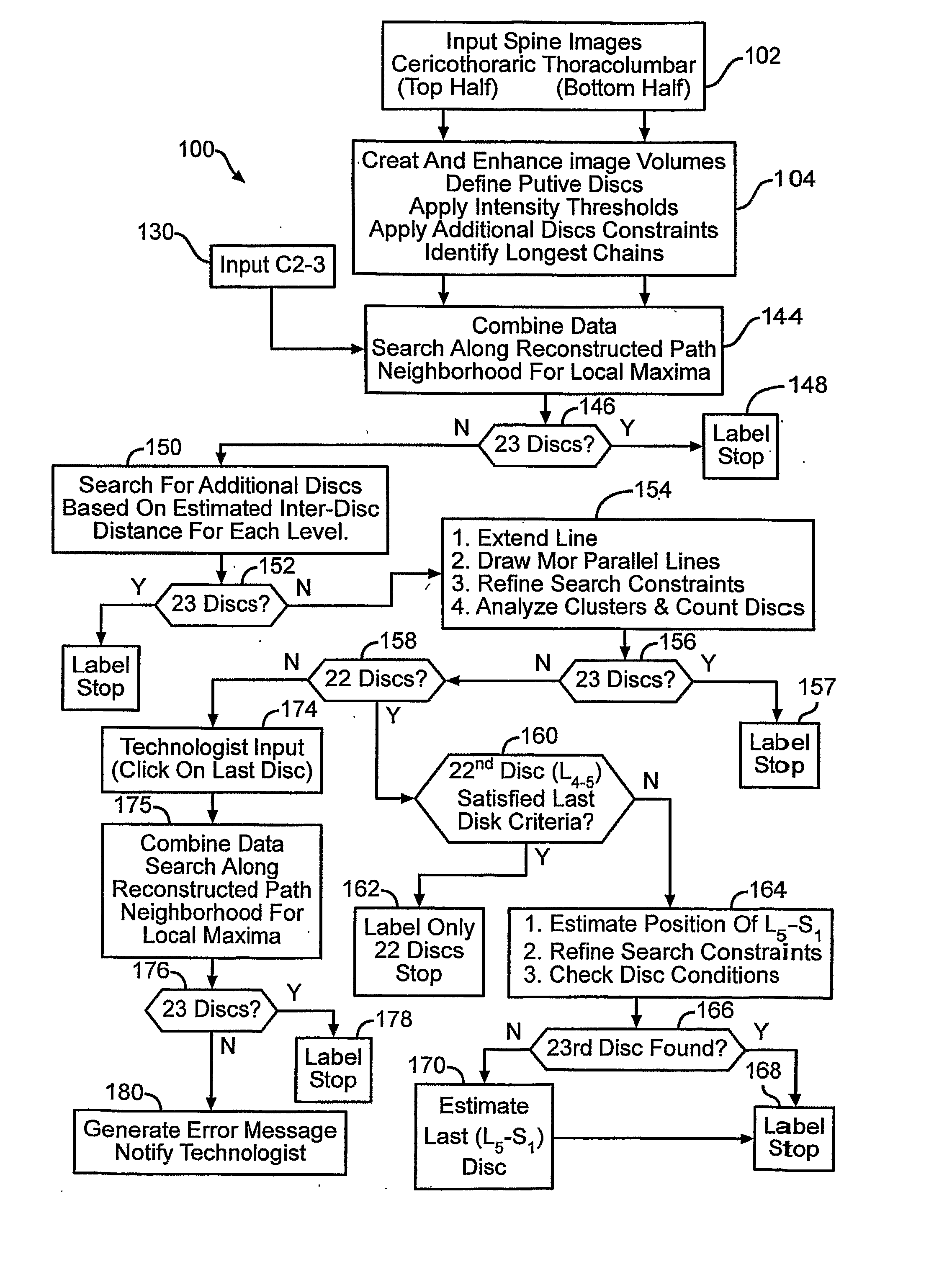
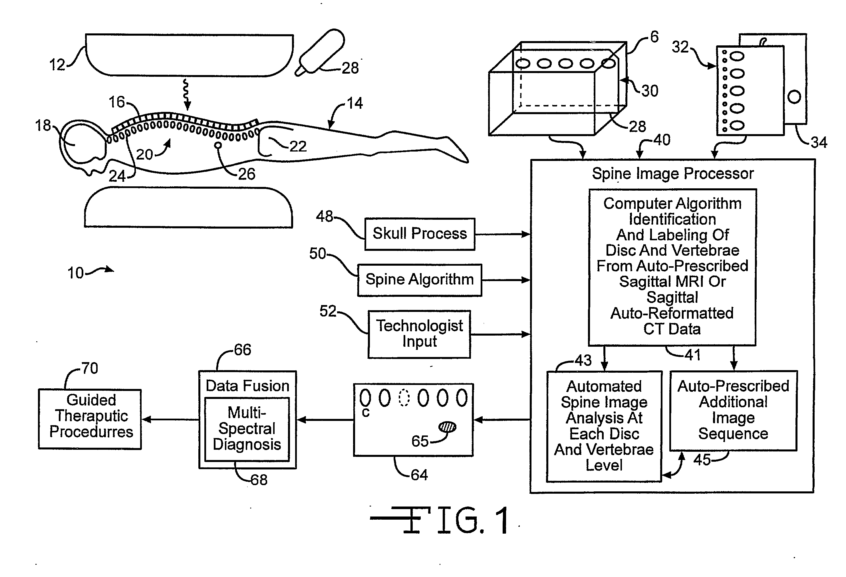
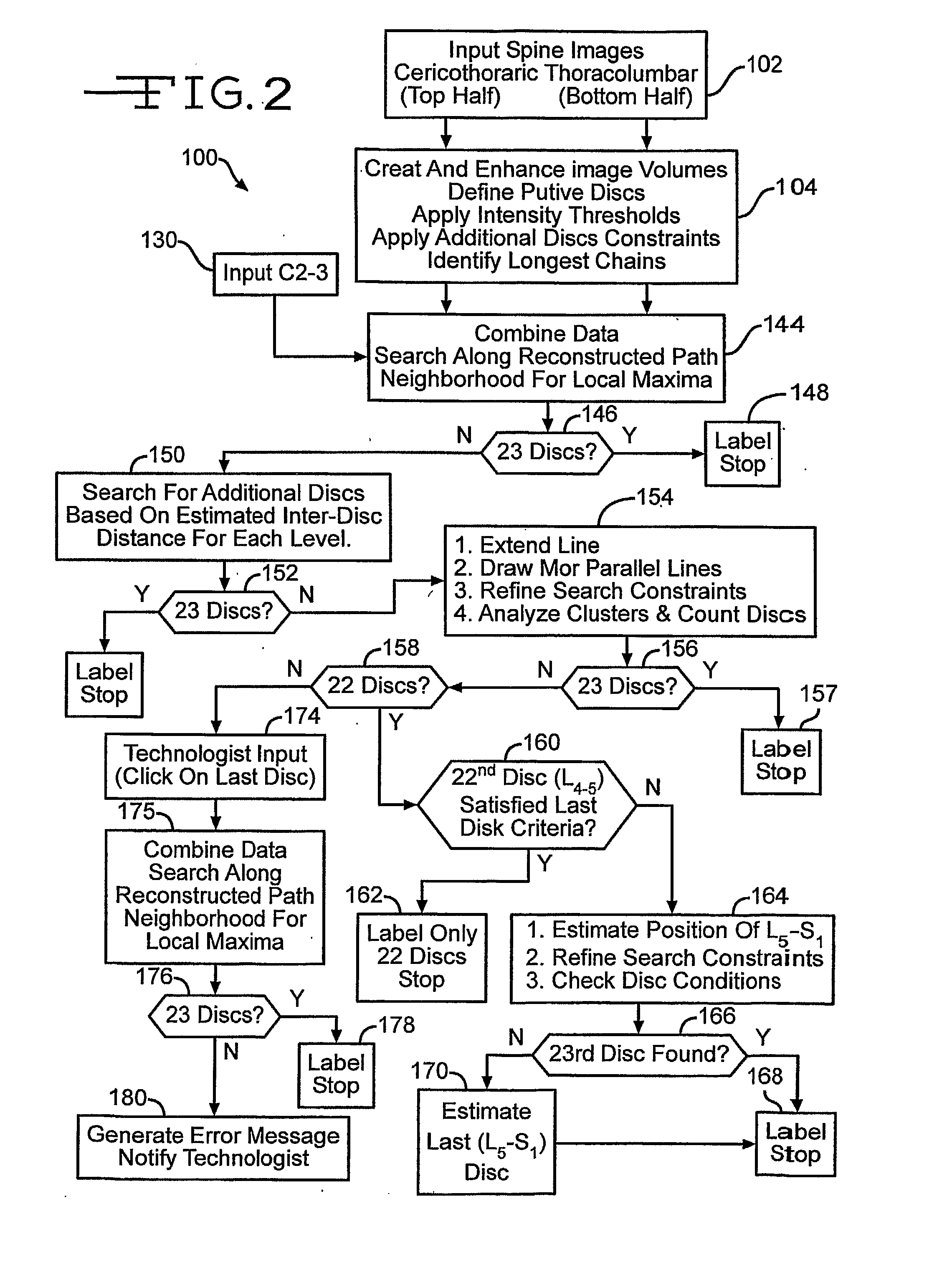
[0038] In FIG. 1, an automated spinal diagnostic system 10 includes a diagnostic imaging system 12 (e.g., MRI, CT) that is used to image a torso of a patient 14 that is advantageously covered by a skin / surface marking system 16 that serves as an integrated multimodality, multi-functional spatial reference. The diagnostic imaging system 12 may include scanning of the skull 18, the full spine 20, and pelvic bones 22. The diagnostic imaging system 12 serves as an automated MRI technique that rapidly surveys the entire spine providing accurate definitive numbering of all discs and vertebrae. In the particular illustrative version, the entire spine can be effectively surveyed with sub-millimeter in-plane resolution MRI in less than 1 minute. C-T-L vertebrae and discs can be readily identified and definitively numbered by visual inspection or semi-automated computer algorithm (“ASSIST”).

[0039] Correctly identifyi...


PUM


Abstract

Automated spine localizing, numbering and autoprescription system enhances correct location of diseased or injured tissue, even allow multi-spectral diagnosis. Externally located this tissue is facilitated by an integrated self adhesive spatial reference and skin marking system that is designed for a variety of modalities to include MRI, CT, SPECT, PET, planar nuclear imaging, radiography, XRT, thermography, optical imaging and 3D space tracking. The device ranges from a point localizer to a more multifunctional and complex grid / phantom system. The specially designed spatial reference(s) is affixed to an adhesive strip with corresponding markings so that after applying the unit to the skin / surface and imaging, the reference can be removed leaving the skin appropriately marked. The localizer itself can also directly adhere to the skin after being detached from the underlying strip. A spine autoprescription process performs image analysis that is able to identify vertebrae and discs even in the presence of abnormalities.

Description

CROSS REFERENCE TO RELATED APPLICATIONS [0001] The present application claims the benefit of the U.S. Provisional Patent Application Ser. No. 60 / 552,332, filed 11 Mar. 2004, the disclosure of which is hereby incorporated by reference in its entirety.FIELD OF THE INVENTION [0002] The present invention relates, in general, to medical diagnostic imaging devices that perform scout scans for localization and autoprescription. BACKGROUND OF THE INVENTION [0003] Diagnostic imaging of the spine of a patient is often useful for identifying disease or an injury to the spine itself or as a readily locatable landmark for other tissues. Unfortunately, human error may occur due to the variability in the patient population or due to an oversight. The mistake may arise in incorrectly labeling vertebrae and discs in a diagnostic image. The mistake may arise in incorrectly visually identifying the corresponding vertebrae under the skin before performing a surgical or therapeutic (e.g., radiation) tre...

Claims


Application Information

Patent Timeline
Patent Type & Authority Applications(United States)
IPC IPC(8): G06K9/00A61B5/05A61B5/103A61B5/117A61B6/00G06T5/00
CPCB60R25/00Y10T428/24G06T7/0012G06T7/0081G06T2207/10016G06T2207/10072G06T2207/20101G06T2207/20221G06T2207/30012G06T2207/30016H04L2012/40273A61B5/0033G06T2207/10088Y10T428/24744G06K2209/055G06T7/11A61B5/0036G06V2201/033G16H70/60G16H20/10G16H30/40G06T2207/10081G06T2207/30004A61B5/0042A61B5/0263
Inventor WEISS, KENNETH L.STORRS, JUDD M.
Owner ABSIST
Why Eureka
Try Eureka