Supercharge Your Innovation With Domain-Expert AI Agents!

Method to determine a background phase in phase image data sets

a phase image and data set technology, applied in the field of method to determine the background phase of the phase image data set, can solve the problems of no physical or mathematical basis for the use of such polynomials, phase changes that are incorrectly interpreted as temperature changes, and computation of the determination of the polynomial is computationally expensiv

Active Publication Date: 2011-07-21
SIEMENS HEALTHCARE GMBH
View PDF4 Cites 8 Cited by
  • Summary
  • Abstract
  • Description
  • Claims
  • Application Information

AI Technical Summary

Benefits of technology

[0009]An object of the present invention is to provide a method with which the background phase in phase values of an MR image data set can be determined in a simple manner without using reference data sets.
[0017]The iteration can be repeated until the difference in the phase values in the partial region between two iteration steps is smaller than a determined limit value, or when a specific number of iteration steps (that can depend on the size of the partial region, for example) has been implemented. This described iteration has the advantage that it converges for all cases, meaning that the background phases in the partial region converge into a spatial phase curve that corresponds to the MR system-dependent background phase.
[0018]An imaging sequence that is sensitive to local magnetic field differences is advantageously used to create the phase image data set. For example, a fat-suppressed, two-dimensional gradient echo sequence can be used, wherein the fat suppression prevents interfering signal portions of fat and therefore prevents larger susceptibility differences in the pixels.

Problems solved by technology

These methods operating with reference data sets have the disadvantage that movements of the examination subject during the acquisition of the two data sets or other external interferences lead to phase changes that are incorrectly interpreted as temperature changes.
These methods have the disadvantage that information about how the MR system-dependent background phase changes spatially over the image must be present.
However, there is no physical or mathematical basis for the use of such polynomials.
The determination of the polynomial is likewise computationally expensive and can lead to problems in the calculation.
Moreover, the spectral line of the adipose signal is subject to changes from one location to another, which leads to different phase values over the different resonance frequencies, whereby problems occur in the determination of the background phase with the aid of the adipose signal portions.

Method used


Image

  • Method to determine a background phase in phase image data sets
  • Method to determine a background phase in phase image data sets
  • Method to determine a background phase in phase image data sets

Examples


Embodiment Construction

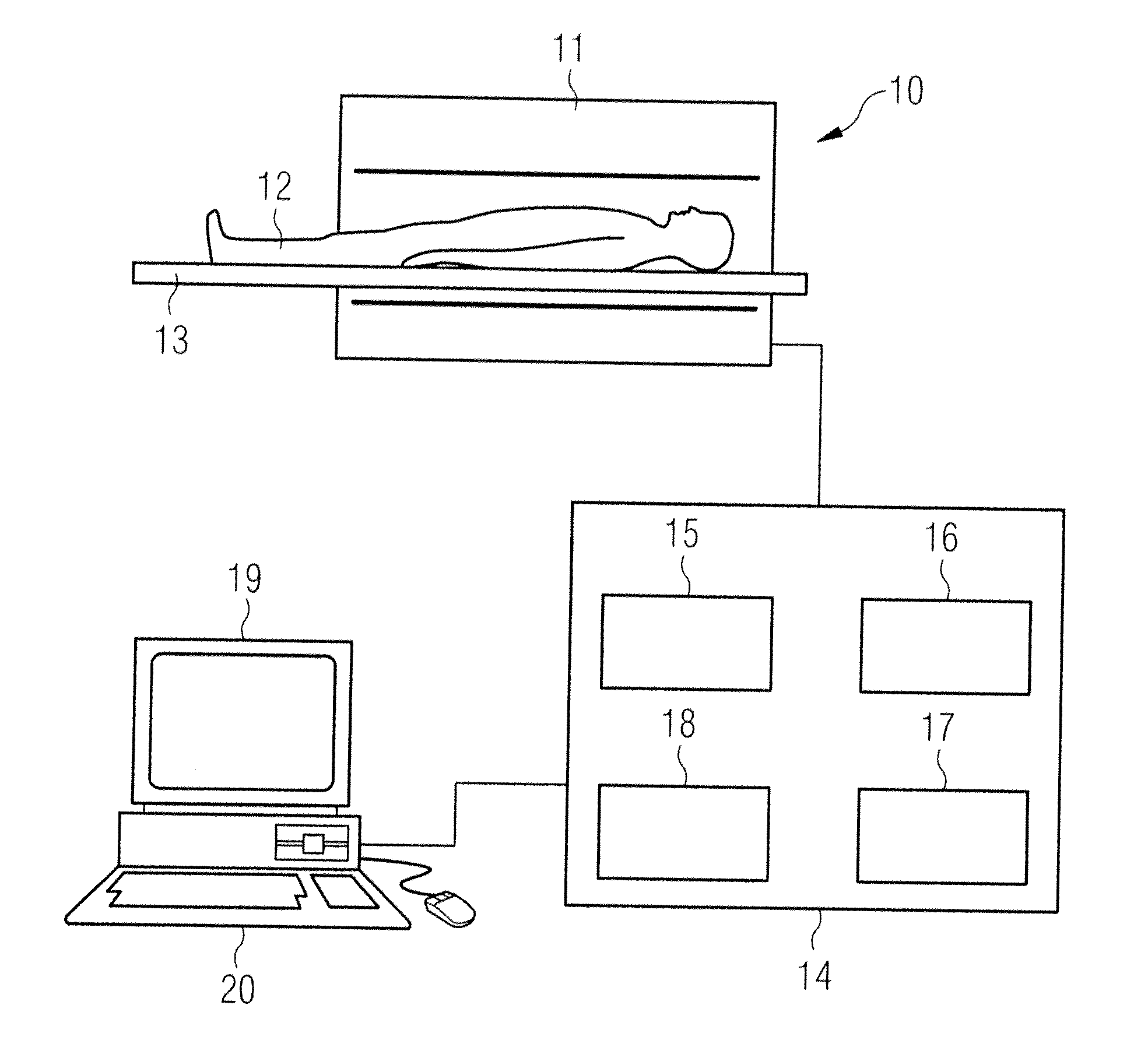
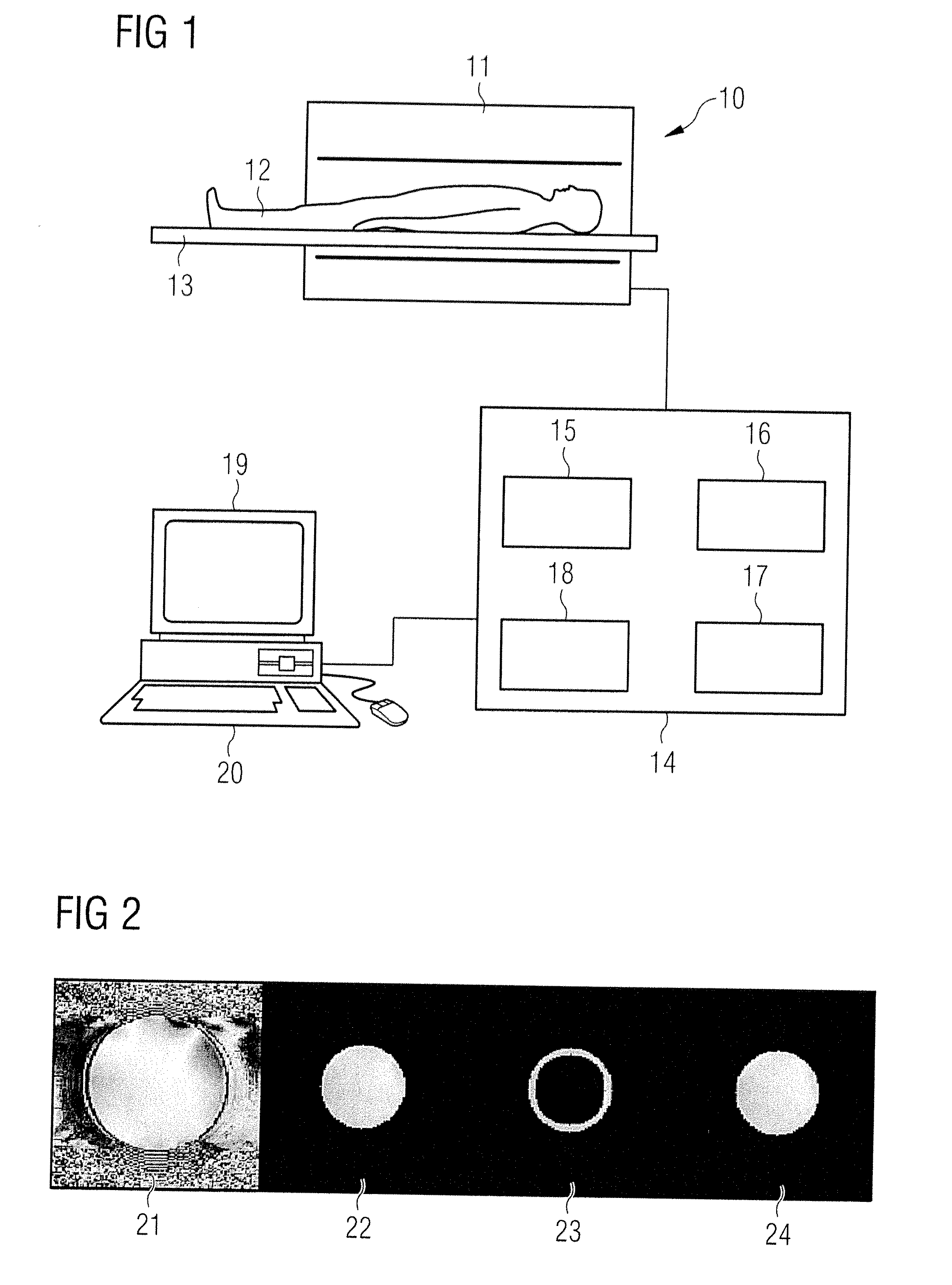
[0035]An MR system with which system-dependent phase information or, respectively, the background phase can be determined is schematically shown in FIG. 1. The MR system has a basic field magnet 11 to generate a B0 field into which an examined person 12 (arranged on a bed 13) can be inserted. The shown MR system can be used, for example, in combination with a thermotherapy in which individual regions of the examined body are heated (with ultrasound, for example) in order to destroy localized tumor tissue in a heated region. The temperature development in the shown tissue can be non-invasively checked in multiple dimensions with the acquisition of MR phase images of a gradient echo sequence and the presentation of phase images. The MR system has a central control unit 14 with which the control of the MR system is possible. Since the fundamental mode of operation to generate MR images is known to the man skilled in the art, only a few system components are schematically dealt with in ...


PUM


Abstract

In a method to determine a background phase in phase values of a phase image data set that is acquired from an examination subject, wherein the background phase is determined in a partial region of the phase image data set, the phase image data set of the examination subject is acquired, and a substantially closed, planar contour is established in the phase image data set around the partial region, the planar contour having a contour area with a width of at least one pixel of the phase image data set. The phase values in the partial region are determined with the assumption that the spatial curve of the background phase is a harmonic or quasi-harmonic function, the phase values of the pixels in the partial region being determined based on the phase values in the planar contour.

Description

BACKGROUND OF THE INVENTION[0001]1. Field of the Invention[0002]The present invention concerns a method to determine a background phase in phase values of a phase image data set and a device to determine the background phase.[0003]2. Description of the Prior Art[0004]The phase information of the magnetic resonance signal that describes the position of the magnetization perpendicular to the B0 field direction can be used in many regards in magnetic resonance tomography (MRT). For example, the phase information contained in the MR signal can be used to separate adipose and aqueous tissue, for flow measurement, in susceptibility-weighted MRT and to determine temperature. In a method known as thermotherapy the temperature in tumor cells is specifically increased in order to kill the tumor cells or to make these more sensitive to accompanying therapy measures. For example, the tissue heating can hereby ensue via focused ultrasound or with the aid of lasers. A temperature monitoring of th...

Claims


Application Information

Patent Timeline
IPC IPC(8): G06K9/00
CPCG01R33/56509G01R33/4804
Inventor SALOMIR, RARESVIALLON, MAGALIEROLAND, JOERGGROSS, PATRICK
Owner SIEMENS HEALTHCARE GMBH
Features
  • R&D
  • Intellectual Property
  • Life Sciences
  • Materials
  • Tech Scout
  • Unparalleled Data Quality
  • Higher Quality Content
  • 60% Fewer Hallucinations
Learn More