Spectral contrast for glaucoma imaging

a glaucoma and glaucoma imaging technology, applied in the field of spectral contrast for glaucoma imaging, can solve the problems of optic nerve fiber microscopic damage that is difficult to detect in vivo, cannot be restored, and cannot be seen

Active Publication Date: 2011-09-22
UNIV OF MIAMI
View PDF4 Cites 19 Cited by
  • Summary
  • Abstract
  • Description
  • Claims
  • Application Information

AI Technical Summary

Problems solved by technology

Untreated glaucoma may lead to permanent damage of the optic nerve resulting in vision loss, which can progress to blindness.
Once vision has been lost due to glaucoma, it can never be restored.
However, early, microscopic damage to the optic nerve fibers is difficult to detect in vivo.
Damage to the RNFL is usually only readily detectable after vision loss has already occurred.
However, there is no set threshold of intraocular pressure that causes glaucoma.
Detection of glaucoma through direct measurement of intraocular pressure is therefore not reliable.
However, as explained above, measuring the IOP alone is an inaccurate indicator of early onset glaucoma.
Other eye exams that measure changes in size or shape of the eye, anterior chamber angle, or include visible examination of the optic nerve using a slit-lamp microscope, also lack the precision to determine microcellular damage to the RNFL.
But none of these techniques can accurately detect early microscopic signs of RNFL damage.
In advanced glaucomatous disease, the RNFL decreases in thickness due to optic neuropathy and becomes difficult to visually discern from the surrounding tissue.
This makes segmentation difficult and imprecise.
Such segmentation of the RNFL may be difficult due to the varying of thickness within the RNFL.
This can cause variable intensity contrast between tissue layers and thus additional difficulties in segmentation.
This also causes reduced signal strength in nasal retina.
Because segmentation is based solely on intensity, differences in the RNFL become difficult to discern when the RNFL becomes thin Segmentation algorithms will frequently follow retinal tissue boundaries to produce a layer identified as RNFL.
However, these algorithms break down and become inaccurate when applied to retinas having damaged RNFLs.
This leads to inaccuracies and makes identifying damaged tissue unreliable.

Method used


Image

  • Spectral contrast for glaucoma imaging
  • Spectral contrast for glaucoma imaging
  • Spectral contrast for glaucoma imaging

Examples


Embodiment Construction

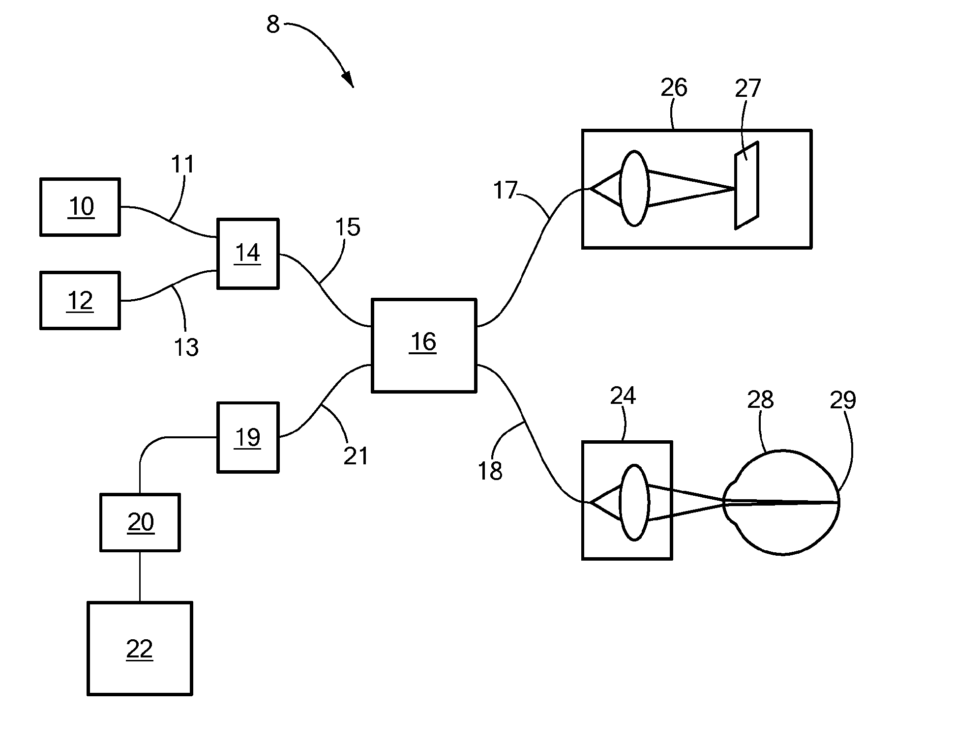
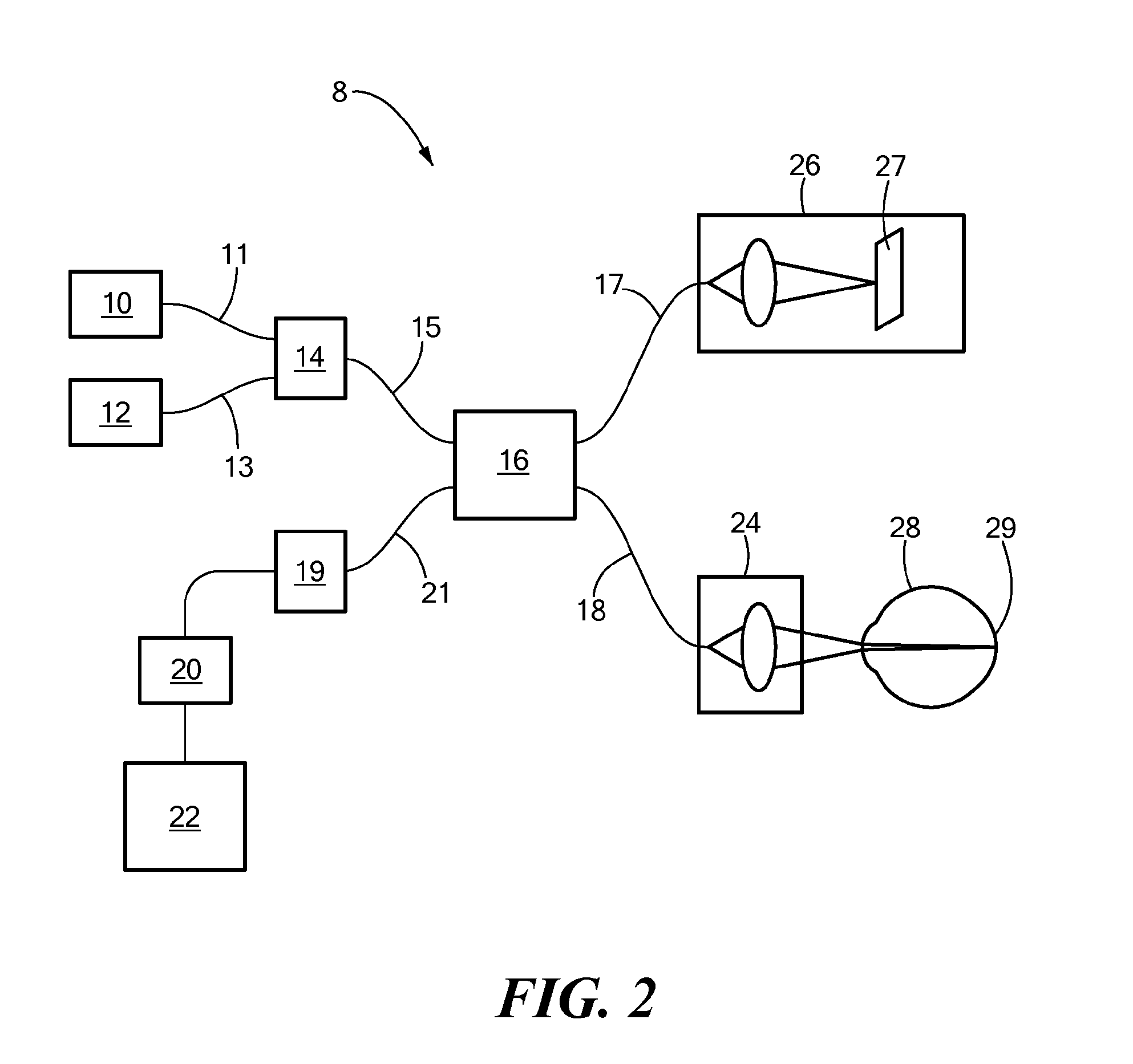
[0020]The present invention advantageously provides a method and system for early stage detection of glaucoma comprising a dual-band optical coherence tomography (OCT) system that utilizes both short and long wavelengths of light to detect early damage to the RNFL. The method also includes an OCT wavelength contrast method to discriminate between the RNFL and surrounding tissue, which improves the detection of RNFL tissue loss in advanced glaucoma disease.

[0021]The present invention also provides for a method of measuring retinal tissue loss resulting from glaucoma by calculating the spectral reflectance from a dual-band OCT. The measured spectral reflectance data may then be correlated to reveal tissue loss resulting from glaucomatous optic neuropathy.

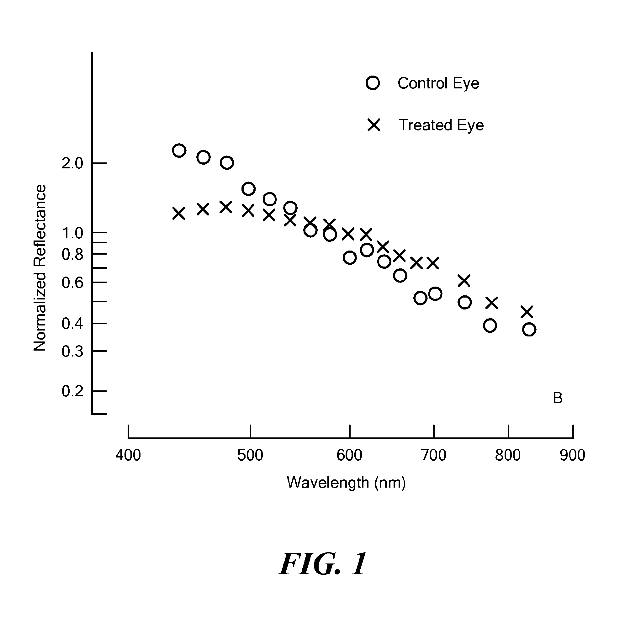
[0022]The reflective properties of the RNFL differ from those of surrounding tissue. Specifically, a healthy RNFL exhibits relatively high reflectance of visible light. This increased reflectance by the RNFL of visible light, ranging ...


PUM


Abstract

A system for analyzing and detecting early stage damage to the retina related to glaucoma. The reflectance of different wavelengths of light by the retinal nerve fiber layer are compared. Changes in relative reflectance values indicate damage to the retinal nerve fibers and indicate early glaucomatous optical neuropathy.

Description

FIELD OF THE INVENTION[0001]The present invention relates to a method and system for contrasting the spectral reflectance of the retinal nerve fiber layer (RNFL) using optical coherence tomography (OCT).BACKGROUND OF THE INVENTION[0002]The optic nerve is a bundle of over a million nerve fibers that connect the retina to the brain. These nerve fibers are formed by the axons of retinal ganglion cells that extend from the retina where they detect light, to the brain where they transmit visual information. Glaucoma is a class of ocular diseases resulting from damage to the optic nerve. According to the National Eye Institute, among the United States population 40 years and older there are over 2.2 million glaucoma patients (290,000 between ages 40-49). Owing to the rapid aging of the United States population, this number may increase to more than 3 million by 2020.[0003]Glaucoma occurs when the retinal ganglion cells disposed in the optic nerve degenerate in a characteristic and identif...

Claims


Application Information

Patent Timeline
Patent Type & Authority Applications(United States)
IPC IPC(8): A61B3/12A61B3/10A61B3/14
CPCA61B3/12A61B3/102
Inventor JIAO, SHULIANGHUANG, XIANGRUNKNIGHTON, ROBERT W.
Owner UNIV OF MIAMI
Why Eureka
Try Eureka