MicroRNAa (miRNA) AS BIOMARKERS FOR DIAGNOSING DIFFERENT GRADES OF GLIOMAS AND PATHWAYS OF GLIOMA PROGRESSION

a technology of glioma progression and micrornas, which is applied in the field of micrornas (mirna) as biomarkers for detecting glioma progression pathways, can solve the problems of chemotherapy, insufficient understanding of astrocytoma development, and insufficient prognosis of gbm patients, etc., to achieve accurate classification of different grades of glioma

Inactive Publication Date: 2011-10-20
DEPT OF BIOTECHNOLOGY MINIST OF SCI & TECH GOVERNMENT OF INDIA +1
View PDF0 Cites 6 Cited by
  • Summary
  • Abstract
  • Description
  • Claims
  • Application Information

AI Technical Summary

Benefits of technology

[0015]Another object is to develop a process which

Problems solved by technology

Current treatments, including surgery, radiation therapy, and chemotherapy, unfortunately have not changed the natural history of these incurable neoplasms; and the prognosis of patients with GBMs has not improved significantly in the past 30 years (Davis et al., 1998).
Despite all this information about astrocytoma, our understanding of astrocytoma development is not sufficient enough to improve prognosis for GBM patients.
But this has serious limitations, because tumors with similar histopathological appearance can follow significantly different clinical courses and show different responses to therapy.
However, differential protein expression (GFAP, vimentin, synaptophysin, nestin) has not helped to improve therapeutic approaches.
Predi

Method used


Image

  • MicroRNAa (miRNA) AS BIOMARKERS FOR DIAGNOSING DIFFERENT GRADES OF GLIOMAS AND PATHWAYS OF GLIOMA PROGRESSION
  • MicroRNAa (miRNA) AS BIOMARKERS FOR DIAGNOSING DIFFERENT GRADES OF GLIOMAS AND PATHWAYS OF GLIOMA PROGRESSION
  • MicroRNAa (miRNA) AS BIOMARKERS FOR DIAGNOSING DIFFERENT GRADES OF GLIOMAS AND PATHWAYS OF GLIOMA PROGRESSION

Examples


example 1

[0066]Tissue Collection

[0067]Tumor samples were collected from patients who were operated at National Institute of Mental Health and Neurosciences and. Sri Satya Sai Institute of Higher Medical Sciences, Bangalore, India. A portion of the non-dominant anterior temporal cortex resected during surgery for intractable epilepsy served as normal control brain sample. A total of 73 samples of malignant astrocytoma and control brain tissue were used in this study. For microarray hybridization, a set of 39 samples of malignant astrocytoma comprising 13 AA, 13 sGBM and 13 pGBM tissues and 7 controls were used. For subsequent real time RT-PCR validation of selected miRNAs, we used an independent set of 24 samples of GBM and 3 controls. Tissues were bisected, and one half was snap-frozen in liquid nitrogen and stored at −8020 C. until RNA isolation. The other half was fixed in formalin and processed for paraffin sections. These were used for histopathologic grading of astrocytoma and immunohis...

example 2

[0068]Total RNA was extracted from the frozen tissue or cell lines by using the TRI reagent (Sigma) as per the manufacturer's instructions. The RNA samples were quantified by measuring the absorbance using a spectrophotometer and visualized on a MOPS-formaldehyde gel for quality assurance. For the RNA samples used in microarray experiment, the quality and quantity was verified by using Bioanalyzer 2100 (Agilent).

[0069]MicroRNA Array Profiling

[0070]One μg total RNA from sample and reference were labeled with Hy3™ and Hy5™ fluorescent label, respectively, using the miRCURY™ LNA Array power labeling kit (Exiqon, Denmark) following the procedure described by the manufacturer. The Hy3™-labeled samples and a Hy5™-labeled reference RNA sample were mixed pair-wise and hybridized to the miRCURY™ LNA array version 10.0 (Exiqon, Denmark), which contains capture probes targeting all miRNAs for all species registered in the miRBASE version 10.0 at the Sanger Institute. The hybridization was perf...

example 3

[0071]Data analysis

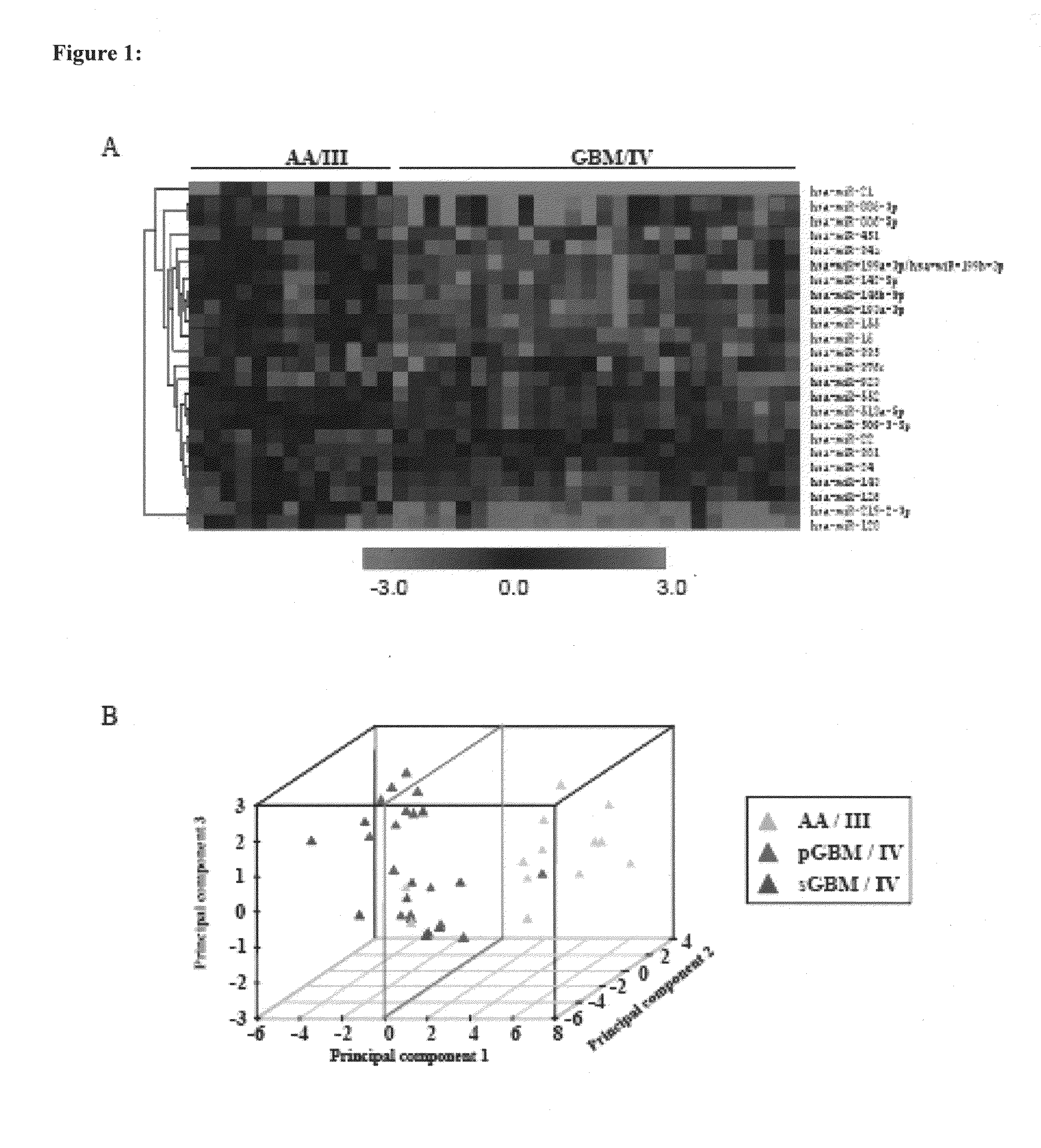
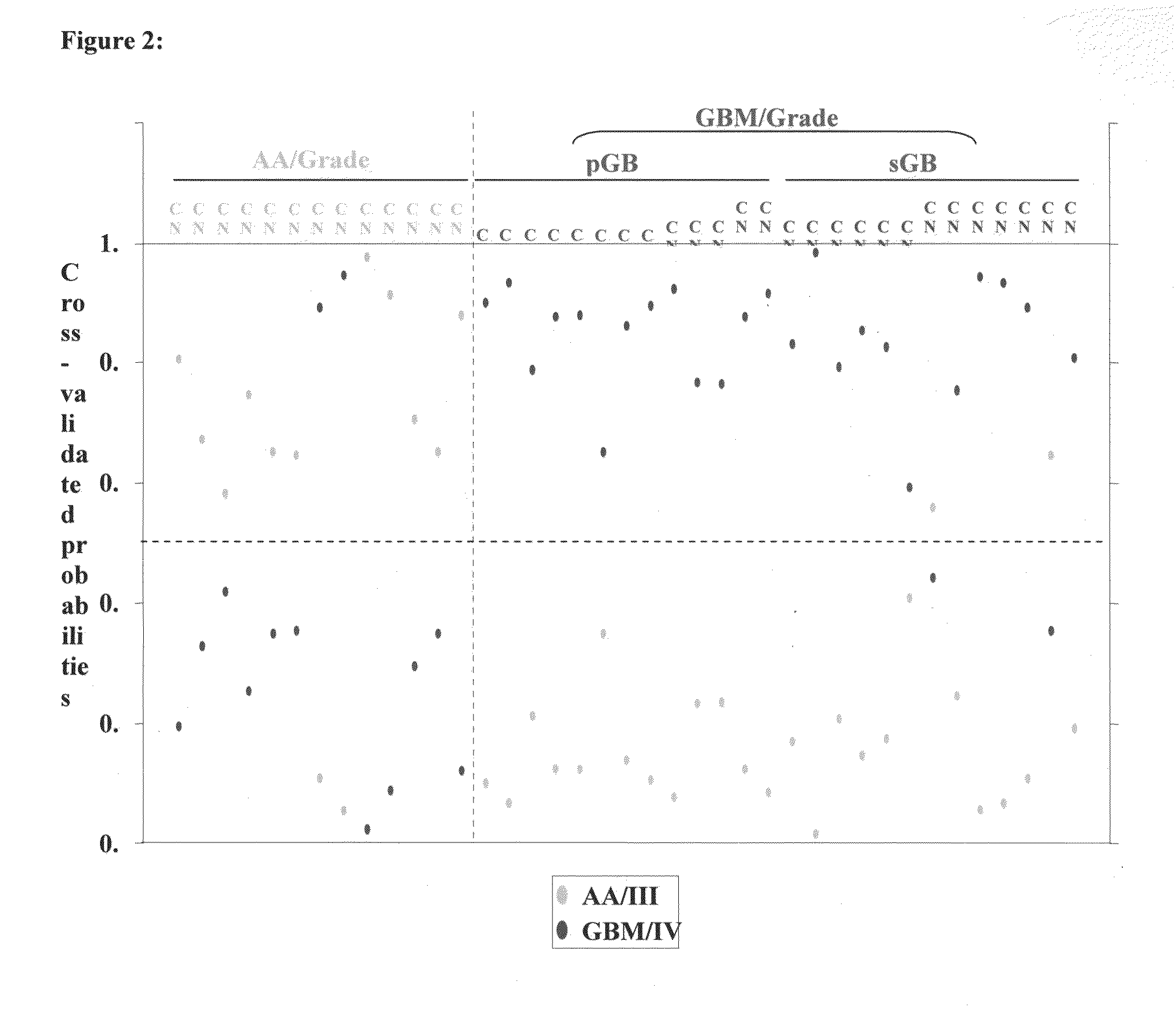
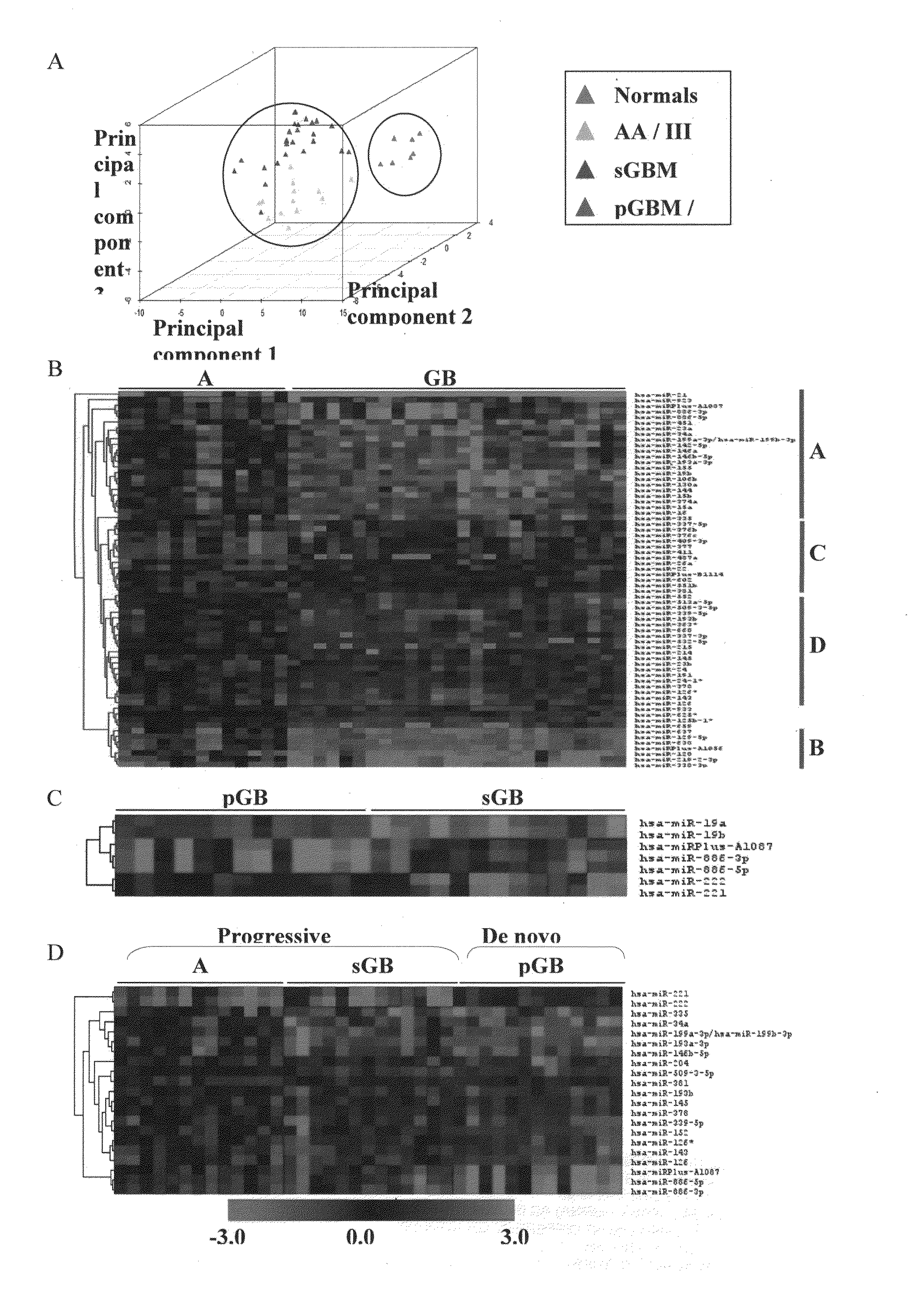
[0072]The median ratio of Hy3 / Hy5 intensity for replicative spots for each miRNA was obtained after normalization. The ratio Hy3 / Hy5 values were log2 transformed. Difference in log2 ratio for each miRNA is obtained by subtracting the log2 ratio of a given sample from the average log2 ratio of normals. The difference in log2 ratio was considered in subsequent analyses. We have analyzed 7 normals, 13 AA (Grade III), 13 primary GBM (Grade IV) and 13 secondary GBM (Grade IV) by microarray hybridization. The miRNAs having values in at least 70% of the samples in each group were considered for further analysis. To find the significantly differentially regulated miRNAs between normal brain and astrocytoma and between the groups of malignant astrocytoma, data were analyzed by Significance Analysis of Microarrays (SAM). In SAM analysis, P values were obtained from permutation tests (1000 permutations in each analysis). The significant genes that were identified by SAM were...


PUM

PropertyMeasurementUnit
Fractionaaaaaaaaaa
Fractionaaaaaaaaaa
Fractionaaaaaaaaaa

Abstract

This invention relates to a method for detecting, (i) malignant astrocytoma from normal brain tissue (ii) glioblastoma and anaplastic astrocytoma (iii) primary glioblastoma and secondary glioblastoma (iv) progressive pathway and denovo pathway comprises determining the level of expression of miRNAs listed in table 2, 3, 4, 5, wherein a higher or lower level of expression of miRNAs in the test sample as compared to the control sample differentiates and kit for characterizing a) malignant astrocytoma from normal brain tissue cell comprising reagent capable of specifically detecting the level of expression of the genes of miRNAs and instructions for using said kit for characterizing malignant astrocyoma from normal brain tissue cells b) glioblastoma from anaplastic astrocytoma comprising reagent capable of specifically detecting the level of expression of the genes of miRNAs and instructions for using said kit for characterizing glioblastoma from anaplastic astrocyoma c) primary glioblastoma from secondary glioblastoma comprising reagent capable of specifically detecting the level of expression of the genes of miRNAs and instructions for using said kit for characterizing primary glioblastoma from secondary glioldastoma, d) progressive pathway from denovo pathway comprising reagent capable of specifically detecting the level of expression of the genes of miRNAs and instructions for using said kit for characterizing progressive pathway from denovo pathway.

Description

FIELD OF INVENTION[0001]This invention relates to a MicroRNAs (miRNA) as biomarkers for detecting different grades of gliomas and pathways of glioma progression.BACKGROUND OF INVENTION[0002]Gliomas are the most common primary brain tumors and occur at an incidence of almost 12 per 100,000 people (Landis et al., 1999). Diffuse astrocytoma may be classified (as per WHO classification) as lower-grade diffuse (DA; Grade II), anaplastic (AA; Grade III) and glioblastoma (Grade IV; GBM), in the order of increasing malignancy (Mischel et al., 2001). Currently, these classifications are based on the observed histopathological characteristics of the tumor, which are sometimes subjective and inconsistent. GBM constitutes more than 80% of malignant gliomas (DeAngelis et al., 2001) and patients with GBM have a median survival of less than one year. Current treatments, including surgery, radiation therapy, and chemotherapy, unfortunately have not changed the natural history of these incurable neo...

Claims


Application Information

Patent Timeline
IPC IPC(8): C12Q1/68
CPCC12Q1/6886C12Q2600/178C12Q2600/158C12Q2600/112
Inventor SOMASUNDARAM, KUMARAVELRAO, SOUMYA ALIGA MAHABALASANTOSH, VANI
Owner DEPT OF BIOTECHNOLOGY MINIST OF SCI & TECH GOVERNMENT OF INDIA
Why Eureka
Try Eureka