Non-invasive detection of complement-mediated inflammation using cr2-targeted nanoparticles

a technology of complement-mediated inflammation and nanoparticles, which is applied in the field of non-invasive detection of complement-mediated inflammation using cr2-targeted nanoparticles, can solve the problems of limited risk-freeness, tissue destruction, and cell lysis of target structures of complement-mediated cells,

Active Publication Date: 2011-11-24
UNIV OF COLORADO THE REGENTS OF
View PDF3 Cites 19 Cited by
  • Summary
  • Abstract
  • Description
  • Claims
  • Application Information

AI Technical Summary

Benefits of technology

[0011]In one aspect, provided herein are methods of detecting complement-mediated inflammation in an individual comprising: (a) administering to the individual an effective amount a composition comprising an effective amount of CR2-targeted ultrasmall superparamagnetic iron oxide (“USPIO”) nanoparticles or aggregates thereof; and (b) taking a magnetic resonance image of the individual. In certain embodiments, the CR2-targeted USPIO nanoparticles comprise a targeting moiety comprising an antibody or fragment thereof directed to C3 or a portion thereof, for example but not limited to C3b, iC3b, C3dg, C3d and the like. In some embodiments, the antibody is a polyclonal antibody. In some embodiments, the antibody is a monoclonal antibody. In some embodiments, the antibody is a monoclonal antibody or fragment thereof directed to C3d. In certain embodiments, the individual is a mammal. In certain embodiments, the mammal is a human, a mouse, or a rat. In certain embodiments, the composition is administered by injection. In certain embodiments, the injection is parenteral, intravenous, subcutaneous, or intramuscular.

Problems solved by technology

Inappropriate complement activation and its deposition on host cells can lead to complement-mediated cell lysis of target structures, as well as tissue destruction due to the generation of powerful mediators of inflammation.
While biopsies are in many respects routine, they have their limitations and are not risk-free.
Because commonly used needle or punch biopsies sample only a small portion of the target organ, there is a risk of sample error leading to an incorrect diagnosis.
Furthermore, although biopsy is a generally safe procedure, major complications such as internal bleeding may occur in a significant number of cases.
Although renal biopsy is generally a safe procedure, complications may occur in 6% or more of biopsies and intra-renal bleeding and hematuria are common.
Patients requiring repeat biopsies are at concomitantly greater risk of complications.

Method used


Image

  • Non-invasive detection of complement-mediated inflammation using cr2-targeted nanoparticles
  • Non-invasive detection of complement-mediated inflammation using cr2-targeted nanoparticles
  • Non-invasive detection of complement-mediated inflammation using cr2-targeted nanoparticles

Examples


example 1

Non-Invasive Detection of Alternative Complement-Mediated Inflammation in MRL / lpr Mice, an Animal Model of Lupus Nephritis Associated with Systemic Lupus Erythematosus

Materials and Methods

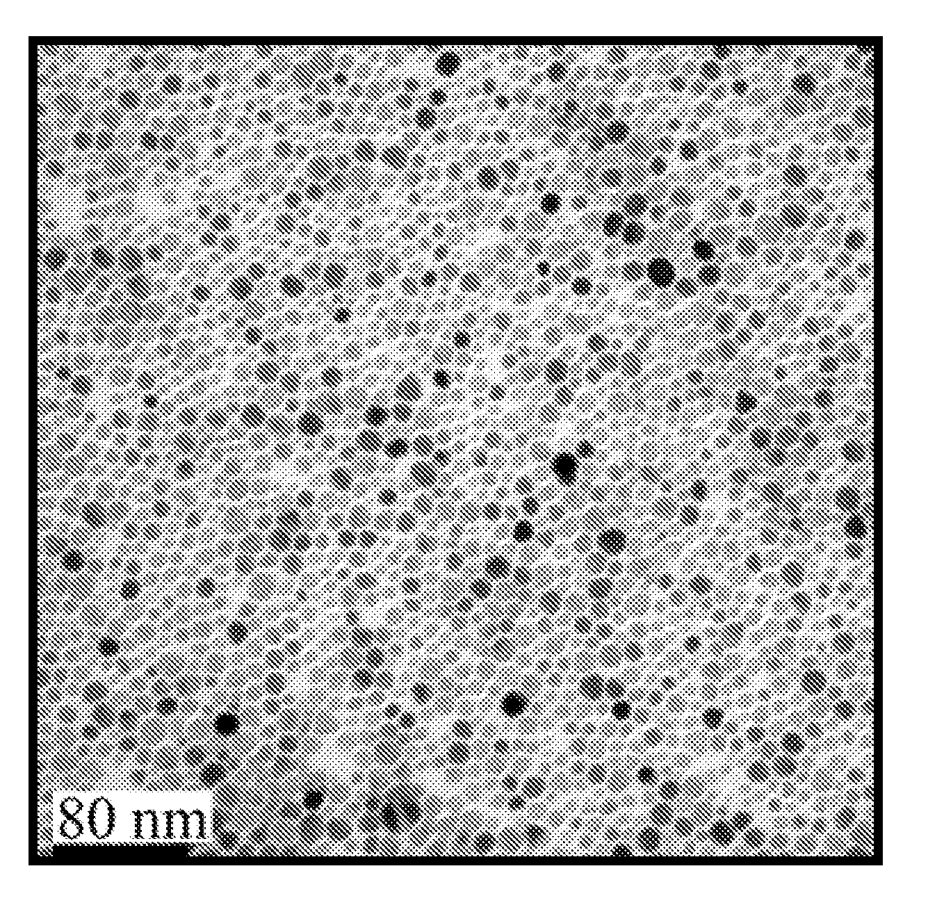
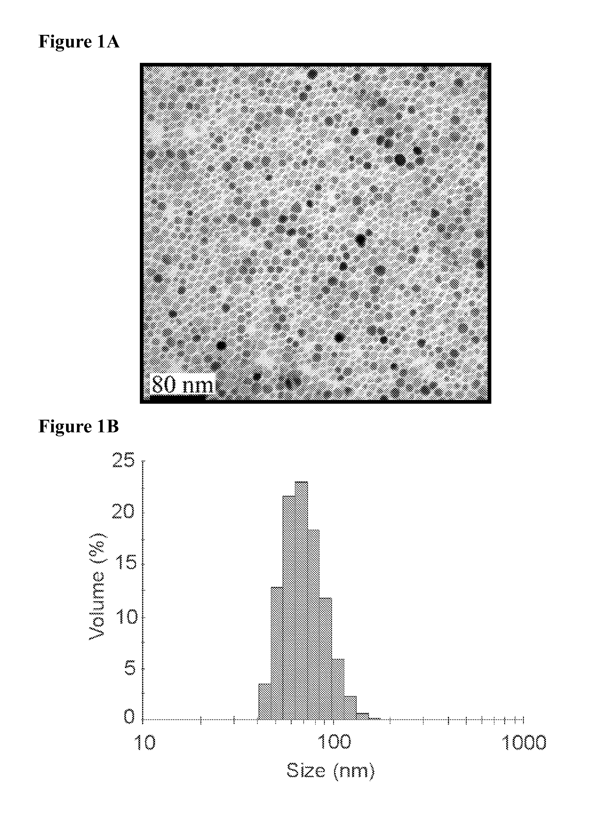
[0234]Synthesis of iron oxide nanoparticles. Ultrasmall superparamagnetic iron oxide nanoparticles were generated and functionalized for conjugation to proteins as previously described. See e.g., A. J. Barker et al., 2005, J. Appl. Physics 98:063528; B. A. Larsen et al., 2008, Nanotechnol. 19:265102. Briefly, USPIO were synthesized by a solvothermal method using an Iron (III) Acetylacetonate precursor with trioctylamine and heptanoic acid (Sigma-Aldrich, St. Louis, Mo.) as surfactants, yielding ˜10 nm magnetite nanoparticles (FIG. 1A) with a hydrophobic heptanoic acid surface termination. The as-synthesized USPIO nanoparticles were resuspended in tetrahydrofuran (THF) and titrated with a 1% (v / v) solution of acetic acid until the desired level of aggregation (˜75 nm) was reached. The acetic acid pa...


PUM

PropertyMeasurementUnit
diameteraaaaaaaaaa
diameteraaaaaaaaaa
diameteraaaaaaaaaa

Abstract

Methods of non-invasive imaging of complement-mediated inflammation are provided. Compositions including CR-targeted ultrasmall superparamagnetic nanoparticles or aggregates thereof for use with those methods are also provided.

Description

CROSS-REFERENCES TO RELATED APPLICATIONS[0001]This application is a National Stage filing of International Application No. PCT / US2010 / 023201, filed Feb. 4, 2010, and claims the benefit of U.S. Provisional Application No. 61 / 149,975, filed Feb. 4, 2009, each of which is incorporated herein by reference in its entirety and for all purposes.STATEMENT AS TO RIGHTS TO INVENTIONS MADE UNDER FEDERALLY SPONSORED RESEARCH OR DEVELOPMENT[0002]This invention was made with government support under grant number R01 CA53615 awarded by the National Institutes of Health. The Government has certain rights in the invention.BACKGROUND OF THE INVENTION[0003]This application pertains to methods and compositions for non-invasive detection of complement-mediated inflammation using CR2-targeted nanoparticles.[0004]Complement is the collective term for a series of blood proteins that constitute a major effector mechanism of the immune system. Complement plays an important role in the pathology of many autoi...

Claims


Application Information

Patent Timeline
Patent Type & Authority Applications(United States)
IPC IPC(8): A61K49/16A61P37/00A61K49/08
CPCA61K49/1812A61K49/1863A61K49/1866A61K49/16A61K49/1887B82Y5/00A61K49/1875A61P37/00
Inventor THURMAN, JOSHUA M.SERKOVA, NATALIESTOLDT, CONRADLARSEN, BRIAN
Owner UNIV OF COLORADO THE REGENTS OF
Why Eureka
Try Eureka